Rheumatology Flashcards
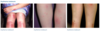
What are the signs of synovitis?
Boggy swelling, warm and erythema of joint with associate effusions, pain and stiffness Often relieved by activity and NSAIDs
Give examples of an enthesitis
- inflammation of tendon and ligament attachment - plantar faciitis - achilles tenditis - costochondritis
Inflammatory rheumatological pain vs non-inflammatory
- inflammatory pain often relieved by exercise and worse at rest - non inflammatory pain generally exacerbated by activity and relieved at rest
Describe the etg suggested pattern of recognition of articular MSK presentations
- Inflammatory vs non-inflammatory 2. Acute vs chronic 3. Mono/oligo/polyarticular
Define mono/oligo and polyarticular
Mono = 1 Oligo = up to 5 Poly = >5
Give examples of acute INFLAMMATORY articular pain Mono (4) Oligo (5) Poly (5)
Mono: gout, CPPD, reactive arthritis, septic arthritis Oligo: gout, reactive arthritis, enteropathic arthritis, psoriatic arthritis, rhematoid arthritis Poly: viral arthritis, rheumatoid arthritis, gout, drug reaction, reactive arthritis
What is the other name for pseudogout?
Calcium pyrophosphate dihydrate crystal deposition disease
Give examples of chronic INFLAMMATORY articular pain Mono (3) Oligo (4) Poly (6)
Mono: septic arthritis with atypical pathogen, psoriatic arthritis, reactive arthritis Oligo: psoriatic arthritis, reactive arthritis, gout, CPPD Poly: gout, psoriatic arthritis, reactive arthritis, rheumatoid arthritis, SLE, CPPD
What is the classical non-inflammatory arthritis?`
OA, can be mono/oligo/poly
Patern of recoginition of articular MSK presentations. Etg flowchart
Pattern of recognition of peri-articular MKS presentation, ETG flow chart.
How would you classify periarticular MSK presentations?
- Inflammatory vs non-inflammatory
- Acute vs chronic
- Focal vs diffuse
Give examples of ACUTE and CHRONIC inflammatory peri-articular conditions.
Focal: calcific tendinitis of shoulder, de Quervain tenosynovitis, flexor tendosynovitis, preptaella bursitis, plantar fasciitis
Diffuse: PMR (shoulder and hip involvement)
What are the specific investigations for the following…
a) septic arthritis
b) gout/CPPD
c) inflammatory myopathies/giant cell arteritis
a) gram stain and culture of synovial fluid
b) polarised light microscopy of synovial fluid
c) tissue biopsy
What rate of patients with SLE have positive ANA?
90%
But also 5% of the healthy population
What antibodies would you test for in suspected Sjogren’s?
Ro (SS-a) and or La (SS-B) antibodies on enzyme link immunosobrent assay
Anti-ENA antobodies
What are the differences in ESR and CRP?
Both are useful indicators of acute phase response
- ESR is affected by age and hyperviscosity, unlike CRP
- CPR also responds more rapidly than ESR
However a normal ESR/CRP does not exclude inflammatory pathology
What findings on iron studies would suggest anaemia of chronic disease?
- mild normochromic, normocytic anaemia with raised ferritin and low rion
Testing for ANA
DO: young women with intermittent small joint arthralgia, alopecia, paynaud or serositis
DON’T: in patients with non specific long standing history of fatigue and constant pain
Note the higher the ANA titre the higher the likelihood of connective tissue or autoimmune disease (i.e thyroid)
Outline murtagph’s diagnostic model
- What is the probability diagnosis
- What are the serious disorders not to be missed
- WHat are the pitfalls?
- Seven masquerades checklist
- Is the patient trying to tell me something
What are the 7 masquerades?
Depression
Diabetes
Drugs
Anaemia
Thyroid disease
Spinal dysfunction
UTI
When should HLA-B27 be performed?
Inflammatory back pain with strong suspicion of spondyloarthritis
What are potential contraindications to joint aspiration in monoarthritis?
- overlying infection
- overlying psoriasis
- prosthetic joint
Define the findings in synovial fluid analysis for…
A) normal aspirate
B) non-inflammatory
C) non-infective inflammatory
D) septic
Normal: clear, 0-200 WCC, <10% PMN
Non-inflammatory: slightly turbid, 200-2000 WCC, <20% PMN
Non-infective inflamm: slightly turbid, 2000-50,000 WCC, 20-70% PMN
Septic: turbid/purulen, >50,000 WCC, > 70% PMN
Name some non-inflammatory causes of diffuse arthralgia and myalgia.
- fibromyalgia
- benign hypermobility syndrome
- OA
- hypothyroidism
- oestomalacia
- multiple myeloma
Name some inflammatory causes of diffuse arthralgia and mylagia
- PMR
- giant cell arteritis
- SLE
- Sjogren syndrome
- Polymyositis
What baseline investigations should be performed for diffuse mylagia and arthralgia?
FBC, ESR, CRP, CK, thyroid function, serum electrolytes, creatinin, ferritin, BSL, liver biochemistry
Assessment of undifferentiated polyarthritis
- DDx: rheumatoid or reactive arthritis
- Most cases of viral arthritis resolve spontaneously within 12 weeks
- Investigations: FBC, ESR/CRP, RF, anti-CCP, ANA, serum uric cadi, liver biochem, creatinine and urinalysis, viral serology
Management of undifferentiated polyarthritis
- smoking cessation
- NSAID + paracetamol
- fish oil 2.7g daily (may take up to 3 months to take effect)
- if severe may need to start prednisolone prior to specialist review however this may delay diagnosis (5-15mg daily)
What is the first line management of “tennis elbow”
- lateral epicondylitis
- first line = referral to physio
- steroid injection may assist analgesia for 4-6 weeks however worse outcomes at seen than in comparison with the “wait and watch”/physio group
- generally a self limiting inflammation which can last up to 12 months
- occurs from chronic micro trauma
DDx of anterior knee pain in active children
- most common = patellofemoral pain syndrome
- usually due to poor foot biomechanics, patella alta or variation in femoral trochlea shapes
- DDx: osgood schlatter, sindig larssen johansen, patella tendon pain, synovial plica pain
XR is always warranted
What is Sindig-Larsen-Johansson syndrome?
Sinding-Larsen-Johansson (SLJ) syndrome is pain at the bottom of the kneecap (patella). It is caused by swelling and irritation of the growth plate there.
What is the other name for tibial tuberosity apophtysitis?
Osgood Schlatter
What is sever’s disease?
Calcanela apophysitis
Inflammation of growth plate in heel
Treatment is avoiding activity!
Name the common growth plate/bony injuries sustained in the following sports:
a) gymnasts
b) throwers
c) rowers
d) volleyball
e) sprinters/runners
A) distal radiaus epiphyseal pain
B) medial epicondyl avulsion or osteochronrosis of elbow capitellum (Panner’s disease)
C) rib stress factures
D) pars interarticularis stress fractures
E) pelvic apophyses irritation, sever’s disease
What is osteochondritis dissecans?
- painful effused knee
- in severe forms cartilage may seperate with or without associated bone and become a losse body in the knee
- commonly associated with medial condyl of femur
“Joint condition in which bone underneath the cartilage of a joint dies due to lack of blood flow. This bone and cartilage can then break loose, causing pain and possibly hindering joint motion”
Which drugs effect urate levels?
- increase: aspirin, diuretics, especially thiazides
- lower: fenofibrate, losartan, atorvastatin, CCB
If able avoid HCT in patients with gout and hypertension
Assessment of undifferentiated monoarthritis
- common causes: infection, trauma and crystals
- in setting of septic arthritis XR no role as destructive changes only become apparent after 10-14 days
What diagnoses are suggested by oligoarthritis?
Spondyloarthritis!
- ankylosing spondylitis
- psoriatic arthritis
- reactive arthritis
- enteropathic arthritis
- seronegative arthritis
What must not be missed as a diagnosis for migratory polyarthralgia?
ACUTE RHEUMATIC FEVER
Useful investigations for chlidren with suspectec inflammatory rheumatological disease
- FBC, ESR, CRP
- antistreptolysin-O titre (ASOT)
- ferritin, if > 1000 likely systemic arthritis
- e/LFT
- connective tissue: ANA, dsDNA, ENA
- urinalaysis
- plain XR if bony changes suspected: perthes, OM,
- US if suspect synovitis
What are the 4 groups of immunomodulatory drugs used for rheumatological disease?
- systemic corticosteroids
- conventional synthetic DMARDs
- biological DMARDs
- targeted synthetic DMARDs
Give examples of csDMARDs (conventional synthetic)
- azathioprine
- cyclophosphamide
- hycroxychloroquine
- methotrexate
- sulfasalazine
Given examples of bDMARDs (biological)
Abacept, adalimumab, infliximab, rituximab
What genotype test must be performed before starting azathioprine?
csDMARD
- TPMT genotype testing because 1:300 individuals have homozygous genetic mutation which leads to severe myelosuppression if dosing is not reduced
Note: FBC should be done veery 2 weeks during dose changes and then every 4-6 weeks once stable dose achieved
Precautions for NSAID use
- peptic ulcer disease or UGIB
- GRF 30, avoid long term use if GFR 30-60
- liver cirrhosis
- avoid if possible in patients with established CVD
Name the 3 selective COX-2 inhibitors.
Celecoxib
Meloxicam
Etoricoxib
Which NSAID has the highest and lower cardiovascular risks
Highest = diclofenac
Lowest = naproxen and potentially low dose celecoxib
What serological tests would you request for suspected SLE if ANA positive?
Anti-dsDNA, anti-phospholipid antibodies, C3 & C4 complement levels
What is first line management for olecranon bursitis?
Naproxen 500mg BD for 7 days
Common diagnosis for the limping child
a) 0-4
b) 5-10
c) >10
A) transient hip synovitis, acute myositis, toddler’s fracture, DDH
B) transient hip synovitis, acute myositis, DDH, Perthe’s
C) Stress fractures and sprains, traction apophysitis (OGS or Sever’s), SUFE
ALL AGES
Infection: OM/septic arthritis, bursitis, discitis, epidural collection
Trauma
NAI
Malignancy: bone, haematological, soft tissue
Immuno/Rheum: reactive arthritis, autoimmune, HSP, vasculitis, GBS
Functional
Intraabdominal pathology
Red flags for a child with a limp
- > 7 days duration
- history of fall, trauma or injury
- severe localised joint pain (septic)
- change to urinary or bowel habits
- inability to walk/weight bare
- nocturnal pain/symptoms
- systemic symptoms: fever, sweats, rash
- constitutional symptoms: fatigue, weight loss, anorexia
- recent viral infection
RCH flow chart for management of child with limp
What are the Kocher criteria for septic arthritis in children?
4 criteria = fever > 38.5, non weight baring, WCC > 12, ESR>4- or CRP >20
0 = low
1 = very low
2= moderate (refer)
3 or 4 = very high (refer)
What is BACM?
Benign acute childhood myositis
- seen in school afted children recovering from URTI
- more commonly seen in boys
- can present in tip toe walking
- rhabdo uncommon but serious complication, should always due urine analysis
Dermatomyositis
- idopthaic inflammatory myopathy characterised by muscle weakness and skin changes
- uncommon but up to 25% of cases are associated with malignancy
- triggers: medications, malignany, viral infections, silica exposure
- clinical features: face rash is common
- “heliotrope rash”: biltera lilac discolouration of eyelids and welling
- “shawl sign” - fixed redness affecting chest/neck and back
- Gottron papules: purple papules and plaques on top of small joints of the hands
- Gottron sign: redness ove rback of hands, elbows and knees
Name 4 rashes associated with dermatomyositis
- heliotrope rash of face
- shawl sign on shoulders/chest
- gottron papules
- gottron sign
How is dermatomyositis diagnosed?
Muscle enzymes:: CK, AST and LH
Antibodies: anti-Jo, anti-OJ, enti EK , anti KS, antzo
DDx - SLE or connective tissue disorder
Management - steroids or immunosuppression
What is meralgia paresthetica?
- symptoms: burning sensation to outter side of thigh
- involving entrapment/compression of lateral femoral cutaneous nerve
- treatment options may include weight loss and wearing loose clothing
Describe the management of a 1st metatarsal shaft #
- short leg walking cast or boot for 6 weeks, with follow up every 2-4 weeks
- healing time up to 6 weeks
Describe management of lesser metatarsal shaft #
- short leg walking cast or moonboot for 6 weeks, follow up every 2-4 weeks
- healing time of 6 weeks
Fracture of the 5th metatarsal tuberosity management
Short leg walking boot for 2 weeks with progressive mobility and ROM as tolerating
- follow up every 2-4 weeks
- healing time 4-8 weeks
What is a Jones # and how is it managed
- proximal fifth metatarsal #
- short leg NON weightbaring for 6-8 weeks
- if evidence of healing at 6-8 weeks start gradual weight baring
- if no evidence of healing continue immobilsation for 4 weeks
- healing time 6-12 weeks
Describe the ottawa foot rules
- bony tenderness at base of 5th
- bony tenderness at naviclar
- inability to weight bare immediately after the injury and at time of examination
Achilles tendinopathy
- often seen in sports with alot of running or jumping
- swelling and pain typically 2-6 cm from tendon insertion into calcaenus
- provocative tests: single heel raise (low load), hopping on affected leg (high load)
Patella tendinopathy
- often in sports with jumping suhc as volleyball, basketball, netball
- patella tendon origin is at lower power of patella and this is where tenderness is felt
- provocative testing: squat on feet on declining surface (low load), high single leg jump or landing from height (high load)
General management principles for tendinopathy
- avoid complete rest but also avoid high loads on tendon i.e. jumping
- substitute isometric exercises and increase load as pain improves
- nil good evidence for PRP at this stage
- can take up to 4 mnoths or longer to improve
ITB syndrome
- seocnd most comon running injury
- most common cause of lateral knee pain in runners
- associated with cycling
- due to friction not a tendinopathy
- focal tenderness over condyle, place pressure over condyle and move knee through 30 deg of flexion will reproduce pain
- management = avoidance of aggravating activities for 4-6 weeks, ice and NSAIDs may help
- ITB stretches to be held for 30 sec and repeated 5 x each session 3 times a day
What is medial tibial stress syndrome?
Shin splints!
- exercise induced pain along miedle to distal posterio-medial aspect of tibia
- pain may be particularly related to heel strike
- pain is felt on hopping or jumping
- management: reduction in training load until pain resolves
- alternative activities = cycling, swimming, deep water running
- DDc; chronic exertional compartmet syndrome of deep posterior compartment can present with medial tibial pain
What are the target serum urate levels for patients with gout and gouty tophi?
Gout - <0.36
Gouty tophy <0.3
How long after serum urate levels have been achieved in gout can you expect flares to begin to settle?
May have ongoing flares for 12-18 months despite having target urate level
What is the first line management for gout?
- NSAIDs
OR
- cholchicine (1.2mg followed by 0.6mg 6 hours later and then 0.6mg daily or BD for 2-3 days
- steroids should be reserved for those who cannot have NSAIDs (0.5mg/kg prednisolong for 5-10 days or intraarticular steroid injection)
Should allopurinol be stopped during acute flare?
NO!
This means therapeutic benefit will be lost and urate level will increase again
Does starting allopurinol during acute flare lengthen or worsen the flare?
No - 2 x small recent studies suggest allopurinol can be started during an acute flare with symptomatic management
What does should allopurinol be started at?
5-100mg per day, less in renal impairment
- should uptitrate this to reach urate targets
- should titrate every 2-5 weeks
Example: 100mg daily for 2 weeks increase to 200mg daily for 2 weeks and then 200mg daily for 2 weeks with a repeat urate at this stage
What is the maximum allopurinol dose in Australia?
900mg daily
Who would be high risk for AHS? What is AHS?
allopurinol hypersentivity syndrome
- patients of Asian descent
- renal impairment
- presence of human leucocyte antigen (HLA)-B*5801
- rare and potentially fatal: desquamating rash, fever, hepatitis, eosinophilia and deteriorating renal function
- often seen within first 3 months of treatment
What should be used as prophylaxis when starting allopurinol?
- NSAID, naproxen: 250mg BD
OR
- Colchicine: 0.5 - 1mg per day
American College of Rheumatology suggests prophylaxis sfor at least 6 months duration or three months after reaching target serum urate if nil tophi (or 6 months after if tophi present)
How often should urate be checked in patients with gout?
- 6 monthly urate tests to ensure adequate management and adherence
Management of gout in patients with severe CKD
- low dose allopurinol 1.5mg/ml of GFR with very gradula up titration (25-50mg per month)
- could consider low dose febuxostat
Describe treatment for asymptomatic hyperuricaemia
There isn’t any!
What causes increased serum uric acid?
- purine rich foods (meat/seafood), alcohol, fructose containing drinks
- disorders of high cell turnover (haematological malignancy, psoriasis) can also increase serum ruic acid
- drugs: thiazides, aspirin, loop diurectics, ciclosporin
What is required to make a definative diagnois of gout?
- joint aspiration with identification of monosodium urate crystals
ETG guide to managing acute gout
- local corticosteroid injection
- NSAID for 3-5 days
- prednisolone 15-30mg daily for 3-5 days
- colchicine 1mg orally and then 500mcg 1 hour later as a single day course
Etg guideline to starting allopurinol
What age group is pseudogout seen in ?
Calcium pyrophosphate deposition is seen in older patinets with men age between 65=75
- causes crystallisation in joints and is the most common cause of chondrocalcinsosis
- most commonly affects knee and wrist
- can be accompanied with fever and mimic septic arthritis
- diagnosed through joint aspiration
- management: intraarticular steroid, consider NSAIDs, steroids and colchicine if needed
Lifestyle managment of OA.
- aerobic exercise with functional and progressive lower limb muscle strengthening is recommended
- strengething and aerobic exercsie should be undertaken at least 3 times a week
- also suggest balance traning to reduce falls
- there is evidence for land based exercises including Ta Chi and water based exercise like aqua aerobics
- weight loss is recommended, tager 5% reduction
Non-pharmacological management of OA
- Evidence suggests that acupuncture, transcutaneous electrical nerve stimulation (TENS), lateral heel wedge insoles, manual therapy, magnets and valgus braces are not effective in the management of osteoarthritis.
- CBT and psychology may be helpful in assisting patients to manage their pain
Pharmacological management of OA
- topical NSAID QID
- capsaicin application 3-4 times a day
- paracetamol and NSAID if appropriate
- duloxetine may reduce pain - 30mg daily for 1 week and increase to 60mg (max 120mg daily)
Complementary medicine for OA
- glucosamine, fish oil and chondroitin
- recent study has show fish oil may have some role in pain relief
- nil evidence to support glucosamine or chondroitin
Which viruses cause symmetrical polyarthritis?
- alphaviruses: RRV, BFV, chikungunya
- flaviviruses: dengue, zika, yellow fever
- parvovirus B19
- rubella
- hepatitis B and C
- HIV
Most causes of viral arthritis are self limiting and don’t require investigation
Diagnosis of viral arthritis
- largely clinical diagnosis
- generally presents as symmetrical polyarthritis
- typically will also have myalgia, fever, headache and red eyes
- maculopapular rashes also common
- generally arthritis symptoms peak ~1-2 weeks and are largely resolved by 6 weeks
- main ddx is early rheumatoid, however this will persist >6 weeks
- monoarthritis is very rarely viral!
What is a spondyloarthritides?
- inflammatory arthropathies
- ankylosing spondylitis, reactive arthritis, enteropathic arthritis, psoriatic arthritis
- these all share clinical and radiological features
- clinical features in peripheral joints, spine, enthese, skin, GIT tract
Enthesitis is HALLMARK of all spongyloarthritides (inflammation of insertion sites), common in achilles, plantar fasciitis, anterior chest wall and pelvis
What gene is linked to spondyloarthritides?
HLA-B27
What are the clinical features associated with spondyloarthritides?
Articular/periarticular
- enthesitis: inflammation of attachment sites
- spondylitis: inflammation of spine with sacroillitis
- peripheral arthritis: oligo, asymmetrical, lower limbs and large hoints
- dactylitis: sausage digits
Possible extra-articular features
- psoriasis like skin and nail lesins
- conjunctivitis or acute anterior uveitis
- chronic GIT or GU inflammation
Anklyosing spondylitis
- onset: adolescent to 40
- predominantly males
- gradual onsent
- > 90% have HLA-B27
- 100% involvement of spine with up to 20% peripheral joint involvement
- associated with anterior uveitis in 30% of cases
Reactive arthritis
- onset: childhood to middle age
- predominantly males but 1:1 after GIT infection
- acute onset
- 75% prevalence of HLA-B27
- less than 50% involvement of spine, 90% involvement of peripheral joints
- 33% of cases will be associated with conjunctivitis
- strep infection of skin can trigger skin lesions
- bacterial gastroenteritis is a common trigger
Enterpathic arthritis
- onset: childhood to middle age
- 1:1 Male:Female
- variable onset
- spine and peripheral joints commonly involved
- diagnosis of IBD usually preceeds athritis
Psoriatic arthritis
- onset: childhood to middle age
- 1:1 gender distribution
- 95% involvement of peripheral joints, up to 20% spinal involvement
- 50% prevalence of conjunctivitis
- commonly associated with skin and nail changes
- increased risk of IBD
Clinical features of ankylosing spondylitis
- gradual onset before age of 40
- duration of symptoms > 3 months
- prolonged morning stiffness and night pain
- improvement with physical activity or exercise, failure to improve with rest
- response to NSAIDs
- spinal osteoporosis is common hence need to be suspicious for # in patients with acute back pain
- inflammation (spondylitis) leads to stiffness (ankylosis)
What is the most common extra-articular feature of ankylosing spondylitis?
Acute anterior uveitis, seen in up to 30% of patients with Ank Spond
- symptoms: unilateral eye pain, photophobia, increased lacrimation, conjunctival injection around rim of iris is characteristic
- treatment: corticosteroid drips and mydiatrics to reduce inflammation
- less common extra-articular features: aortic insufficiency secondary to aortitis, cardiac conduction defes and apical pulmonary fibrosis
Diagnosis of anklyosising spondylitis
- inflammatory markers: ESR and CRP
- plain XR
- MRI: sensitive for detecting axial inflammation, good to be used as an adjunct but typical ank spond lesions can be noted in health population
Poor prognostic factors in ank spond
- hip involvement
- age <16 at onset
- presence of 3 or more of the following within 2 years of symptom onset
1. ESR > 30, CRP > 6
2. limitation of spinal movement
3. dactylitis
4. peripheral oligoarthritis
5. inadquate response to NSAIDs
Management of ank spond
- smoking cessation
- exercise
- NSAIDs
- education/bio/psycho/social approach
- local corticosteroid injections may be used for peripheral enthesitis
- bDMARDS are effective to treat axial inflammation and enthesitis, used by specialists in severe disease
Reactive arthritis
- post infective disease which usually presents 1-3 weeks after infection
- following GU infections occurs predominantly in males between 20-40 (generally following chlamydia)
- reactive arthritis following GIT infections affects males and females equally
What are the most common enteric pathogens to cause reactive arthritis?
- Salmonella
- Shigella
- Yersinai
- Campylobacter
How long does reactive arthritis take to settle?
6 months in 80% of cases however 20% of cases will havce chronic reactive arthritis
What is the common presentation of reactive arthritis?
- triad: arthritis, conjunctivitis, urethritis
- commonly inflammatory peripheral arthropathy with asymmetrical oligoarticular distribution
- generally affects lower limbs and commonly causes dactylitis
- enthesitis can also occur
- extra-articular features: conjunctivitis in 33% of caes, urethrethritis, prostatitis, balantitis, keratoderma blennorhagica (pustular rash on palms and soles of feet)
What is enteropathic arthritis?
- inflammatory arthropathy occurs inconjunction with IBD, specifically Crohn’s disease and ulcerative colitis
- enteropathic arthritis is the most common extra-intestinal feature of IBD
- tends to be oligoarticular, asymmetrical and predominantly affect the lower limbs, entersitis may be seen
- other extra intestinal features of IBD: acute anterior uveitis, conjunctivitis, episcleritis, cutaneous lesions
What cutaneous lesions are seen in Crohn’s disease?
Erythema nodosum
- characterised by red and tender lumps, most commonly on the shins
Which cutaneous manifestation is seen in ulcerative colitis?
Pyoderma gangrenosum
- inflammatory skin disorder that is characterized by small, red bumps or blisters (papules or nodules) that eventually erode to form swollen open sores (ulcerations)
Psoriatic arthritis
- typically presents in young to middle aged adults
- 1:1 gender distribution
- associated with increased cardiovascular risk
- generally affects peripheral joints and may present with dactylitis oe enthesitis
- most cases with present with oligoarticular peripheral arthritis (50%) however with time will progress to polyarticular disease (>5 joints)
What is athritis mutilans?
Affects 5% of patients with psoriatic arthritis
- presents as osteolysis or dissolution of bone affecting small joint sof hands and feel resulting in shortening of digits and flail joints
Management of psoriatic arthritis
- NSAIDs
- corticoid steroid injections
- csDMARDs: methotrexate, sulfasalazine, leflunomide
Describe the pathophysiology of rheumatoid arthritis.
Involved cytokines, effector cells and signalling pathways
TNF, IL6 and 1 appear to be key cytokine mediators
Features suggestive of rheumatoid arthritis
- family history of inflammatory arthritis
- early morning stiffness lasting > 1 hour
- swelling in 5 or more joints (polyarthralgia)
- symmetry of areas affected
- bilateral compression tenderness of metatarsophalangeal joints
- RF positivity
- anti-CCp antibody positivity
- symptoms present for > 6 weeks
- bony erosions evident on XRs of wrists, hands or feet (uncommon in early disease)
- raised inflammatory markers CRP, ESR (in absence of infection)
- presece of rheumatoid nodules
Indicators of poor prognosis in rheumatoid athritis
- high RF titre and or positive anti-CCP
- sustained raised inflammatory markers
- swelling in more than 20 joints
- impaired function early in disease
- bony erosions on XR early in disease
- smoking
Complications of rheumatoid arthritis
- atherosclerosis
- osteoporosis
- depression
- vasculitis
- peptic ulcer disease
- lung disease
- neuropathy
- atlanto-axial involvement
Inducing and maintaining rheumatoid arthritis remission
- remission = symptom relief, normal inflam markers, absence of joint swelling
- convential synthetic DMARDs are used to induce remission in combination with steroids
- if csDMARDs fail may require b or tsDMARD
- remission can be achieved in ~40% of patients
What is the drug of choice for rheumatoid arthritis
Methotrexate 10mg weekly, increasing to 25mg plus 5-0mg folic acid per week (not on same day as methotrexate)
Methotrexate can be used in combination with other csDMARDS (leflunomide, sulfasalazine, hyroxychloroquine)
Should see responde to csDMARDs within 12 weeks
What is a precaution with bDMARDS?
Risk of infection without usual signs of fever
Management of residual joint pain in patients with RA in clinical remission
NSAIDS
Fish oil 2.7g daily
Lifestyle management of RA
Exercise: land and water based aerobic exercises, regular aerobic exercise improves physical function, strengething exercise is also recommended to prevent muscle wasting
Diet: contraversial, some small evidence to suggest gluten free/strict vegan and mediterranea diet may slightly reduce RA symptoms
Smoking cessation
What is juvenile idiopathic arthritis?
The name given to inflammatory arthritis beginning before age 16, present for at least 6 weeks with nil underlying cause found after appropriate investigation
Most commonly oligoarthritis with 4 or fewer joints affected (50-60%)
Investigations: ANA, RF, HLA B27
Important mimics of juvenile arthritis in the lower limbs
Perthes - occurs in children 3-12 and more common in boys, present with hip/groin pain and limp but may also have referred knee pain, hip abduction and internal rotation are limited, confirm on XR
SCFE - adolescent boys, limp and groing pain which may radiate to knee, leg is externally rotated and abducted with limited internal rotation, confirm on XR
Irritable hip - transient synovitis of help, self limiting, usually seen between 3-8 years, often in setting of recent URTI, pt will appear well and have normal XR, FBC and ESR
SLE
Systemic lupus erythemtosus
- chronic autoimmune disease
- peak incidence in women of child bearing age
- clinical course generally remission and relapse
- diagnosis: need 4/11 of American College of Rheumatology classification
- investigation: ANA, dsDNA, complement, phospholipid
Malar rash in SLE
- malar erythema, flat or raised
- “butterfly rash”
Discoid rash in SLE
- erythematous raised patches with ketartotic scaling and follicular plugging
What are the 11 features of the American College of Rheumatology classification criteria for SLE?
- Malar/butterfly rash
- Discoid rash
- Photosensitivity
- Oral ulcers - usually painless
- Arthritis
- Serositis: pleurisy or pericarditis
- Renal features: proteinuria or casts
- Neuro: seizures or psychosis
- Haematological: haemolytic anaemia, lecopenia, lymphopenia, thrombocytopenia
- Immunological: dsDNA antibodies, anti-SMith antibody or antiphospholipid antibodies
- ANA antibody postive
Sjogren syndrome
- chronic autoimmune disease associated lymphoid infiltration of exocrine glands
* especially slivary and lacrimal glands causes dysfunction and sicca symptoms
- can be primary or secondary when occuring with rheumatoid or SLE
- symptoms of sicca + fatigue, arthralgia and arthritis + Raynaud phenomenon
- can rarely transform into a lymphoid malignancy of B cell origin
Diagnosis of Sjogren syndrome
- significant and persistent sicca symptoms (i.e dry eyes requiring lubricant several times a day)
- polyclonal hypergammaglobulinaemia
- positive ANA
- presence of Ro (SS-A) and La (SS-B)
- rasied ESR, positive RF and anaemia of chronic disease are also common in Sjogren’s
Management of Sjogrens
Symptomatic relief and patient education
No cure
Systemic sclerosis
- systemic form of scleroderma
- characterised by vascular abnormalities, inflammation and fibrosis of skin and rang of other organs
- Raynaus phenomenon is generally first symptoms and usually ANA positive
What are the 2 variants of systemic sclerosis?
- Limited cutaneous disease: calcinosis, Raynaud, oesophgeal dysmotility, sclerodactyly, tlangiectasis and positive nati-centromere antibody (CREST)
- Diffuse cutaneous disease: rapidly progressive, more severe, interstitial lung disease, scleroderma renal crisis, antibodies to Scl-70 or RNA polymerase III may be present
What nail bed changes would you seen in dermatomyositis?
Polymyalgia rheumatica
- inflammatory condition characterised by bilateral aching stiffness of shoulders and hip-girdle causes by low grade synovitis
- morning stiffness if hallmark feature
- unable to turn over in bed and difficulty getting out of bed
- occurs almost exclusively in patient > 50
- diagnosis: elevated ESR and or CPR
DDx: fibromyalgia, drug induced, parkinsons
Should display rapid response to corticosteroids
PMR and giant cell arteritis
15% of patients with polymyalgia have giant cell arteritis
Symptoms: jaw claudication, severe headache, visual symptoms, scalp tenderness, malaise
Management of PMR
- long course of steroids is required with tapering
- prednisolone 15mg daily for 4 weeks, reduce daily dose by 2.5mg every 4 weeks to 10mg daily
- then reduce daily dose by 1mg every 4-8 weeks to stop
Do not reduce dose if patient has signs of active disease
Giant cell arteritis
- previously known as temporal arteritis
- vasculitis which affects cranial arteries including opthalmic artery
- peak incidence 70-79
- jaw claudication, headache, tenderness, PMR, visual changes
- do not need to delay treatment to wait for biospy, but will need a biopsy within ~1 week
- management: prednisolone 40-60mg daily for minimum for 4 weeks and then taper when markers have normalised and symptoms have settled
- if hx of visual loss given methylprednisolone
- use aspirin to prevent ischaemic events
Raynaud phenomenon
- vasospasm of digits in repsonse to cold exposure or stress
- episodic blanching cyanosis and then erythema of digits
- often painful
- can be primary or secondary to systemic sclerosis
- secondary raynaud’s can cause digital ischaemia
- advised to avoid cold exposure, wear gloves, cease smoking and avoid beta blockers which can worsen symptoms
- if severely affected: amlopdipine 5-10mg daily
Name the 4 muscles of the rotator cuff
Supraspinatus
Infraspinatus
Subscapularis
Teres minor
What is the clinical course of rotator cuff disease?
25% of cases will resolve within 1 months
50% of cases resolve within 3 months
Many partial thickness tears progress to full thickness tears over time
Adhesive capsulitis
- frozen shoulder
- most common in patients aged 50-60 and more common in diabetics
- self limiting course but can last 2-3 years
- initial phase: painful for 2-9 months
- intermediate phase: frozen phase 4-12 months
- recovery phase: gradual return to movemet 5-24 months
- can use prednisolone in initial painful phase (30mg daily for 3 weeks and then taper)
- avoid concurrent use of NSAIDs and steroids
Tennis and golfers elbow
- lateral = tennis
- medial = golfers
- pain, tenderness over epicondyl and pain with resisted movements, tennis elbow: pain with resisted dorsiflexion, golfers elbow: resisted volar flexion of wrist
- generally self limiting and recover within 12 month
- management: analgesia, avoiding aggrevating activities, physiotherapy for progressive loading progam
- limited evidece to support ice/heat, bracing, strapping, splinting or acupuncture.
De Quervain tenosynovitis
- affects abductor pollicis longus and extensor pollicic brevis at distal end of the radius
- pain and swelling occur over radial aspect of the wrist extending to the thumb
- seen in patients who perform repetitive manual tasts and in pregnancy
- treatment: steroid injection, NSAID, splinting of thumb for 6 weeks
What is finkelstein test used for?
To diagnose De Quervains tenosynovitis which affects EPB and APL
What a special tests are used to diagnose carpal tunnel?
Phalen’s - wrists held at full flexion and pushed together, should reproduce carpal tunnel symptoms (reverse prayer)
Hoffmann-Tinel sign - tap over the carpal tunnel to reproduce symptoms
What is the median radial and ulna innervation of the hand?
ulna - 4th and 5th fingers
median - thumb, 2nd + 3rd finger (Mockingjay)
What other conditions are carpal tunnel associated with?
Pregnancy
Diabetes
Hypothyroidism
Rheumatoid arthritis
Overuse of forearms
Conservating mx of carpal tunnel
- NSAIDs
- elevation of wrist at night
- splinting and bracing
- corticosteroid injection
What flexor tenosynovitis?
Trigger finger
- commonly seen between aged 50-60
- ddx: dupuytren contracture or underlying inflammatoyr disease
- management: NSAIDs, splinting, local CSI injection at A1 pulley has 50-70% success rate
- surgery is suggested in patients with ongoing symptoms despite conservative management
Greater trochanteric pain syndrome
- trochanteric bursitis, gluteus medius/minimums tendonitis all collectively are known as greater trochanteric pain syndrome
- physio for exercise program to strengthen glutes
- avoid compression of gluteal tendons over GT (cross legged sitting, hip hanging, side lying i n bed)
- NSAIDs are suggested
- consider ICS if severe symptoms but shouldn’t be done more than twice a year
Which antibotic can predispose to achille tendon rupture?
Ciprofloxacin (fluoroquinolone antibiotics)
Plantar fasciitis
- pain at anteromedial aspect of calcaneus
- worst during first few steops in the morning and improves during the day
- may reperesent enthesitis associated with spondyloarthritis
- most cases that aren’t associated with spondyloarthritis will resolve within 12 months
- heel spurs are common incidental finding and not related to the experience of pain!
- need to modify aggrevating activities, stretch the fascia and calf muscles and ice massage, ICS may be of use
- refer if not imroved after 6 month rehab program
Cervical radiculopathy
- due to compression or irritation of nerve roots in cervical spine
- C6/7 most commonly involved
- rare non-compressive causes: shingles, malignancy, nerve root infarction, demyelination
- cervical radiculopathy may be associated with spinal cord compromise
- incidence peaks 50-54
- favourable prognosis with most improving within 4 weeks and many recovered within 6 months
- MRI preferred
Management of cervical radiculopathy
- wearing a semi hard collar during the day with rest for 3-6 weeks or supervised graded exercise program with home exercises for 6 weeks may benefit acute symptoms (<1 month)
- limited evidence for short course steroids to reduec pain and function (30mg for 5-10 days and the taper over 1-3 weeks)
- surgical consult is needed for all aptients with progressive neurological deficit, may consider referring if ongoing pain for 6-8 weeks despite conservative mx
Cervical myelopathy
- results from midline disc herniation compressing the spinal cord
- requires surgical decompression to prevent progressive neurological damage
- will present with difficulty walking, unsteady gait, loss of proprioception, positive ROmberg’s
- lhermitte phenonmeon: paracesthis occuring with neck flexion may be present
- may have weakness, hyper or hyporeflexia, upgoing pkantars with sensory loss
- MRI is preferred
Neck pain in rheumatoid arthritis
- atlanto-axial instability arises due to erosive damage to complex ligament and joint structures which link the atlas (c1) to the axis (C2)
- in severe cases the peg moved backwards related to C1 and compresses the upper spinal cord
- with use of DMARDs incidence is declining
- investigation: XR in flexion and extension, MRI
Symptomatic lumbar disc herniation
- often asymptomatic
- most commonly at L4/L5
- herniated portion of disc may regress over time, most patients will improve rapidly within 2 weeks, 80% of patienst with recover within 8 weeks and 95% recover witin a year
- assessment: positive straight leg test in highly sensitive (91%) will reproduce symptoms if positive
- early imaging (before 6-8 weeks) if unncesesarry unless severe or progressive deficit
- MRI preferred but otherwise CT if not available
Symptoms and signs of nerve root involvement in different spinal levels
What symptoms or signs would you expect in
a) L3/4 nerve root involvement
b) L5
c) S1
d) cauda equina
a) L3/L4: reduce knee extension, reduced sensation anterior and lateral aspects of thight, reduced or absent knee jerk
b) L5: reduce dorsiflexion of big toe and ankle, reduced sensation dorsum of foot, reduced or absent ankle jerk
c) S1: reduced plantar flexion of ankle, reduce lateral aspect of foot, reduced or absent ankle jerk
d) cauda equina: progressive bilateral foot or leg weakness, saddle parasthesia, altered bladder and/or bowel function
Management of symptomatic lumbar disc hernia
- conservative management recommended for first 6-8 weeks
- imaging not suggested at this stage unless concerns
- neuropathic agents could be used for radicular pain
- consider referral if ongoing severe pain in same distribution as disc herniation
Symptomatic spinal canal stenosis
- presents with low back pain, radiating down buttocks and legs
- usually bilateral, may only present with leg pain
- pain may be aggrevavted by walking or standing, relieved by sitting or leaning forward
- radicular leg pain in single dermatomal distribution is more suggestive of single nerve root compression
- MRI indicated or CT if unable to access MRI
- surgical referral indicated
Define neurogenic claudication or psuedoclaudication. How does this differ from arterial claudication?
- pain in bilateral legs aggrevated by walking or standing and relieved by sitting or leaning forward
- arterial claudication will be relieved by standing/resting
Pharmacological management of fibromyalgia
- amitriptyline 10-25mg nocte or dosulepin 25mg nocte
- can trial gabapentin or pregabalin second line
Assessment of fibromyalgia
- pain is typically widespread and in both tissues and joints
- severity fluctuates but doesn’t follow consistent diurnal pattern of inflammatory disorders
- chronic widespread musculoskeletal pain is hallmark but may be associated with cognitive clouding (fibrofog), fatigue, impaired concentration, sleep dysfunction, depression, GIT and GU dysfunction
- allodunia should be sought in history and examination, good test would be pain with syphmomanometer cuff
Scheuermann disease
- unknown aetiology
- affects developing intervertebral disc, typically in thoracic spin and associated with kyphosis
- XR: end plate irregularity, schmorl nodes in thoracic vertebar
- treat as non specific thoracic spine pain
First line management of olecranon bursitis
NSAID
- naproxen 500mg BD for 5 days
What is the most specific special test for meniscal tears?
Thessaly