Respiratory Pathology Flashcards

1
Q

Describe the normal lung anatomy.

A
  • Right - 3 lobes
  • Left - 2 lobes
  • Airways - have cilia
  • Bronchioles - pulls out into the alveolar spaces
2
Q

What are the common non-neoplastic and neoplastic lung diseases?

A
3
Q

Define asthma

A

A condition in which breathing is periodically rendered difficult by widespread narrowing of the airways that changes in severity over short period of time.

4
Q

How has the prevalence of asthma changed?

A

Increased

5
Q

What are the causes and association of astham?

A
  • Allergens and atopy
  • Pollution
  • Drugs - NSAIDs
  • Occupational - inhaled gases/fumes
  • Diet
  • Physical exertion - “cold”
  • Intrinsic

Underlying genetic factors

6
Q

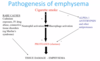
Describe the pathogenesis of asthma

A
  1. Pt exposed to allergen
  2. Antigens absorbed by APC
  3. Sit in respiratory epithelium presents to T cell
  4. Antibody response and recruits cells
  5. Reintroduction - magnified reaction
  6. IgE binds to mast cell
  7. Mast cells in airways activate and release mediators
    1. Secretion of mucus
    2. Leaky capillaries
    3. Acute spasm of bronchiole muscles
  8. Overtime:
    1. Tissue damage
    2. Increased mucous production
    3. Muscle hypertrophy
7
Q

What does this image show?

A
  1. Mucus plug
  2. Overinflated lungs
  3. Mucus plug in-situ

Macroscopic features of asthma

8
Q

Describe the histology in asthma.

A
9
Q

Define COPD

A
  • Chronic cough productive of sputum
  • Most days for at least 3 months over at least 2 consecutive years
10
Q

Casues of COPD

A

• Smoking • Air pollution • Occupational exposures

11
Q

Describe histology of COPD

A
12
Q

What do patients present with in COPD?

A
13
Q

Define bronchiectasis

A

Permanent abnormal dilatation of bronchi

14
Q

What are the common causes of bronchiectasis

A
15
Q

What are the complications of bronchiectasis?

A
16
Q

Define CF

  • prevalence
  • inheritance pattern
  • mutation
  • result
A

in lung - thick dense mucous

17
Q

What organ systems do CF affect

A
18
Q

What treatment is available for CF?

A
19
Q

Define pulmonary oedema

What are the causes

Describe the pathology

A
20
Q

Define diffuse alveolar damage

What is the pathogenesis (in adults and neonates)
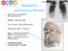
A
21
Q

Describe.

A
  1. Fluffy white infiltrates in all lung fields (“white out”)
  2. Lungs are expanded and firm, plum coloured, airless, often weigh >1kg

Diffuse alveolar damage

22
Q

Describe the histology of diffuse alveolar damage.

A
23
Q

What are the clinical outcomes of DAD

A
24
Q

What are the infective agents causing pulmonary infections? What symptoms does it cause?

A
25
What are the variety of patterns of bacterial pneumonia
Most common - bronchopneumonia lobar pneumonia - not seen as much - aggressive
26
What is the pattern difference in bronchopneumonia and lobar pneumonia?
Lobar - massively consolidated pattern
27
Describe this image. Who is prone to get this?
**Bronchopneumonia** * Compromised host defence - elderly * Often low virulence organisms - Staphylococcus, haemophilius, streptococcus, pneumococcus * Pathology - patchy bronchial and peribronchial distribution, often lower lobes
28
Describe lobar pneumonia.
29
What does this image show and what is it indicative of?
Hyperaemia → asthma
30
What is the pattern seen here?
31
What are the complications of infection?
- alcoholics and immunocompromised get abscesses
32
Define Emphysema - what causes it?
33
Describe the pathogenesis of emphysema
34
Where would smoke and alpha-1 antitrypsin present ephysema
* Smoking → loss centred on bronchiole - CENTRILOBULAR * Alpha-1 antitrypsin deficiency → diffuse loss of alveolar - PANACINAR
35
What are the complication of emphysema?
36
Define granulomas. What causes granulomatous diseases?
37
Define fibrosing lung disease. What are the types?
38
Describe idipathic fibrosing lung disease.
39
Define pulmonary thromboembolis. What are the common sites of formation. How do thrombi form? What is the effect of the embolus size?
40
Describe the presentation of small emboli.
41
Describe the presentation of large emboli.
42
Name non-thrombotic emboli
43
What causes pulmonary hypertension. Define PH.
44
Describe the lung tumours: benign and malignant. What is characteristic about them? How do we divide them?
45
Where do tumours arise
large airways or peripheries of the lung
46
Describe the aetiology of lung cancer
47
What advice should you give smokers?
Stop. It's never too late
48
What are the other risk factors (apart from smoking) of lung cancer
49
Common lung cancer types
- Squamous cell carcinoma
50
Describe the pathway of development off SCC
51
What is the pathway of development in adenocarcinoma?
52
Describe frequency, RFS, SIte, Behaviours of adenocarcinoma
53
Describe small cell carcinoma: frequency, RFs, Site, Behaviour and Histology?
54
How do we divide types of lung cancer. How would this affect treatment, prognosis?
55
What has changed about categorising of lung cancers? Why?
We now subtype non small cell carcinomas
56
How do patient present with lung carcinoma
* WL * Cough * Hemoptysis * Recurrent infections * Asymptomatic *
57
What is the role of pathologist? How do it affect management and prognosis.
- increasingly sending for molecular abnormalities - EGFR pathway
58
What molecular changes have we identified in lung cancer
* EGFR * KRAS * PTEN * FGR1 * AKT1 * DDR2 *
59
What is the role of immunotherapy in lung cancer?
60
What are the current standards of care for lung cancer?
61
What does this image show and what is it indicative of?
Eosinophilic inflammation and goblet cell hyperplasia → asthma
62
What does this image show and what is it indicative of?
Hypertrophic constricted muscle → asthma
63
What does this image show and what is it indicative of?
Mucous plugging and inflammation → asthma
64
What does this image show and what is it indicative of?
Dilatation of airways → COPD
65
What does this image show and what is it indicative of?
Hypertrophy of mucous glands → COPD
66
What does this image show and what is it indicative of?
Goblet cell hyperplasia → COPD
67
What does this image show and what is it indicative of?
* Massively dilated airways * Low normal lung parenchyma * Erosions to adjacent blood vessels - blood * Chronic hypoxia ## Footnote **CF**
68
What does this image show and what is it indicative of?
* Pink material - plasma that has leaked out of the capillaries ## Footnote **Pulmonary oedema**
69
What does this image show and what is it indicative of?
Iron laden macrophages, fibrosis → Chronic phase of pulmonary oedema
70
What does this image show and what is it indicative of?
Iron laden macrophages, fibrosis → Chronic phase of pulmonary oedema
71
What does this image show and what is it indicative of?
Large spaces in the alveoli Alveolar walls been destroyed (usually by smoking) **Emphysema**
72
What does this image show and what is it indicative of?
* Central necrotic area * Giant cells and histiocytes surrounding it ## Footnote **Necrotic granuloma**
73
What does this image show and what is it indicative of?
**Non-necrotising granuloma**
74
What does this image show and what is it indicative of?
* Honey comb changes ## Footnote **Idiopathic pulmonary fibrosis**
75
What does this image show and what is it indicative of?
* Tiny embolus * Local haemorrhage infarct * Usually irritates pleural surface - feels painful to breathe SMALL EMBOLUS
76
What does this image show and what is it indicative of?
Large emboli - can occlude main pulmonary trunk (saddle embolus)
77
What is the prognosis of having a large embolus?
78
What does this image show and what is it indicative of?
This is a bone marrow embolus
79
What does this image show and what is it indicative of?
This is a talc embolus in an IVDU
80
How frequent are invasive SCCs?
35% pulmonary carcinomas
81
What are the risk factors of SCCs
Closely associated with smoking
82
What site do invasive SCCs arise from?
Traditionally centrally located arising from bronchial epithelium, however increasing number of peripheral SCCs
83
What is the typical behaviour of SCCs?
Local spread, metastasise late.
84
What does this image show and what is it indicative of?
SCCs
85
What does this image show and what is it indicative of?
Indicative of Adenocarcinoma in the lung Precursor lesion: **atypical adenomatous hyperplasia** Proliferation of atypical cells lining the alveolar walls.
86
What does this image show and what is it indicative of?
Progression of Atypical Adenomatous Carcinoma → Non-mucinous adenocarcinoma-in-situ → Mixed pattern invasive adenocarcinoma (vessels and further sites)
87
What does this image show and what is it indicative of?
Indicative of INVASIVE ADENOCARCINOMA
88
What sites do large cell carcinoma arise from? How well differentiated are they? What is the prognosis?
89
What does this image show and what is it indicative of?
Large cell carcinoma
90
What does this image show and what is it indicative of?
small cell carcinoma No differentiated at all
91
What is EGFR?
Tyrosine kinase inhibitor that regulated several pathways involved in transcription, proliferation, migration and angiogenesis. Variety activating mutations.
92
What is Crizotinib?
Molecular targeted therapy against ALK1 mutation positive tumours.
93
What percentage of lung cancers in the UK develop in non-smokers?
10-20%