Respiratory Anatomy Flashcards
Define:
Cellular respiration
At the cellular level, O2 and CO2 exchange
Define:
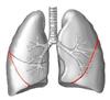
Respiratory system
Organ system (lungs, trachea, etc)
Define:
Respiration
Organismal*
Define:
Pulmonary ventilation
Movement of air into and out of lungs
Define:
External respiration
Exchange between air and blood
Define:
Transport of respiratory gases
Cardiovascular system
Define:
Internal respiration
Transfer between blood and tissues

External nares
(Nostrils)
6

nasal vestibule

nasal vestibule

nasal septum

inferior nasal conchae

middle nasal conchae

- superior nasal conchae
- middle nasal conchae
- inferior nasal conchae

sphenoid sinus


Hard and soft palate

Cleft palate
Pharynx consists of three parts
nasopharynx
oropharynx
laryngopharynx

pharyngeal tonsils (adenoids)

Pharyngeal tonsils (adenoids)
otitis media
Otitis media is the presence of fluid, typically pus, in the middle ear with symptoms of pain, redness of the eardrum, and possible fever.

opening of auditory (pharyngotympanic) tubes