Reproductive Radiology Flashcards

Coronal T2 MRI
NORMAL
Sagittal T2 MRI
NORMAL
Diagnosis?

Bicornuate Uterus
(Coronal T2 MRI)
Diagnosis?

Pelvic Mass
R = rectum (in B it should be farther up, right)
(A) Axial, (B) sagittal T2W, (C) post-contrast T2W axial images

Ovarian Cancer
diagnosis?

Mucinous Cystadenoma of the ovary
diagnosis?

Ovarian Teratoma

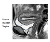
Normal Uterus in Transabdominal Transverse View

Tubo Ovarian Abscess

Vaginal U/S of normal ovary w/ follicle

Vaginal Ultrasound of Ovarian Cysts
CASE: 28 year old female with excessive hair growth on her face and abdomen presents with irregular vaginal bleeding.
“string of pearls” visible around ovary.
Diagnosis?

POLYCYSTIC OVARIAN SYNDROME
(PCOS)
Note the multiple follicle in the same stage of development, and lack of a dominant follicle

Stein-Leventhal Syndrome
(PSOS)

Fibroids

Normal vaginal ultrasound uterus- longitudinal view
endometrial stripe normally present in the uterus of a women who is not pregnant
normal

Dark structures to the right of ovary are uterine vein & artery
Abnormal hysterosonogram showing an endometrial polyp within the uterine cavity.

–A: polyp
–B: saline
–C: muscular wall of the uterus

Ovarian Torsion
Hysterosalpingogram
(normal)

A: right tube
B: uterine cavity
C: left tube
D: catheter with balloon tip

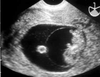
pseudosac
(ectopic pregnancy)

ruptured ectopic pregnancy

Ruptured ectopic with free fluid in pouch of Douglas

Cornual Ectopic Pregnancy

Cornual Ectopic Pregnancy