Red Eye Flashcards

1
Q

What are causes of subconjunctival haemorrhage?

A
  • Trauma
  • Spontaneous
  • Haemorrhagic disorders
  • Valsalva pressure spikes
2
Q

What investigation might you consider doing in somoene with a subconjunctival haemorrhage?

A

BP check

3
Q

What is episcleritis?

A

A benign, self-limiting inflammatory disease affecting part of the eye called the episclera. The episclera is a thin layer of tissue that lies between the conjunctiva and the connective tissue layer that forms the white of the eye (sclera)

4
Q

What causes episcleritis?

A

No cause in 70%, but can be:

  • Rheumatic fever
  • SLE
5
Q

What are features of episcleritis?

A
  • Pain
  • Discomfort
  • Sectoral redness
6
Q

How would you manage someone with episcleritis?

A
  • Systemic/topical NSAIDs
  • Topical Steroids
  • Lubricants
7
Q

What is scleritis?

A

“Vasculitis of the Sclera”

A serious inflammatory disease that affects the white outer coating of the eye, known as the sclera

8
Q

What are causes of scleritis?

A
  • Autoimmune conditions (Wegener’s, polyangitis)
  • Infections
9
Q

What are symptoms of scleritis?

A
  • Severe Pain
  • Redness
  • Photophobia
  • Decreased vision
10
Q

What are signs of scleritis?

A
  • Generalised inflammation
  • Conjunctival oedema
  • Scleral thinning
  • Decreased visual acuity
11
Q

What tests might you consider doing in someone presenting with scleritis?

A

Check for autoimmune disorder

  • ESR
  • ANCA
12
Q

How would you manage someone with scleritis?

A

Refer to a specialist

  • Oral steroids
  • Immunosuppression
13
Q

What is the following?

A

Corneal foreign body

14
Q

How would you manage a corneal foreign body?

A
  • Remove foreign body under magnification - Cotton bud or needle
  • Remove rust ring
  • Treat corneal abrasion
15
Q

What is the following?

A

Rust ring

16
Q

What is a corneal abrasion?

A

Breach in the epithelium of the eye - occurs without keratitis

17
Q

What is keratitis?

A

Inflammation of the cornea - marked by white area on the cornea, indicating a collection of white cells on the corneal tissue

18
Q

What is important to do when examining someones eyes for a corneal foreign body?

A

Invert the upper lid to look for additional FBs

19
Q

What clinical features would point towards a corneal abrasion?

A
  • Pain
  • Watering
  • Photophobia
  • Conjunctival injection
  • Swollen lids
20
Q

How would you investigate for a corneal abrasion?

A

Stain with flourescin and bright blue light (shone tangentially across the globe)

21
Q

How would you manage a corneal abrasion?

A
  • Look for conjunctival foreign bodies +Evert eye lid
  • Topical antibiotics - chloramphenicol
  • Cycloplegics
  • Pressure pad and patch
22
Q

When would you not use a pressure pad and patch in someone with a corneal abrasion?

A

If there is suspected infection

23
Q

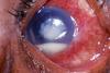
What is a corneal ulcer?

A

Also known as ulcerative keratitis - an inflammatory, or more seriously, infective condition of the cornea involving disruption of its epithelial layer with involvement of the corneal stroma.

Image - corneal ulcer with hypopyon

24
Q

What organisms cause of corneal ulcers?

A
  • Bacteria
  • Herpes viruses
  • Fungi - candidia, aspergillus
  • Acanthoemeba
  • Vasculitis - RA,
25
How would you manage a corneal ulcer?
* **Bacterial** - do not patch * **Topical steroids** * **Refer to opthalmologist**
26
What are symptoms of a corneal ulcer?
* **Pain** * **Redness** * **Photophobia** * **Watery** * **Discharge**
27
What are features of viral keratitis?
* **Discomfort** * **Foreign Body sensation** * **Watering** * **Photophobia**
28
What is viral keratitis most commonly caused by?
Herpes simplex/zoster
29
What are signs of viral keratitis?
* **Epithelial dendrites** * **Stromal keratitis** * **Central ulceration**
30
How would you manage viral keratitis?
* **Aciclovir (3%) 5x per day, for 3 weeks** * **Topical cycloplegics** * **May need topical steroids** * **Refer to opthalmologist**
31
What is uveitis?
Inflammation of the uvea, the pigmented layer that lies between the inner retina and the outer fibrous layer composed of the sclera and cornea. The uvea consists of the middle layer of pigmented vascular structures of the eye and includes the iris, ciliary body, and choroid.
32
What are the different types of uveitis?
* **Anterior uveitis** * **Intermediate uveitis** * **Posterior uveitis**
33
What are symptoms of anterior uveitis?
* **Pain** * **Blurred vision** * **Photophobia** * **Redness** * **Watering**
34
What conditions is uveitis associated with?
* **Positive HLA-B27** - Ankylosing spondylitis * **Arthritis** * **Inflammatory bowel disease** * **Sarcoid** * **Tuberculosis** * **Syphilis** * **Toxoplasmosis** * **Behçet’s syndrome** * **Lymphoma** * **Viruses -** herpes, CMV and HIV infection
35
What are signs of anterior uveitis?
* **Circumcorneal redness** - ciliary flush * **Keratic precipitates on corneal epithelium** * **Cells/flare in anterior chamber** * **Miosis** - due to sphincter spasm * **Hypopyon** * **Posterior/Peripheral anterior Synechaie/Festooned pupil** * **Iris atrophy** * **Fibrinous membrane in the pupillary**
36
How would you manage anterior uveitis?
* **Topical steroids** * **Cycloplegics** * **Topical anti-glaucoma meds** - if raised IOP * **Steroid ointment** - for night
37
What are cycloplegics?
Used to paralys the ciliary muscle of the eye, resulting in a loss of accommodation. Because of the paralysis of the ciliary muscle, the curvature of the lens can no longer be adjusted to focus on nearby objects. Often used to prevent synechiae, and reduce pain from ciliary muscle spasm
38
Which type of uveitis is photophobia and pain most commonly associated with?
Anterior uveitis
39
What type of uveitis are floaters associated with?
Posterior uveitis
40
What factors contribute to the recurrence of herpes keratitis?
* **Ultraviolet light** * **Stress** * **Menstruation**
41
What parts of the eye are affected in anterior uveitis?
Iris with or without ciliary body inflamed
42
What parts of the eye are affected in intermediate uveitis?
Ciliary body inflamed - leaks proteins and cells
43
What parts of the eye are affected in posterior uveitis?
Choroid
44
What is the pathophysiology of uveitis?
Inflammation of the uveal tract, which includes the iris, ciliary body and choroid. Inflammation causes plasma and WBCs to leak into the aqueous humour * **Hazy anterior chamber** * **Cells depositied at the back of the cornea** * **Cell in AC may settle inferiorly - hypopyon**
45
What is the pathophysiology of acute closure glaucoma?
Aqueous humour produced by ciliary bodies enters the anterior chamber via the pupil. This drains via the trabecular network at the angle to enter Schlemm's Canal. In acute angle closure glaucoma, the iris obstructs the trabecular meshwork, obstructing the drainage of aqueous humour.
46
What can untreated increased Intraocular pressure lead to?
Optic nerve damage and permanent visual field loss
47
What is the normal intraocular pressure of the eye?
10-21 mmHg
48
What are glaucomas?
Group of diseases in which the pressure inside the eye is sufficiently elevated to cause optic nerve damage and result in visual field defects
49
What can IOP rise to in acute angle closure glaucoma?
\>50mmHg
50
Who are those most at risk of acute angle closure glaucoma?
* **Hypermetropes** * **Women**
51
When is acute angle glaucoma most likely to occur?
When pupil is dilated - e.g. low level light/while sleeping
52
Which sex does acute angle glaucoma most commonly occur in?
Women
53
What are symptoms of acute angle closure glaucoma?
* **Red eye - painful** * **Blurred vision** * **Unwell with nausea and vomiting** * **Headache** * **Tender, hard eye** * **Semi-dilated pupil**
54
What are signs of acute closure glaucoma?
* **Hard Eye** * **Vision** - counts finger -\> hand motion visible * **Circumcorneal congestion** * **Corneal oedema** * **Shallow anterior chamber** * **Semidilated pupil** * **IOP** - 40-70 mmHg
55
How would you manage someone with acute angle closure glaucoma?
Reduce IOP * **G. Timilol** **(0.5%)** * **G. Pilocarpine (1%)** - every 10 mins x3 * **IV acetozolamide** 500mg stat * **IV Mannitol** - 20%, 250-500 ml over 30-45 mins * **Oral glycerol**
56
Describe the following features of Anertior Uveitis: * **Pain** * **Redness** * **Discharge** * **Vision** * **Photophobia** * **Pupil**
* **Pain** - mild/moderate * **Redness** - pericorneal/none * **Discharge** - minimal/none * **Vision** - blurred * **Photophobia** - ++ * **Pupil** - Constricted
57
Describe the following features of bacterial conjunctivitis: * **Pain** * **Redness** * **Discharge** * **Vision** * **Photophobia**
* **Pain** - itching/gritty * **Redness** - peripheral/diffuse * **Discharge** - yellow * **Vision** - normal * **Photophobia** - +
58
Describe the following features of viral conjunctivitis: * **Pain** * **Redness** * **Discharge** * **Vision** * **Photophobia**
* **Pain** - itching/gritty * **Redness** - peripheral/diffuse * **Discharge** - watery * **Vision** - normal * **Photophobia** - +
59
Describe the following features of scleritis: * **Pain** * **Redness** * **Discharge** * **Vision**
* **Pain** - severe/boring * **Redness** - sectoral/diffuse * **Discharge** - none * **Vision** - normal
61
Describe the following features of acute angle glaucoma: * **Pain** * **Redness** * **Discharge** * **Vision** * **Photophobia** * **Pupil size**
* **Pain** - severe, often accompanied by headache * **Redness** - pericorneal * **Discharge** - yes/no * **Vision** - lost * **Photophobia** - none * **Pupil size** - Dilated
62
Describe the following features of keratitis/corneal ulcer: * **Pain** * **Redness** * **Discharge** * **Vision**
* **Pain** - severe, particularly on eye closing * **Redness** - pericorneal * **Discharge** - yes/no * **Vision** - lost
63
Describe the following features of orbital cellulitis: * **Pain** * **Redness** * **Discharge** * **Vision**
* **Pain** - severe periocular pain * **Redness** - none * **Discharge** - yes/no * **Vision** -normal
64
What is the differential diagnosis for Red eye?
* **Trauma** * **Foreign body** * **Acute glaucoma** * **Keratitis/Corneal ulcer** * **Conjunctivitis** * **Scleritis/Episcleritis** * **Orbital cellulitis** * **Anterior uveitis** * **Spontaneous conjunctival haemorrhage**
65
What are red flag features of red eye on history and examination?
* **Severe pain** * **Photophobia** * **Reduced vision, particularly if sudden** * **Coloured halos around point of light in a patient’s vision** * **Proptosis** * **Smaller pupil in affected eye**
66
What are red flag features of red eye on medical assessment?
* **High intraocular pressure** * **Corneal epithelial disruption** * **Shallow anterior chamber depth** * **Ciliary flush**
67
What are common causes of keratitis?
* **Herpes** * **Contact lenses** * **Blepharitis**
68
What is the following?
Bacterial conjunctivitis - purulent discharge
69
What is the following?
Conjunctivits - Most likely viral as no purulent discharge
70
What is the most common cause of viral conjunctivitis?
Adenovirus
71
What might precipitate a viral conjunctivitis?
URTI
72
What is conjunctival injection?
**Engorged conjunctival vessels**
73
What are symptoms of bacterial conjunctivitis?
Ususally only 1 eye, but can spread * **Discomfort** * **Purulent discharge** - eye may be difficult to open in the morning
74
What are signs of bacterial conjunctivitis?
* **Mucopurulent discharge** * **Normal vision** - after discharge has been blinked clear * **Uniform engorgement of all the conjunctival blood vessels**
75
How would you manage a bacterial conjunctivitis?
* **Topical antibiotic eye drops** - chloramphenicol * Every 2 hours for first 24 hrs, then decreasing to 4x per day for 1 week. * **Chloramphenicol ointment** - at night * **Advised about general hygiene** - e.g. not sharing face towels.
76
What are symptoms of viral conjunctivitis?
Normally both eyes * **Gritty and uncomfortable** * **Associated symptoms of a cold and a cough.** * **Watery discharge** * **Photophobia** - if they develop discrete corneal opacities.
77
What are signs of viral conjunctivitis?
Both eyes * **Red with diffuse conjunctival injection** * **Clear diacharge** * **Small white lymphoid conjunctival aggregations** * **May be head and neck lymphadenopathy**
78
How would you manage viral conjunctivitis?
Usually self limiting - Warn that may take a few weeks * **Consider Chloramphenico**l - provide symptomatic relief and help prevent secondary bacterial infection. * **Strict hygiene measures** - very contagious * **Chronic infection** - steroid eye drops may be indicated, under opthalmology supervision
79
What are symptoms of allergic conjunctivitis?
Both eyes * **Itching** * **Clear discharge** * **History of atopy** - similar may have occured in previous seasons
80
What are signs of allergic conjunctivitis?
* **Diffusely injected conjunctivae** * **Chemosis** * **Clear and stringy dsicharge** * **Papillae** - round oedoematous swelling in the eyelid caused by fibrous septa that tether the eyelid
81
How would you manage allergic conjunctivitis?
* **Topical/oral antihistamine** * **Vasoconstrictor eye** * **Steroids** - with opthalmology supervision
82
What is iritis?
Inflammation of the iris
83
What is cyclitis?
Inflammation of the ciliary body
84
What is iridocylitis?
Anterior uveitis
85
What are signs of a corneal ulcer?
* **May be decreased visual acuity** - depends on the location and size ulcer, * **Discharge** * **May be a watery** - due to reflex lacrimation * **May be mucopurulent** - bacterial ulcers. * **Conjunctival injection** - generalised or local * **May be hypopyon**
86
What is a dentritic corneal ulcer indicative of?
Herpes simplex virus
87
Why does photophobia occur in anteriro uveitis?
Inflammation coupled with ciliary muscle spasm
88
What can worsen photophobia experienced in anterior uveitis?
When the patient is reading and contracting the ciliary muscle
89
What is the diagnosis?
Viral conjunctivitis
90
What is the diagnosis?
Bacterial conjunctivitis
91
What is the diagnosis?
Corneal ulcer with hypopyon
92
What is the diagnosis?
Episcleritis
93
What is the diagnosis?
Scleritis
94
What is the diagnosis?
Acute angle glaucoma
95
What is the diagnosis?
Anterior uveitis
96
What is the diagnosis?
Orbital Cellulitis