Radiology Flashcards

1
Q

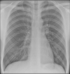
What does this CXR show? Where is the air bronchogram?

A

Right middle and lower lobe pneumonia. Air bronchogram circled.

Opacification in RLZ. Hemidiaphragm (lower lobe) and right heart border (middle lobe) are obscured.

2
Q

What kind of pneumonia causes lobar opacification?

A

CAP

3
Q

How can you differentiate consolidation from lung collapse?

A

Lung collapse will display volume loss- a shift in mediastinal structures, diaphragm, hila, fissures.

Consolidation may also have air bronchograms

4
Q

What does this CXR show? What is circled in red and green?

A

Shows left lower lobe pneumonia. Red: air bronchogram. Green: stomach and splenic flexure.

Increased opacification in left lower lobe, left diaphragmatic border reduced.

5
Q

What is upper lobe diversion? What causes it?

A

Increased pulmonary vasculature in upper lobe, caused by heart failure.

6
Q

What does this CXR show? What is circled in green?

A

Bronchopneumonia with NG tube (green) in wrong position.

Multifocal opacification, more so in L than R. Air bronchograms present.

7
Q

Where should a NG tube be positioned on CXR?

A

Should cross the carina and end below the diaphragm in the stomach

8
Q

What does this CXR show?

A

Left lower lobe collapse

Triangular opacity that is obscuring the diaphragm and descending thoracic aorta. Depressed left hilar. Smaller left lung. Darker left lung.

9
Q

What kind of pneumonia causes bronchopneumonia?

A

HAP

10
Q

What does this CXR show?

A

Left upper lobe collapse.

Patient also has breast implant. Increased opacification in upper zone from collapsed lung. Left visible heart border and mediastinal shift. Left hilar point pulled up.

11
Q

What does this CXR show?

A

Complete left lung collapse. Entire hemithorax opacification.

Could also be due to pneumonectomy

12
Q

How do you differentiate a white out due to lung collapse from pleural effusion?

A

There would be mediastinal shift in lung collapse.

13
Q

What else should you look for if pleural effusion is present?

A

Lobulated pleural thickening due to malignant mesothelioma

14
Q

What does this CXR show?

A

Multifocal well defined round opacifications. Cannonball mets due to pulmonary mets.

15
Q

What are the common sources of pulmonary mets?

A

bronchus, breast, prostate, colon, kidney

16
Q

Are nodules from infection ill defined or well defined?

A

Ill-defined e.g. miliary TB

17
Q

What does this CXR show?

A

Lung cancer- ill defined, spiculated opacity in the LUL

18
Q

What is an opacity >3cm and <3cm?

A

>3cm is a mass, <3cm is a nodule

19
Q

What does this CXR show?

A

Left pneumothorax. Darker lung and visible lung border

20
Q

What does this CXR show?

A

Right pneumothorax

21
Q

What does this CXR show?

A

Tension pneumothorax

Increased lung lucency on left side, mediastinal shift and diaphragm pushed down. MEDICAL EMERGENCY

22
Q

What does this CXR show?

A

Giant bulla

Darker right hemithorax but not a tension pneumomthorax as septal lines are present in the lung field

23
Q

What does this CXR show?

A

Pneumomediastinum

Can see air in the mediastinum. Air present going up to neck as well. Likely due to oesophageal perforation.

24
Q

What does this CXR show?

A

Obstructive lung disease e.g. emphysema or asthma

Large volume lungs, flattened diaphragm

25
Q

What does this CXR show?

A

Pulmonary oedema

Lungs look opacified on both sides, increased pulmonary vasculature, distension of upper lobe veins, perihilar opacification

26
Q

What does this CXR of a young patient without cardiac disease show?

A

Non-cardiac pulmonary oedema

ECG leads and ET tube present. Increased opacification and air bronchograms. Apices and lung bases look normal. Heart size normal. Dense perihilar consolidation.

Due to ARDS, smoke inhalation, certain drugs, head injury

27
Q

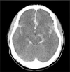
What does this CT head show?

A

Sub arachnoid haemorrhage

Small ventricles, loss of celsi, fissures filled with white fluid (blood as CSF is black on CT)

28
Q

What structure is highlighted here?

A

stomach

29
Q

How do you differentiate small bowel from large bowel on AXR?

A

Small bowel have valvulae conniventes (folds stretch across bowel), colon has haustral folds (folds go half way across)

30
Q

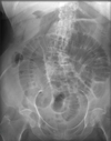
What does this AXR show?

A

Small bowel obstruction

Cluster of dilated loops of small bowel.

31
Q

Common causes of small bowel obstruction?

A

Most commonly adhesions.

Also: hernia into hernial orifices

32
Q

What does this AXR show?

A

Descending colon obstruction

Dilated large bowel in the transverse colon and caecum.

33
Q

Causes of large bowel obstruction?

A

Colonic adenocarcinoma, toxic megacolon, ischaemia if dilation in vascular territory

34
Q

What does this AXR look like?

A

Football sign- suggests intestinal perforation.

Also look for air under the diaphragm in CXR

35
Q

What is Rigler’s sign?

A

When there is air on both sides of the intestinal wall.

36
Q

What does this AXR show?

A

Sigmoid volvulus

37
Q

What does this AXR show?

A

Staghorn calculus

38
Q

What % of gallstones and renal stones calcify?

A

10% gallstones 90% renal stones

39
Q

What does this AXR show?

A

Chronic pancreatitis

Calcification crosses over spine

40
Q

What does this AXR show?

A

Gallstones

41
Q

What does fat stranding indicate?

A

inflammation

42
Q

Causes of pneumoperitoneum?

A
  • Post-op
  • Vaginal aspiration
  • Pneumothorax
  • Pneumomediastinum
  • Trauma
43
Q
A