Cardiology Flashcards
Physical signs of infective endocarditis.
Changing murmur (90%), Pyrexia (90%), Microscopic haematuria (70%), Petechiae (50%), Roth spots, Janeway lesions, Osler’s nodes, splinter haemorrhages, clubbing
What are the main organisms that cause Infective Endocarditis?
Staphylococcus from IVDU (aureus, epidermidis), Strep viridans (50-70%)
What increases risk of infective endocarditis due to strep viridans?
Damaged valve e.g. rheumatic heart disease, older age. Strep viridans is usually wiped out by immune system. Increases risk of other valvular disorders as well.
What organism causes acute rheumatic fever?
Group A strep causes strep throat. A few weeks later you get an autoimmune reaction that attacks self tissue.
What causes chronic rheumatic heart disease?
Fibrosis of heart valves (MS, AR) occurs ~20 years later.
Why do IVDUs get infective endocarditis? What side of the heart is mostly affected?
Get Staph infection entering the venous drainage which enters the right heart (tricuspid and pulmonary valves affected)
What side of the heart is commonly affected by strep viridans in infective endocarditis patients?
Left side. Older patients as they have damaged mitral valves as it’s pummelled by the highest amount of pressure.
Features of acute rheumatic fever? (major criteria for Duckett Jones criteria)
Carditis, polyArthritis, Sydenham’s chorea, Erythema marginatum, Subcutaenous nodules along the extensor surfaces
Which murmur causes mitral facies?
Mitral stenosis
What murmur causes a loud second HS?
Mitral stenosis
Why might you not hear mitral stenosis? What can you do to hear it?
Raised cardiac output is required to hear the murmur. Won’t hear it if inactive. Exercise the patient to hear the murmur.
What signs would be present in mitral stenosis?
malar flush, usually middle aged woman, AF, tapping apex (palpable heart sound), non-displaced apex, right ventricular heave, blowing mid diastolic murmur with presystolic accentuation
What signs would be present in mitral regurgitation?
Displaced apex, apical thrill, S1 quiet, pansystolic murmur radiating to axilla. S3 Present.
What signs would be present in aortic regurgitation?
Collapsing pulse, Corrigan’s sign, De Musset’s sign, Quincke’s sign, dynamic apex, early diastolic murmur at left sternal edge, systolic flow murmur, Luetic/Marfans/Ank spondylitis/Reiter/endocardiis/old rheumatic fever signs
What does this image show?

Malar flush
What does this image show?

Erythema marginatum- acute rheumatic fever
What does this ECG show?

atrial fibrillation
Causes of AF?
- Ischaemic heart disease
- Rheumatic heart disease
- Thyrotoxicosis
- PE
- Cardiomyopathy
- Ca bronchus
- Alcohol
- lone AF
What does this ECG show?

R wave progression which is normal and means there’s unlikely to be PMH of MI
What does this ECG show? Why is not pericarditis? Where is the pathology likely to be?

Full thickness acute MI. V1-V6 ST elevation.
Not pericarditis as the ST elevation is not global.
LCA occlusion causes this pattern.
What does this ECG show?

ST elevation in leads II, III and aVF. This suggests an inferior MI.
There is also ST depression in leads aVL, V1, V2, V3. Some ST change in V4 and V6. These are non-specific so suggest ischaemic changes.
The bradycardia could be due to arrhythmia (issue in SAN) or on beta blockers for past MI.
What does this ECG show?

ST depression in leads aVL, and V2 leads. This is actually an upside down ST elevation of the posterior wall. Likely due to an infarction with true posterior extension. Posterior infarction by itself is unlikely.
What does this ECG show? It is a 47 year old man with severe central crushing chest pain.

ST elevation MI with Type I heart block
How do you manage complete heart block? Is it regular or irregular?
Needs pacing, attach an external pacemaker. It is regular as there is a new ventricular origin of heart beat.
What is the main complication of a RCA occlusion? and LCA occlusion?
RCA: atria are affected so can get arrhythmias: heart block or abnormal atrial rhythm.
LCA: heart failure as you lose the pump.
Main principles of management in acute MI?
- Call for help
- Take A-E approach
- Assuming airway patent/managed, start on morphine 5mg orally. Don’t forget antiemetics (metoclopramide 10mg). Can also give diamorphine IV 2.5-5mg but harder to get hold of.
- Start them on nitrates sublingual
- Aspirin 300mg, then second antiplatelet (ticragelor 180mg or clopidogrel but ticragelor better)
- Streptokinase 1.5 MU over 1hr
- Beta blockage if not in heart failure
- Aspirin and nitrates reduce tissue death so must be done rapidly.
- If NSTEMI can heparinize them (fondaparinux subcut), don’t heparainize if STEMI as going to PCI and they can do controlled heparin.
If patient with acute MI goes into VF what do they need rapidly?
Shock them, if you wait more than ~4minutes then its difficult to recordinate tissue.
Complications of MI?
- Arrhythmia (inc VF and death)
- Cardiac failure
- Embolism
- Rupture/aneurysmal dilatation
- Pericarditis
When would you get early pericarditis following MI?
Full thickness anterior MI (common), presents as positional chest pain day after MI better sitting forward. Use NSAIDs
What is Dressler’s syndrome?
Pericarditis developing ~6 weeks after MI due to immune response
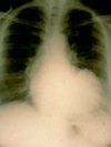
What does this CXR show? What ECG changes would you expect?

Enlarged LV- LV aneurysm in someone who had an MI a year ago. These patients have persistent ST elevation in LV segment.
What does this pulmonary angiogram show?

Massive pulmonary embolism. There is blockage of pulmonary vasculature to the right lung and the upper left lung.
What changes are present on the ECG of this lady with a PE?

S1Q3T3- suggests severe PE
- Large S wave in lead I
- Q wave in lead III
- Inverted T wave in lead III
What ECG changes could you expect in a person with PE? (common to uncommon)
- Completely normal
- Sinus tachycardia
- Right ventricular strain
- Inverted T waves in V1 to V4
- S1Q3T3


