Radiologie Flashcards
Diagnose?

Pneumothorax
Diagnose?

Spannungspneumothorax
- Entsprechender Hemithorax ist vergrößert.
- Das ipsilaterale Zwerchfell steht niedriger.
- Das Zwerchfell ist zur Gegenseite verlagert.
- Der Lungenrand ist sichtbar.
- Der Pneu ist eine strukturlose Aufhellung.
- Die Lunge ist in diesem Fall verschattet, mit ovalären Aufhellungen: Einblutungen (Alveolarraumverschattung) und Pneumatozelen nach Unfall.
Diagnose?

Jefferson Fraktur
- Fraktur des Wirbelbogens HWK 1 (erstes Bild bilateral)
- Axiales Kompressionstrauma Instabile Fraktur
- Prävertebrales Hämatom + caudale Verletzung A. vertebralis

Diagnose?

Densfraktur
- Schnelle Ante/Retroflexion, Osteoporose
- Typ 1: Densspitze → stabil
- Typ 2: Densbasis → instabil
- Typ 3: Basis und Corpus HWK 2
- Prävertebrales Hämatom, Cave: Myelonverletzung

Diagnose?

Hangman-Fraktur!
- Bilaterale Fraktur vom Pars interarticularis HWK 2
- Hyperextention/flexion + axiale Kompression
- Stabile oder instabile Fraktur
- Typ 1: keine Dislokation
- Typ2: ≥3mm anterior HWK2
- Typ 3: disloziertes Fragment, Subluxation Facettengelenke
- Prävertebrales Hämatom
- Zusätzlich HWK 1 Frakturen
- Cave: Myelonverletzung, Verletzung A. vertebralis

Diagnose?

Kompressionsfraktur: Bruch ant. Wirbelkörper
Berstungsfraktur: Bruch vordere + mittlere Säule

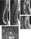
Diagnose?

Myelonverletzung = Schädigung der Axone
- Sturz aus großen Höhen
- MRT: Kulkarni Klassifikation
- Typ 1: T1 inhomogen = Hämatom
- Typ 2: T2 hyperintens = Ödem
- Typ 3: T2 zentral hypointens
Diagnose?

Dissektion A.vertebralis
- Überdehnungstrauma
- CT/MRT -> Angiographie
- T1 hyperinteses semizirkuläres Wandhämatom

Diagnose?

Spinale Metastasen
- Hämatogen aus CA der Lunge, Mamma, Prostata, Niere, GIT
- Osteolytisch → CT hypodens
- Osteoblastisch → CT hyperdens

Diagnose?

Plasmozytom / Multiples Myelom
- BWS > LWS > HWS
- Pathologische Fraktur häufig

Diagnose?

Schwannom
- WHO Grad I Tumor von Schwannzellen

Diagnose?

Meningeom
- WHO Grad I Tumor
- Ausgehend von der Arachnoidea
- BWS > HWS > LWS
Diagnose?

Emendymom
- WHO Grad II Tumor, aus Ependym des Zentralkanals
- HWS > BWS > Konus
Diagnose?
Astrozytom
- WHO Grad I/II Tumor, aus Astrozyten des Myelons

Diagnose?

Spondylodszitis/Osteomyelitis
- Bakterielle Entzündung von Wirbelkörper + Bandscheiben
- S.Aureus, E.coli
Diagnose?

Morbus Bechterew = Spondylitis ankylosans
- Inflammatorische Arthopathie + Erosion Iliosakralgelenk
- Bambusartige Deformierung der Wirbelsäule

Diagnose?

Virale Myelitis
- Viele Segmente mit T2 hyperintenser Auftreibung
Diagnose?

Multiple Sklerose
- Autoimmune, mehrzeitige Demyelinisierung des zentralen Nervensystems

Diangose?

Bandscheibenvorfall
- Herniation: < 50% der Bandscheibenzirkumferenz
- Protrusion → breitbasiger Kontakt zur Bandscheibe
- Extrusion → schmaler Kontakt zur Bandscheibe
- Sequester → freies Fragment

Diagnose?

Lumbale Spinalkanalstenose
- Multifaktorielle degenerative Veränderungen der Bandscheiben, Endplatten, Facettengelenke, Ligg. flava
- < 12 mm relative Stenose
- < 10 mm absolute Stenose
Diagnose?

Spodylolyse
- Repetitive Stressfraktur im LWK 5
- Knochenmarksödem angrenzend

Diagnose?

Myeloninfarkt
- Arterieller Verschluss einer Arterie
- Meist radikuläre Arterie, Aortendissektion
- Geringe Schwellung in Akutphase
- T2 hyperintens der grauen Substanz = Eulenaugen
- Blutung → T2 hypointens KM-Anreicherung in Subakutphase

Diagnose?

Spinale durale arteriovenöse Fistel
- Arteriovenöse Kurschlussverbindung im Spinalkanal
- Intramedullär, intradural, extradural
- Evtl. Stauung oder Blutung
- Myelonödem → T2 hyperintens Blutung → T2 hypointens, T1 hyperintens
- Erweiterte Venen → „flow voids“, KM- reiche „Schlangen“

Diagnose?

Protrusion

Diagnose?

Kindliches Medulloblastom
- Infratentoriell
- Zysten, inhomogene KM-Aufnahme
- Häufigster kindlicher Hirntumor
Diagnose?

Ependymom
Diagnose?

Akustisneurinom = Schwannom
Diagnose?

Bakterielle Meningitis
Diagnose?

Arteriovenöse Malformation
Diagnose?

Basilaristhrombose
Diagnose?

Aneurysma der Arteria cerebri rechts
Diagnose?

A.Carotis interna Stenose rechts
Diagnose?


Diagnose?

Totaletelektase links
Diagnose?

Azinäre Verschattung

Diagnose?

Gut abgrenzbare, einzelne Verdichtung
3-6 cm, von Lungengewebe umgeben
Diagnose?

Multinoduläre Herde = Miliartuberkulose
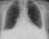
Diagnose?

Spannungspneumothorax
Diagnose?

Emphysem
Diagnose?

Intrazerebrale Blutung - ICB
Diagnose?

Basilaristhrombose
Diagnose?

Bakterielle Meningitis
Eiter DWI und FLAIR hyperintens; KM-Anreicherung der weichen und harten Hirnhäute
Diagnose?

Abgangstenose A.carotis interna
Diagnose?

Aneurysma der Arteria cerebri media rechts

Diagnose?

Arteriovenöse Malformation
Diagnose?

Zerebritis/Abszess:
Diffuses T2-hyperintenses Ödem und fleckige KM- Anreicherung; Abszess: zentrale Diffusionsstörung, T2 hyperintenser Rand mit KM-Anreicherung
Diagnose?

Spannungspneumothorax
Diagnose?

Herpes Enzephalitis
Hypodense Veränderung rechts temporal mit Diffusionsstörung; T2- hyperintense Veränderung rechts temporal und insulär mit gyriformer KM- Anreicherung
Diagnose?

Multiple Sklerose
T2 hyperintense Läsionen im Marklager und infratentoriell; später konfluierend; „hufeisenförmige“ KM-Anreicherung subkortikal
Diagnose?

Eingebluteter Hirntumor
Hyperdense Einblutung links frontal betont und kortikal; multiple kortikale / subkortikale KM- Anreicherungen
Diagnose?

Liquorunterdrucksyndrom
Gering hyperdense subdurale Ergüsse / Hämatome beidseits; CT-Myelographie: KM Austritte entlang von Spinalnerven; CT gesteuerter „Blutpatch“
Diagnose?

Hirnblutung bei Kokain Missbrauch
Hyperdense Blutung rechts frontoparietal; Kalibersprünge von peripheren Arterien
Diagnose?

Subdurales Hämatom
Subakutes SDH beidseits mit Überschreitung der Schädelnähte und Sedimentierung mit Spiegelbildung
Diagnose?

Subdurales Hämatom
Subakutes SDH rechts (linkes Bild), akutes SDH links, sehr schmales SDH rechts (rechtes Bild)
Diagnose?

- Kalter Knoten: malignitätsverdächig
- Heißer Knoten: Hyperthyreose + heißer Knoten = autonomes Adenom, operaive Tx nach Eutyhreose: entsprechend der Läsionen
-> Bild = Kalter Knoten!
Szinitgraphie (99mTechnetium)
Diagnose?

Thyreoiditis
Diagnose?

Heisser Knoten
Diagnose?

Mittellappenatelektase
- Atelektase ist volumensmindernd (Zwerchfellhochstand)
- Oft Aerobronchogramm.
- Die Mittellappenatelektase löscht den Herzrand teilweise aus, da sie vorne liegt.
- “Silhouettenphänomen” zum rechten Herzrand.
Diagnose?

Oberlappenatelektase
Diagnose?

Lungenemphysem
- Vergrößertes Lungenvolumen.
- Verminderte Struktur, hier besonders links basal.
- Tiefe Zwerchfelle
- Abgestumpfte Randsinus.
- Verbreiterter Retrosternalraum.
- Verbreitert erscheinende Zwischenrippenräume.
- Hier bei Alpha-1 Antitrypsinmangel.
Diagnose?

Subdurales Hämatom
Diagnose?

Infarkt A.cerebri anterior
Diagnose?

Lungenödem
- “Aerobronchogramm”: Bronchien sind durch die enthaltene Luft
- negativ kontrastiert, da der Alveolarraum gefüllt ist mit Eiter bei der Pneumonie; mit Blut nach Verletzung; mit Wasser nach (Beinahe-)Ertrinkung.
Diagnose?
Epikarderguss
Diagnose?

Benigner Magenulkus
Diagnose?

Magen-CA
Diagnose?

Meckel Divertikel mit Sellink
Diagnose?

Schlaganfall rx
Diagnose?

Blutung bei arteriovenöser Malformation
Hyperdense intraventrikuläre Blutung, „Knäuel“ rechts dorsal des Seitenventrikels; erweiterte Gefäße in CT Angiographie

Herpes Simplex Encephalitis


Multiple Sklerose

Astrozytom (BILD GRAD IV)
- Grad IV: KM-Anreicherung + KLINIK wichtig + Nekrose
- Grad III: KM-Anreicherung, gut abgrenzbar vom Ventrikel!
- Grad II: Keine KM-Anreicherung!!

Oligodendrogliom -> CT hilfreich weil Verkalkung!

Carotis-sinus-cavernosus Fistel

Meningitis

Kontusionsblutung

Aspergillus

Diffuse axonale Schädigung

Oben: Aneurysmatische SAB
Unten: Hypertensive Massenblutung

Astrozytom Grad II -> Keine KM-Anreicherung

Herpesencephalitis

Arnold-Chiari-Malformation

Tuberkulose
