Pulmonary I Flashcards

1
Q
A

Chronic Inflammation

2
Q
A

Acute Inflammation

3
Q
A

Polymorphonuclear cell (PMN) - Acute inflammation

Neutrophils

4
Q
A

Mononuclear Cells - Chronic Inflammation

5
Q

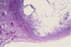
What is a granuloma composed of?

A

Th1 cells, B cells on outside, Multinucleated giant cells surrounding necrotic core and persistent antigens

6
Q
A

Granulomatous inflammation

7
Q
A

Granulomatous inflammation

8
Q
A

Normal Alveolar Tissue

9
Q
A

Normal lung histology

10
Q
A

Normal lung histo

11
Q
A

Normal Lung

12
Q
A

Acute Bronchopneumonia

Patchy infiltrates

Typical organisms: Streptococcus pneumoniae, Staphylococcus aureus, Pseudomonas aeruginosa, Hemophilus influenzae, and Klebsiella pneumoniae (among others)

13
Q
A

Acute Bronchopneumonia (lobular)

Areas of tan-yellow consolidation, firmer and more raised than surrounding lung

Remaining lung is dark due to pulmonary congestion

14
Q
A

Acute bronchopneumonia

Higher magnification, pattern of patchy distribution seen

Consolidated areas closely match pattern of lung lobules (“lobular pneumonia”)

Bronchopneumonia is classically hospital acquired.

Typical organisms: S. aureus, Klebsiella, E. coli, Pseudomonas

15
Q
A

Acute Bronchopneumonia

16
Q
A

Acute Bronchopneumonia Histology

17
Q
A

Acute lobar pneumonia

More extensive than bronchopneumonia, all of one lobe is consolidated

18
Q
A

Lobar pneumonia + fibrinous pleuritis

If there’s exudate on the surface, may become empyema

19
Q
A

Lobar pneumonia + fibrinous pleuritis

May result in empyema (collection of pus in pleural space)

20
Q
A

Lobar pneumonia + Gray-white hepatization of lower lobe

Filled with leukocytes

21
Q
A

Lobar pneumonia + gray-white hepatization

22
Q
A

Acute pneumonia

23
Q
A

Acute Bronchopneumonia

24
Q
A

Lobar pneumonia

25
Bronchopneumonia
26
Lobar pneumonia
27
Lobar pneumonia
28
Acute lobar pneumonia
29
Aspiration pneumonia Usually mixed infection & right airways more vertical, so more common in R. lung Contains multinucleated giant cells Contains neutrophilic exudate and dark blue bacterial colonies suggests aspiration or hematogenous spread of infection to lung. Aspirated material from oral-pharyngeal region has bacterial flora. Hematogenous spread of infection to lungs could occur from septicemia or infective endocarditis involving right side of heart
30
Aspiration pneumonia
31
Aspiration Pneumonia
32
Aspiration Pneumonia
33
2 lung abscesses (one in upper, one in lower lobe) Adjacent areas of tan consolidation w/ bronchopneumonia Abscess = complication of severe pneumonia, most typically from virulent organisms (S. aureus) Usually a complication of aspiration, more frequent in R. posterior lung
34
Lung Abscess + bronchopneumonia Several abscesses w/ irregular, rough-surfaced walls seen w/in areas of tan consolidation If large enough, abscesses will contain liquefied necrotic material & purulent exudate that often results in air-fluid level by chest radiograph in abscess
35
Lung Abscess Purulent exudate has drained following sectioning to reveal abscess cavities Abscesses can be source for septicemia and difficult to treat
36
Lung Abscess
37
Lung Abscess More virulent bacteria and/or more severe pneumonias can be assoc. w/ destruction of lung tissue & hemorrhage Alveolar walls no longer visible because early abscess formation (also hemorrhage)
38
Timeline of Tuberculosis
39
Ghon Complex Small tan-yellow sub-pleural granuloma in mid-lung field on right. In hilum, small yellow-tan granuloma in hilar lymph node next to bronchus (Gohn Complex) - characteristic of primary TB. In most people, granulomatous disease doesn't progress. Over time, decreases in size & can calcify, leaving focal calcified spot on chest radiograph, suggesting remote granulomatous disease.
40
Gohn Complex Closer Gohn Complex. Primary TB is pattern seen with initial infection w/ TB in kids. Reactivation (secondary) TB more common in adults.
41
Secondary (reactivation) tuberculosis Most commonly involves apex of lungs (contain higher O2 content, and mycobacterium is aerobic) Granulomas have areas of caseous necrosis. This is very extensive granulomatous disease. This pattern of mutliple caseating granlumonas primarily in upper lobes most characteristic of secondary (reactivation) TB. However, fungal granulomas (histoplasmosis, crytococcosis, coccidioidomycosis) can mimic pattern.
42
Secondary (Reactivation) Tuberculosis When there is extensive caseation & granulomas involve larger bronchus, it's possible for much of soft, necrotic center to drain out and leave behind a cavity. Cavitation is typical for large granulomas with TB. Cavitation more common in upper lobes.
43
Miliary TB
44
Miliary TB
45
Miliary TB When immune response poor or overwhelmed by extensive infection, can see "miliary" pattern because there are many small tan granulomas 2-4 mm scattered throughout lung parenchyma. Called miliary because looks like small millet seeds.
46
Miliary TB
47
Miliary TB At closer range, miliary pattern seen throughout lung. Dissemination of infectious agent (M. tuberculosis, fungi) may produce a similar pattern in other organs
48
Miliary TB Low power magnification, multiple granulomas.
49
Langhans-type giant cell in granulomatous inflammation At high magnification, granuloma demonstrates that eptihelioid macrophages elongated with long, pale nuclei and pink cytoplasm. Macrophages organize into committees called giant cells. Typical giant cell for infection = Langhan's cell and has nuclei lined up along one edge of cell (Horseshoe). Process of granulomatous inflammation takes place over months to years. Granulomas are composed of transformed macrophages (epithelioid cells) along with lymphocytes, occasional PMNs, Plasma cells, and fibroblasts.
50
Epithelioid response in granuloma
51
Caseous necrosis in TB Edge of granuloma shown here at high magnification. At upper right is amorphous pink caseous material composed of necrotic elements of granuloma as well as infectious organisms. Area ringed by inflammatory component with epithelioid cells, lymphocytes, and fibroblasts.
52
Acid-Fast Bacilli on Ziehl-Neilson Stain In order to find the mycobacteria in a tissue section, a stain for acid fast bacilli is done (AFB). The mycobacteria stain as red rods (seen here). Gold standard is a culture, but that could take weeks.
53
Fungal abscess Here is a fungal granuloma. Note the sharply demarcated borders to this granuloma, which give it a discrete, spherical appearance both grossly and radiographically.
54
Fungal Hyphae of Aspergillus Hyphae = mold form with filament structures, branch at 45 degree angles (but not the only fungus that does so) Has propensity to invade blood vessels
55
What is a dimorphic fungus?
Grows usually as mold in environment, but as yeast in the body (often due to bird/bat feces) 2 examples: Coccidioidomycosis (More common in SW) and Histoplasmosis (more common in Midwest)
56
Coccidioides immitis spherules Well-formed granuloma has large Langhans giant cell in center. Two small spherules of Coccidiodes immitis are seen in the giant cell.
57
Coccidioides immitis spherules At high magnification, thick wall of C. immitis spherule is seen in giant cell in center of image. Spherule contains endospores. In nature C. immitis exists in hyphal form with characteristic alternating arthrospores.
58
Coccidioides immitis spherules high magnification
59
Histoplasma granuloma (calcified)
60
Histoplasmosis within macrophages
61
What are the differences between acute and chronic inflammation? * Duration, inflammatory cells, vascular changes, Edema, Fibrosis, Systemic manifestation?
62
What is granulomatous inflammation? What is a granuloma?
Special form of chronic inflammation seen in response to particular organisms (i.e. Mycobacterium tuberculosis & certain fungi) Granuloma = discrete collectio of epithelioid histiocytes (macrophages) surrounded by a rim of lymphocytes and fibrous tissue. Multinucleated giant cells may also be present.
63
What is bronchopneumonia? How does it present? What organisms cause it?
Patchy consolidation of multiple small areas of lung adjacent to inflamed bronchi and bronchioles ("lobular"). Areas of consolidation appear tan & may be raised. Organisms: S. pneumoniae, S. aureus, E. coli, Klebsiella, Pseudomonas, Haemophilus
64
What is lobar pneumonia? How does it present? What organisms cause it?
Large, confluent areas of consolidation (i.e. entire lobes) Much less common pattern than bronchopneumonia Organisms: Streptococcus pneumoniae (pneumococcus) and Klebsiella
65
What is the pathophysiology of lobar pneumonia due to pneumococcus?
Inhalation -\> Edema -\> Red hepatization -\> Grey hepatization -\> Resolution
66
Compare bronchopneumonia and lobar pneumonia microscopically
Appear similar microscopically. Both characterized by massive infiltration of alveolar spaces by neutrophils, along with proteinaceous material, vascular congestion, and occasionally visible bacterial organisms.
67
What is aspiration pneumonia characterized by?
Fairly localized inflammation centered around airways and/or small foci within alveolar tissue Presence of foreign material in association with foreign body giant cell reaction
68
What are 4 complications of pneumonia?
Abscess formation Empyema: purulent exudates w/in pleural cavity Organization of exudates with pulmonary fibrosis Sepsis
69
What is an abscess?
Focal area of inflammation & necrosis, typically forming a pus-filled cavity due to parenchymal destruction * Uncommon complication bacteria (usually anaerobes) * Abscess may contain liquefied, necrotic material and result in "air-fluid level" on x-rays
70
Describe the progression of TB
71
Primary TB:
* Usually asymptomatic or mild flu-like illness * in 95% of cases, immunity halts disease progression * "Gohn Complex" (parenchymal lung lesion plus nodal involvement)
72
Secondary TB:
* 5% develop complications * Almost always symptomatic (chronic cough, hemoptysis, weight loss, low grade fever, night sweats)
73
What is another type of tuberculosis?
Progressive/miliary TB
74
Describe the significant microscopic findings in TB
Granulomatous inflammation 1. Necrotizing granulomas with "caseous" necrosis 2. Langhans-type giant cells (multinucleated cells with horseshoe arrangement of nuclei) 3. Acid-fast bacilli
75
What are common organisms that cause community-acquired acute pneumonia?
Streptococcus pneumoniae Haemophilus influenzae Moraxella catarrhalis Staphylococcus aureus Legionella pneumophila Enterobacteriaceae (Klebsiella pneumoniae) and Pseudomonas spp.
76
Organisms that cause community-acquired atypical pneumonia
Mycoplasma pneumoniae Chlamydia spp Coxiella burnetti Viruses: RSV, parainfluenza virus (kids), influenza A/B (adults), adenovirus (military recruits), SARS virus
77
Organisms that cause nosocomial pneumonia
Gram negative rods belonging to enterobacteriaceae (Klebsiella, Serratia marcescens, E. coli) & Pseudomonas Staphylococcus aureus (usually penicillin resistant)
78
Organisms that cause aspiration pneumonia
Anaerobic oral flora (Bacteriodes, Prevotella, Fusobacterium, Peptostreptococcus) PLUS aerobic bacteria (S. pneumoniae, S. aureus, H. influenzae, P. aeruginosa)
79
Organisms that cause chronic pneumonia
Nocardia Actinomyces Granulomatous: Mycobacterium tuberculosis, atypical mycobacteria, Histoplasma capsulatum, Coccidioides immitis, Blastomyces dermatitidis
80
Organisms that cause necrotizing pneumonia and lung abscess
Anaerobic bacteria (extremely common) with or w/o mixed aerobic infection S. aureus, K.pneumoniae, S. pyogenes, type 3 pneumococcus (uncommon)
81
Organisms that cause pneumonia in the immunocompromised host
Cytomegalovirus Pneumocytis carinii Mycobacterium avium-intracellulare Invasive aspergillosis Invasive candidiasis "Usual" bacterial, viral, and fungal organisms (see other cards)