Principles Flashcards
Name structure labelled A

Periosteum

Name the structures labelled A

Epiphysis

Name structure labelled A

Hyaline (articular) cartilage

Name structures labelled A

Epiphyseal growth plate

Name structures labelled A

Metaphysis

Name the structure labelled A

Diaphysis

Name the structure labelled A

Coronal Suture

Name the structure labelled B

Sphenoid bone (left)

Name the structure labelled C

Occipital bone

Name the structure labelled A

Sagittal suture

Name structures B and C

Temporal bones (left and right)

Name structure D

Occipital bone

Name structure labelled A

Foramen Magnum

Name stuctures B, C, D, E and F

B&C - Temporal bones
D - Occipital bone
E&F - Parietal bones

Name structure G and H

G - Cribriform plate of ethmoid bone
H - Frontal bone

Name the structure labelled A

Zygomatic bone (left)

Name the structures A,B,C,D and E

A - Condylar process
B - Ramus
C - Mental foramen
D - Mental process
E - Coronoid process

Nane the structures A, B, C, D, E and F

A - Vertebral foramen
B - Transverse process
C - Spinous process
D - Inferior articular process
E - Superior articular process
F - Vertebral arch

Name structures A and B

A - Intervertebral foramen
B - Facet joint

Name structures A, B and C

A - Fibula (right)
B - Tibia (right)
C - (interosseous membrane (right))

Name structures A and B

A - Annulus fibrosus
B - Nucleus pulposus

Name the types of muscle from the examples given

A - Circular
B - Pennate
C - Quadrate
D - Flat (with aponeurosis)
E - Fusiform

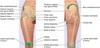
Name the three origins sites (A, B and C) and the one insertion site (D) of the deltoid muscle

A - Spine of scapula
B - Acromium process of scapula
C - Lateral 1/3rd of clavicle
D - Deltoid tuberosity of humerus

Name the layers in the skin from A to E

A - Epidermis
B - Dermis (collagen/elastic fibres)
C - Superficial fascia (adipose tissue)
D - Deep fascia (fibrous tissue)
E - Skeletal muscle

Name structures A to D

A - Cerebrum
B - Cerebellum
C - Brain stem (midbrain, pons and medulla oblongata)
D - Spinal cord

Name structures A, B and C

A - Midbrain
B - Pons
C - Medulla oblongata

Name the regions or structures labelled A to G

A - Cervical region
B - Thoracic region
C - Lumbar region
D - Sacral region
E - Cervical enlargement
F - Lumbosacral enlargement
G - Cauda equina

Name the foramina A to H

A - Cribriform plate of the ethmoid bone
B - Optic canal
C - Superior orbital fissure
D - Foramen rotundum
E - Foramen ovale
F - Internal acoustic meatus
G - Jugular foramen
H - Hypoglossal foramen

Name structures A to E

A - Kidney
B - Ureter
C - Bladder
D - Urethra
E - Pubic symphysis

Name structure A

A - Kidney (right)

Name structures A, B and C

A - Renal artery
B - Renal vein
C - Ureter

Name structures A, B and C

A - Abdominolateral muscles (external oblique, internal oblique and transversus abdominus)
B - Paranephric fat
C - Perinephric fat

Name structures A, B and C

A - Parietal peritoneum
B - Renal (deep) fascia
C - Renal capsule

Name vessels A and B

A - Renal arteries (left and right)
B - Abdominal aorta

Name the veins from A to D

A - Right gonadal vein
B - Right renal vein
C - Left renal vein
D - Left gonadal vein

Name the parts of the kidney A to E

A - Renal papilla
B - Renal pyramid
C - Renal cortex
D - Renal capsule
E - Renal medulla

Name the labelled areas of the nephron A to E

A - Renal corpuscle (glomerulus and Bowman’s capsule)
B - Proximal convoluted tubule
C - Loop of Henle
D - Collecting duct
E - Distal convoluted tubule

Name the parts of the kidney A to E

A - Nephron collecting ducts (make up renal pyramid)
B - Minor calyx
C - Major calyx
D - Renal pelvis
E - Ureter

Name structures A, B and C

A - Ureters
B - Ureteric orifices
C - Urethra

Name structures A to D

A - Urethra
B - External urethral sphincter
C - Internal urethral sphincter
D - Internal urethral orifice

Name structures A to E
A - Bladder
B - Internal urethral orifice
C - Urethra
D - External urethral sphincter
E - External urethral orifice

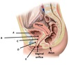
Name structures A, B and C

A - Mandible
B - Pharynx
C - Epiglottis

Name the structures associated with the lungs, A to F
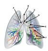
A - Right main bronchus
B - Trachea
C - Bifurcation of bronchi (carina)
D - Left main bronchus
E - Lobar bronchi
F - Segmental bronchi

Name structures A to D

A - Lobar bronchi
B - Segmental bronchus
C - Bronchioles
D - Alveoli

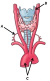
Name the cartilages A and B

A - Thyroid cartilage
B - Cricoid cartilage

Name structures A and B

A - Larngeal inlet
B - Arytenoid cartilages

Name the structures A to E

A - Rima glottidis
B - Vocal cords
C - Thyroid cartilage
D - Arytenoid cartilages
E - Cricoid cartilage

Name structure A

A - Spine of scapula (left)

Name groove A

A - Costal groove

Name the parts of the rib labelled A to D

A - Head
B - Neck
C - Tubercle
D - Shaft (body)

Name the parts associated with the sternum A to D
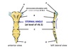
A - Sternoclavicular joint
B - Manubrium
C - Body
D - Xiphoid process

Name the joints A and B, and the structures highlighted by C

A - Sternocostal joints
B - Costochondral joints
C - Costal cartilages

Name structure A

A - Thyroid gland (contains 4 parathyroid glands)

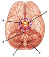
Name structures A to E associated with the brain

A - Spinal cord
B - Medulla oblongata
C - Pons
D - Midbrain
E - Diencephalon (thalamus/hypothalamus)

Name structure A

A - Pituitary gland

These structures are found in the brain, name A, B and C

A - Infundibulum
B - Posterior pituitary
C - Anterior pituitary

Name the vessels labelled A

A - Hypophyseal portal veins

Name vessels A and B

A - Superior, middle and inferior thyroid veins (right)
B - Superior vena cava

Name vessels A, B and C
A - Inferior thyroid artery (right)
B - Superior thyroid artery (left)
C - Aorta (arch)

Name structure A

A - Pancreas

Name vessels A and B

A - Coeliac trunk
B - Superior mesenteric artery

Name vessels A and B

A - Splenic vein
B - Superior mesenteric vein

Name structures A, B and C

A - Major duodenal papilla
B - Bile duct
C - Panceatic duct

Name regions A and B of the adrenal gland

A - Outer cortex
B - Inner medulla

Name structures labelled A

A - Adrenal glands

Name vessels A, B and C

A - Superior suprarenal artery (left)
B - Middle suprarenal artery (left)
C - Inferior suprarenal artery (left)

Name vessels A, B and C

A - Coeliac trunk
B - Superior mesenteric artery
C - Renal vein (left)

Name vessels A, B and C

A - Suprarenal vein (right)
B - Renal vein (left)
C - Suprarenal vein (left)

Name the arteries labelled A

A - Gonadal arteries

Name the veins labelled A and B

A & B - Gonadal veins (right & left)

Name structures A and B

A - Thyroid gland
B - Parathyroid glands

Name vessels A, B and C

A - Brachiocephalic trunk
B - Left common carotid artery
C - Left subclavian artery

Name vessels A and B

A - Common carotid artery (right)
B - Subclavian artery (right)

Name vessel A

A - Subclavian artery (right)

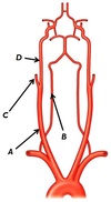
Name vessels A to E

A - Brachiocephalic trunk
B - Subclavian artery (right)
C - Common carotid artery (right)
D - Common carotid artery (left)
E - Subclavian artery (left)

Name vessels A to D

A - Vertebral artery
B - Common cartoid artery (right)
C - External carotid artery (right)
D - Interna caroitid artery (right)

Name vessels A to D
A - Common carotid artery (right)
B - Vertebral artery (right)
C - External carotid artery (right)
D - Internal carotid artery (right)

Name vessels A to E
A - Internal carotid artery (left)
B - Internal carotid artery (right)
C - Basilar artery
D - Vertebral artery (left)
E - Vertebral artery (right)

Name structures A, B and C

A - Right carotid sinus (bifurcation of common carotid artery (right))
B - Braches of glossopharyngeal nerve (CN IX)
C - Internal carotid artery (right)

As a vessel progresses along its path, its name will often change based of its location. Name the vessel at points A to E

A - Subclavian artery (left)
B - Axillary artery (left)
C - Brachial artery (left)
D - Radial artery (left)
E - Ulnar artery (left)

Name the branches and parts of the aorta from A to F

A - Brachiocephalic trunk
B - Coronary arteries
C - Posterior intercostal arteries
D - Abdominal aorta
E - Common carotid artery (left)
F - Subclavian artery (left)

Name arteries A to D

A - External iliac artery (right)
B - Internal iliac artery (left)
C - Common iliac arteries (left & right)
D - Abdominal aorta

Name structures A to E

A - Cervix
B - Fimbriae
C - Infundibulum
D - Isthmus
E - Ampulla

Name structures A, B and C

A - Vas deferens
B - Head of epididymis
C - Rete testis

Name structures A and B

A - Dartos muscle
B - Seminiferous tubules

Name structures A, B and C

A - Spermatic cord (right)
B - Pampiniform plexus (right)
C - Testicular artery (right)

Name structures A to D

A - Seminal vesicle (gland) (right)
B - Ejaculatory duct (left)
C - Prostate gland
D - Urethra

Name structures A to D

A - Seminal vesicle (gland) (right)
B - Prostate gland
C - Vas deferens (right)
D - External urethra meatus

Name structures A to D

A - Vas deferens (right)
B - Ejaculatory duct (right)
C - Prostatic urethra
D - Epididymis (right)

Name vessels A and B

A - External jugular vein (right)
B - Internal jugular vein (left)

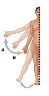
Name the actions demonstrated by A and B
A - Extension
B - Flexion

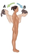
Name the actions demonstrated by A and B

A - Extension
B - Flexion

Name the actions demonstrated by A and B
A - Abduction
B - Adduction

Name the actions demonstrated by A and B
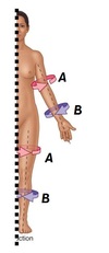
A - Lateral rotation
B - Medial rotation

Name the action demonstrated by A
A - Circumduction

Name the actions demonstrated by A and B
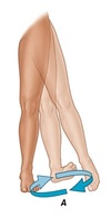
A - Eversion
B - Inversion

Name the actions demonstrated by A and B

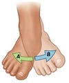
A - Opposition
B - Reposition

Name the actions demonstrated by A and B

A - Elevation
B - Depression

Name structure A

A - Pericardium

Name the layers A, B and C

A - Epicardium (under pericardium)
B - Myocardium
C - Endocardium

Name structures A, B and C

A - Right auricle
B - Left auricle
C - Apex

Name valves A to D

A - Tricuspid
B - Pulmonary
C - Mitral (bicuspid)
D - Aortic

Name muscles A and B

A - Temporalis
B - Masseter

Name muscles A and B

A - Lateral pterygoid
B - Medial pterygoid

Name muscle A

Orbicularis oris

Name this individual

Your Mum

Name muscles A and B

A - Orbicularis oris
B - Buccinator

Name the types of teeth labelled A to D

A - Incisors
B - Canines
C - Premolars
D - Molars

Name the tonsils present based on the locations A to D

A - Adenoid
B - Tubal tonsil
C - Palatine tonsil
D - Lingual tonsil

Name the regions of the colon A to F

A - Appendix
B - Caecum
C - Ascending colon
D - Transverse colon
E - Descending colon
F - Sigmoid colon

Name muscles A to D

A - Stylopharyngeus (longitudinal muscle)
B - Superior pharyngeal constrictor
C - Middle pharyngeal constrictor
D - Inferior pharyngeal constrictor

Name structures A to D

A - Ventral root
B - Dorasal root
C - Dorsal ramus
D - Ventral ramus
E - Spinal nerve

Name structures A, B and C
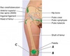
A - Adductor tubercle
B - Medial epicondyle of femur
C - Lateral epicondyle of femur
Name structures A, B and C

A - Medial epicondyle of femur
B - Greater trochanter
C - Lateral epicondyle of femur

Name bony features A and B

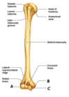
A - Radial groove
B - Deltoid tuberosity

Name bony features A, B and C
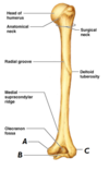
A - Medial epicondyle
B - Trochlea
C - Lateral epicondyle
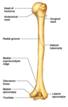
Name bony feature A

A - Olecranon fossa

Name bony features A and B

A - Greater tubercle
B - Lesser tubercle

Name bony features A, B and C

A - Radial fossa
B - Deltoid tuberosity
C - Coronoid fossa
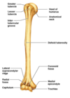
Name bony features A, B and C
A - Capitulum
B - Medial epicondyle
C - Trochlea

