Gastro-Intestinal Anatomy Diagrams Flashcards
Name muscle A
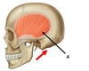
A - Temporalis

Name muscle A

A - Masseter

Name muscle A

A - Lateral pterygoid

Name muscle A

A - Medial pterygoid

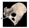
Name the foramen A

A - Foramen ovale

Name structures A and B

A - Hyoid bone
B - Dens of C2 (odontoid process)

Name structures A, B and C

A - Uvula
B - Palatine tonsil
C - Gingiva

Name the different type of papilla A, B, C and D

A - Foliate
B - Vallate
C - Fungiform
D - Filiform

Name foramen A

A - Internal acoustic meatus

Name foramen A

A - Stylomastoid foramen

Name foramen A

A - Foramen rotundum

Name foramen A

A - Jugular foramen

Name the 3 main salivary glands A, B and C

A - Parotid
B - Submandibular
C - Sublingual

Name the 4 extrinsic muscles of the tongue A, B, C and D

A - Genioglossus
B - Palatoglossus
C - Styloglossus
D - Hyoglossus

Name foramen A

A - Hypoglossal canal

Name structures A to E

A & B - CN X, Common carotid artery
C - Thyroid gland
D - Cricopharyngeus
E - CN IX

Name the parts of the stomach A to E

A - Rugae
B - Pyloric sphincter
C - Incisura angularis
D - Cardia
E - Fundus

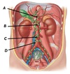
Name structures A to D

A - Gallbladder
B - Lesser omentum
C - Greater omentum
D - Transverse colon

Name abdominal structures A to E

A - Duodenum
B - Jejunum
C - Sigmoid colon
D - Caecum
E - Ileum

Name structures A to E

A - Rectus abdominus
B - External oblique
C - Internal oblique
D - Transversus abdominus
E - Parietal peritoneum

Name structures A to E

A - External oblique
B - Internal oblique
C - Transversus abdominus
D - Parietal pleura
E - Rectus abdominus

Name structures A and B

A - Falciform ligament
B - Greater omentum

Name structure A

A - Lesser omentum

Name structures A, B and C

A - Caecum
B - Mesoappendix
C - Appendix

Name structure A

A - Rectovesical pouch

Name structures A and B
A - Vesico-uterine pouch
B - Recto-uterine pouch

Name structures A to D
A - Right lobe
B - Falciform ligament
C - Left lobe
D - Round ligament of the liver

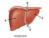
Name the structures A to F

A - Left lobe
B - Caudate lobe
C - Inferior vena cava
D - Right lobe
E - Gallbladder
F - Quadrate lobe

Name vessels A to D

A - Left gastric
B - Hepatic artery proper
D - Common hepatic artery
E - Supraduodenal artery

Name vessels A to C

A - Gastroduodenal artery
B - Splenic artery
C - Superior pancreaticoduodenal artery

Name vessels A to D

A - Right gastric
B - Left gastric
C - Right gastro-omental
D - Left gastro-omental

Name vessels A, B and C

A - Hepatic artery proper
B - Right hepatic artery
C - Left hepatic artery

Name the spaces A and B
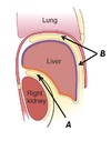
A - Subphrenic recess
B - Hepatorenal recess

Name the vessels A to D

A - Splenic vein
B - Hepatic portal vein
C - Inferior mesenteric vein
D - Superior mesenteric vein

Name structures A, B and C
A - Hepatic portal vein
B - Hepatic artery proper
C - Bile duct

Name structures A, B and C

A - Inferior vena cava
B - Portal hepatis
C - Round ligament of the liver

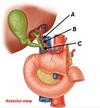
Name the areas of the gallbladder A, B and C, and duct D
A - Fundus
B - Body
C - Neck
D - Cystic duct

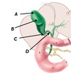
Name structures B, C and D, and the triangle A

A - Cystohepatic triangle (of Calot)
B - Cystic artery
C - Common hepatic duct
D - Cystic duct

Name structures A and B

A - Falciform ligament
B - Round ligament of the liver

Name structures A and B

A - Mesentery
B - Intestine

Name structures A to E

A - Fundus
B - Body
C - Lesser curvature (incisura angularis)
D - Pyloric sphincter
E - Greater curvature

Name structures A, B and C - the three componets of the portal triad

A - Bile duct
B - Hepatic portal vein
C - Hepatic artery proper

Name structures A to E associated with the gallbladder

A - Gallbladder
B - Cystic duct
C - Right hepatic duct
D - Left hepatic duct
E - Common bile duct

Name the three organs which articulate with the spleen at the labelled indentations

A - Stomach (gastric area)
B - Kidney (renal area)
C - Colon (colic area)

Name structures A to F

A - Branch of hepatic portal vein
B - Branch of hepatic artery
C - Biliary duct
D - Sinusoid
E - Hepatocyte
F - Central vein

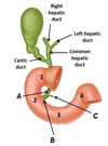
Name structures A to D

A - Cystic duct
B - Right hepatic duct
C - Left hepatic duct
D - Common hepatic duct

Name structures A, B and C
A - Bile duct
B - Hepatopancreatic ampulla (of Vater)
C - Main pancreatic duct

Name structures A and B

A - Pyloric sphincter
B - Duodenaljejunal flexure

Name A, B and C

A - Abdominopelvic splanchnic nerves
B - Superior mesenteric ganglia
C - Periarterial plexus

Name structures A and B

A - Coeliac ganglia
B - Vagus nerve

Name structures A to D

A - Hepatopancreatic ampulla (of Vater)
B - Accessory pancreatic duct
C - Bile duct
D - Main pancreatic duct (of Wirsung)

Name structures A, B and C

A - Major duodenal papilla
B - Minor duodenal papilla
C - Pyloric sphincter

Name structures A, B and C

A - Sphincter of oddi
B - Bile duct sphincter
C - Pancreatic duct sphincter

Name structures A to D

A - Inferior pancreaticoduodenal artery
B - Gastroduodenal artery
C - Splenic artery
D - Dorsal pancreatic arteries

Name vessels A and B

A - Superior pancreaticoduodenal artery
B - Superior mesenteric artery

Name stuctures A and B

A - Ileocaecal valve
B - Caecum

Name the parts of the small intestine A and B
A - Jejunum
B - Ileum

Name the structures A and B

A - Superior mesenteric artery
B - Jejunal and ileal arteries

Name vessels A, B and C

A - Hepatic portal vein
B - Superior mesenteric vein
C - Jejunal and ileal veins

Name the groups of lymph nodes A to D
A - Coeliac nodes
B - Superior mesenteric nodes
C - Inferior mesenteric nodes
D - Lumbar nodes

Name the structure labelled at points A

A - Paracolic gutter

Name structures A to D

A - Teniae coli
B - Haustra
C - Splentic flexure
D - Omental appendices

Name point A

A - McBurney’s point

Name structures A and B

A - Ileocaecal orifice
B - Appendiceal orifice

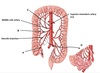
Name vessels A to D

A - Renal arteries
B - Coeliac trunk
C - Superior mesenteric artery
D - Inferior mesenteric artery

Name vessels A to E

A - Gonadal arteries
B - Lumbar arteries
C - Common iliac artery
D - Internal iliac artery
E - External iliac artery

Name vessels A to D
A - Appendicular arteries
B - Right colic artery
C - Inferior pancreaticoduodenal artery
D - Jejunal and ileal arteries

Name vessels A, B and C

A - Ileocolic branches
B - Middle colic artery
C - Superior mesenteric artery

Name structures A and B

A - Arterial arcades
B - Vasa rectae

Name vessels A to D

A - Inferior mesenteric artery
B - Left colic artery
C - Sigmoid arteries
D - Superior rectal artery

Name the large vessel, A, formed from anastomosis

A - Marginal artery of Drummond

Name vessels A to D

A - Superior rectal artery
B - Internal iliac artery
C - Middle rectal artery
D - Inferior rectal artery

Name the key points at A, B and C where the hepatic portal and systemic venous systems meet

A - Skin around umbillicus
B - Distal part of oesophagus
C - Rectum/anal canal

Name vessels A to D

A - Superior rectal vein
B - Middle rectal vein
C - Inferior rectal vein
D - Venous plexus

Name area A and muscle B
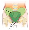
A - Pelvic inlet
B - Levator ani

Name structure A and area B
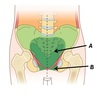
A - Pelvis
B - Perineum

Name structure A
A - Rectal ampulla

Name area A and muscle B

A - Rectovesicle pouch
B - Levator ani

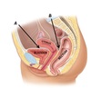
Name structures A and B and areas C and D

A - Vagina
B - Levator ani muscle
C - Vesicouterine pouch
D - Rectouterine pouch

Name muscles A, B and C

A - Iliococcygeus
B - Pubococcygeus
C - Puborectalis

Name structures A to D

A - Pubic bone
B - Puborectalis
C - Anorectal angle
D - Anal canal

Name muscle A

A - External anal sphincter

Name structures A, B and C

A - External anal sphincter
B - Pectinate line (ano-rectal junction)
C - Rectosigmoid junction

Name structures A, B and C

A - Internal anal sphincter
B - Anus
C - Levator ani muscle

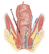
Name nerve A
A - Pudendal nerve

Name the transition at point A

A - Pectinate line (ano-rectal junction)

Name the groups of lymph nodes at points A to D

A - Lumbar
B - Common iliac
C - Internal iliac
D - External iliac

Name arteries A to D
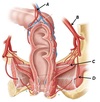
A - Superior rectal artery
B - Internal iliac artery
C - Middle rectal artery
D - Inferior rectal artery

Name veins A to D

A - Venous plexus
B - Superior rectal vein
C - Middle rectal vein
D - Inferior rectal vein

Name vessels A and B

A - Collateral veins (join with portal system)
B - Venous plexuses

Name the pathological structures A and B
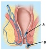
A - Internal haemorrhoid
B - External haemorrhoid

Name region A

A - Ischioanal fossa

Name the pathology at point A

A - Ischioanal fossa abscess

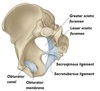
Name the structures and regions at points A, B and C

A - Obturator membrane
B - Greater sciatic foramen
C - Lesser sciatic foramen

Name the structures and regions at points A, B and C

A - Obturator foramen
B - Sacrospinous ligament
C - Sacrotuberous ligament
Name structures A, B and C

A - Liver
B - Splenic artery
C - Spleen

