Practical #1 Flashcards

1
Q
A

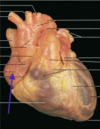
Right Ventricle

2
Q
A

Left Ventricle

3
Q
A

Right Atrium

4
Q

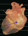
Posterior-Lateral View

A

Left Atrium

5
Q
A

Aorta

6
Q
A

Pulmonary Trunk

7
Q
A

Superior Vena Cava

8
Q
A

Inferior Vena Cava

9
Q
A

Pulmonary Veins

10
Q
A

Pulmonary Valve

11
Q
A

Aortic Valve

12
Q
A

Mitral Valve (left AV)

13
Q
A

Tricuspid Valve (right AV)

14
Q
A

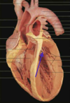
Papillary Muscle

15
Q
A

Chordae tendineae

16
Q
A

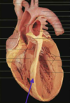
Interventricular Septum

17
Q
A

Right Coronary Artery. comes off of aorta right above aortic valve

18
Q
A

Left Coronary Artery. Comes off aorta right above valve.

19
Q
A

Coronary Sinus. “connects” with the right atrium and lies in the AV groove posteriorly.

20
Q

Smooth muscle that forms ridges in the heart. Easy to see in the atria.

A

Trabeculae carneae

21
Q

First Heart Sound

A

Due to the closing of the AV valves which happens just as the ventricles start to contract.

22
Q

Second Heart Sound

A

Due to the closing of the aorta and pulmonary valves and occurs as the ventricles begin to relax.

23
Q

Murmurs Due to Valves

A

Turbulence. Stenosis is when valves do not open properly. Insufficiency is when valves do not close properly.

24
Q

Where do you hear the aortic valve?

A

Right of the sternum in the 2nd intercostal space.

25
Q

Where do you hear the pulmonary valve?

A

Same level as the aortic just to the left of the sternum in the 2nd intercostal space.

26
Q

Where do you hear the left AV valve?

A

5th left intercostal space near the heart apendex (just below the nipple)

27
Q

Where do you hear the right AV valve?

A

4th left intercostal space next to the sternum.

28
Q

AV stenosis - Diastolic or Systolic?

A

Diastolic

29
Q

AV insufficiency - Diastolic or Systolic?

A

Systolic

30
Q

Aortic or pulmonary stenosis - Diastolic or Systolic?

A

Systolic

31
Q

Aortic or pulmonary insufficiency - Diastolic or Systolic?

A

Diastolic

32
Q

cuff to measure systolic and diastolic blood pressure

A

sphygmomanometer

33
Q

turbulent vs laminar flow

A

laminar flow makes no noise. turbulent flow is what you hear.

34
Q

Pulse Pressure

A

Difference between systolic and diastolic. = SP-DP.

35
Q

Mean Arterial Pressure (MAP)

A

average pressure over the entire cardiac cycle. = DP + 1/3PP (pulse pressure). Estimate.

36
Q

auscultatory gap

A

period of time where sounds are inaudible just below the systolic level when measuring blood pressure. Common in elderly and hypertensive.

37
Q

Oscillometric Devices

A

Use the twitching of the needle when measuring blood pressure. Measures the waveform of the pressure fluctuations.

38
Q

claudication

A

pain on walking due to arterial disease.

39
Q

Ankle-Brachial index

A

measure systolic pressure in lower legs using the posterior tibial artery or the dorsalis pedis artery. The ankle-brachial index is the systolic pressure in the ankle divided by the systolic pressure in the brachial artery. Want a ratio above 1.0

40
Q

ECG

A

As action potential spreads over the heart towards thte second lead it becomes more positive and as it moves away it becomes more negative.

Standard Lead II: recording axis runs from the right shoulder to the left hip

41
Q

Mean Electrical Axis

A

Draw vector to determine the degree of the heart. Negative degrees indicate left ventricular hypertrophy or left bundle block. Positive degrees over 90 indicate right ventricular hypertrophy or right bundle branch block.

42
Q
A

Arteries

  1. ascending aorta 2. aortic arch. 3. left subclavian 4. Don’t need to know. 5. left common carotid 6. brachiocephalic 7. right common carotid 8. don’t need to know 9. right subclavian.

Veins

  1. Superior vena cava 2. left subclavian 3. left external jugular 4. left internal jugular 5. right internal jugular 6. right external jugular 7. right subclavian
43
Q

Renal artery/vein

A

Vein: T at the top of the gonadal on the left side only. connects to the descending vena cava. Artery is just above the vein and slightly deep

44
Q

Popliteal artery/vein

A

only seen on prone cadaver and is behind the knee. vein and artery present.

45
Q

dorsalis pedis

A

only artery in foot. seen on supinated cadaver and is on medial part of foot.

46
Q

Superficial veins

A
  1. basilic: stringy thing on inside of arm not connected to muscle. 2. cephalic: stringy thing on outside running along deltoid and biceps 3. greater saphenous: inside of thigh and stringy. more medial than the femoral group.
47
Q
A

Brain

  1. anterior cerebral arteries
  2. anterior communicating artery
  3. pretend that is the middle cerebral arteries
  4. internal carotid arteries
  5. posterior communicating arteries
  6. basilar artery
  7. vertebral arteries
48
Q

Sinuses on the brains

A

Superior sagittal sinus, transverse sinus, sigmoid sinus

49
Q

pleural Cavities

A

Parietal pleura: against chest wall.

Visceral Pleura: against lung.

Interpleural space: between parietal and visceral.

Mediastinum: Space between the two lungs

50
Q

Muscles of Respiration

A

Diaphragm

External Intercostals (same directions as external obliques)

Internal Intercostals