Porphyria Flashcards

1
Q

Porphyrias result from a mutation in WHAT

A

one of the enzymes in the heme biosynthetic pathway

2
Q

Clinical manifestations of porphyrias results from WHAT

A

Accumulation of toxic metabolites (porphyrins) that cannot be cleared, due to deficient enzyme action.

The porphyrins have no useful function and act as highly reactive oxidants and damage tissues

3
Q

Inheritance pattern of porphyrias:

A

These enzyme deficiencies are inherited as autosomal dominant, autosomal recessive, or X-linked traits, with the exception of porphyria cutanea tarda (PCT), which usually is sporadic.

4
Q

Heme is part of hemoglobin, ____, _____, ____, and ____

A

Heme is part of hemoglobin, myoglobin, catalases, peroxidases, and cytochromes

5
Q

Heme is made in what cells?

A

Heme is made in every human cell (85% in erythroid cells & much of the rest in the liver, where it is used to make the P450 cytochromes)

6
Q

What is the rate-limiting step of heme synthesis?

A

First (and rate-limiting) enzyme in heme synthesis pathway is Aminolevulinic acid (ALA) synthetase (ALAS)

7
Q

Increased demand for ____ induces ALAS

A

heme

8
Q

The heme molecule downregulates ___ by feedback inhibition

A

ALAS

9
Q

ALAS:

A

ALAS is a mitochondrial enzyme that catalyzes the conversion of glycine and succinyl CoA to form delta-aminolevulinic acid. This requires pyridoxal-5’-phosphate as a cofactor.

10
Q

How is ALAS1 induced? Why are these important clinically?

A
  • Depletion of the hepatic pool of heme
  • Drugs, hormones which induce CYPs (and ALAS1)
  • Caloric and carbohydrate restriction
  • Metabolic stress, which may induce hepatic heme oxygenase and accelerate heme destruction

These are the same things that can induce a flare of the porphyria AIP

11
Q

If there’s a downstream block of heme synthesis, then inducing ____ feeds raw materials into the ____ cycle, leading to backup accumulation of the toxic porphyrins—giving symptoms of the respective porphyria

A

If there’s a downstream block, then inducing ALAS feeds raw materials into the porphyria cycle, leading to backup accumulation of the toxic porphyrins—giving symptoms of the respective porphyria

12
Q

Acute Intermittent Porphyria is caused by a deficiency of _____

A

hepatic PBG deaminase (aka hydroxymethylbilane synthase)

13
Q

What is the inheritance pattern of AIP?

A

Autosomal dominant with incomplete penetrance

14
Q

Affected individuals with AIP have a 50% reduction in _____ activity

A

erythrocyte PBG deaminase activity

15
Q

When does AIP appear clinically?

A

Generally around/after puberty

16
Q

What populations are affected by AIP?

A

Symptoms more common in females than males

17
Q

How is AIP diagnosed?

A

Test urine ALA and PBG during crisis - increased levels suggest AIP.

18
Q

In addition to low levels of PGBD activity, disease expression requires _____

A

induction of ALAS1

19
Q

Describe the clinical symptoms of an acute AIP attack:

A

GI symptoms - esp. abdominal pain (distinct from e.g. appendicitis because no inflammatory signs)

Peripheral neuropathy - sensory and motor neuropathy may precede abdominal pain. Prolonged attacks can result in bulbar paralysis, respiratory impairment, and death.

Increased catecholamines

Elevated heart rate and BP

Seizures

SIADH - hyponatremia

Dark or reddish-brown urine

20
Q

What are factors that can exacerbate an acute attack of AIP?

A

Drugs that increase demand for hepatic heme (especially cytochrome P450 enzymes)

•Crash diets (decreased carbohydrate intake)

•Endogenous hormones (progesterone)

•Cigarette smoking (induces cytochrome P450)

•Metabolic stresses (infections, surgery, psychological stress)

21
Q

How is AIP diagnosed?

A

•Send urine for PBG and ALA during an acute exacerbation

•If these are not elevated, then stop.

•If these are markedly elevated, then send PBG deaminase enzyme activity (decreased in 90% of patients, even in asymptomatic periods)

•Genetic testing is available.

22
Q

A slight elevation of urine or stool prophyrins with a normal urine ALA and PBG IS/IS NOT porphyria:

A

IS NOT!

  • These are relatively non-specific, especially if only minimally elevated.
  • Unlike AIP (or other acute porphyrias), other causes of abdominal pain do not cause elevations of urinary PBG. However, other causes of abdominal pain may be associated with elevations in urinary porphyrins (eg, hepatobiliary disease) or ALA (lead poisoning).
23
Q

How should an acute AIP attack be treated?

A

•Hospitalization to control/treat acute symptoms:

•Withdraw all unsafe medications

•Monitor respiratory function, muscle strength, neurological status

•Intravenous 10% glucose at least 300 g per day

•Intravenous hematin ASAP (can give IV glucose while waiting for IV hematin)—hematin is heme, which can feedback inhibit ALA-synthase.

  • Cimetidine for treatment of crisis and prevention of attacks
  • In rare instances where pts have severe, unremitting symptomatic disease, liver transplantation has been done
  • There is a clinical trial using siRNA to shut off synthesis of ALAS1 for patients with frequent relapses
24
Q

Hematin mechanism of action:

A

Reduces production of ALA /porphyrins by negative feedback inhibition on ALA synthetase

Derived from outdated PRBCs from community blood banks

25
Q

What are the adverse reactions of hematin?

A

(Due to degradation products binding to endothelial cells, platelets, & coagulation factors)

  • Thrombophlebitis
  • Anticoagulation (transient ↑ PT, bleeding may occur)
  • Thrombocytopenia
  • Iron overload with repeated use
26
Q

Porphyria Cutanea Tarda is caused by a deficiency of ___

A

Uroporphyrinogen decarboxylase (UROD)

27
Q

Is porphyria cutanea tarda inherited or acquired?

A

Can be either -

Acquired - type I

Inherited - type II

28
Q

Describe the patient population with Cutanea Tarda:

A
  • 60% of PCT patients are male, most of whom ingest excess alcohol.
  • Women who develop PCT are often on estrogen-containing medications.
  • Most patients are ≥ 40 years, and 66% have evidence of iron overload.
29
Q

Uroporphyrinogen decarboxylase (UROD):

A

This enzyme catalyzes the sequential decarboxylation of the four acetyl side chains of uroporphyrinogen to yield coproporphyrinogen.

30
Q

Pathogenesis of cutanea tarda:

A

•Iron overload leads to reduced activity of the UROD enzyme which leads to elevated porphyrin levels, in particular uroporphyrins.

•Hepatic iron levels are correlated with clinical expression of disease.

  • Dysregulation of hepcidin, leading to increased iron absorption.
  • PCT does not appear to be associated with marked upregulation of delta-aminolevulinic acid (ALA) synthase (ALAS1)
  • Associated disorders :
  • Alcoholism.
  • Hemochromatosis.
  • Hepatitis C
31
Q

Describe the skin findings associated with cutanea tarda:

A

•Uroporphyrinogen is made in the liver, but deposits in the skin. Upon exposure to sunlight, reactive oxygen species are generated which further damage the skin, leading to the skin findings

  • Blisters, bullae, increased fragility, scarring and hyper- and hypopigmentation affecting sun-exposed areas of the body, such as the backs of the hands, forearms, face, ears, neck, and feet.
  • Bullae contain porphyrin-rich serous or serosanguinous fluid, and may be painful and become infected
  • Hirsutism is also common, especially on the cheeks and forearms
32
Q

How is the liver affected by PCT?

A
  • Liver enzymes (AST and ALT) are almost always elevated
  • Liver damage is multifactorial:
  • Porphyrins damage the liver
  • Associated disorders are also associated with liver damage
    • Hepatitis C
    • hemochromatosis

•Increased risk for cirrhosis and hepatocellular carcinoma

33
Q

How is PCT diagnosed?

A
  • Screening test - Total plasma porphyrin levels
  • ALA and PBG will be normal, since ALAS1 is not induced
  • Confirmatory testing - More specific levels of urinary porphyrins to speciate the porphyria
34
Q

How is PCT treated?

A

•First, remove all offending agents

  • No alcohol
  • Withhold estrogen
  • Stop smoking

•Cover sun-exposed areas and fanatic application of sunscreen

•Next, start phlebotomy every week or 2:

  • This removes iron sequentially—do this until the ferritin falls to <20
  • Hold for anemia
  • Almost everyone achieves remission
35
Q

What levels are considered lead toxicity?

A

Blood levels >10mcg/dL for adults

Blood levels >5mcg/dL for children

36
Q

What are common sources of lead in adult patients?

A

Workplace exposure - manufacturing batteries, solder, radiators, some cosmetics, ammunition, etc.

Leaded gasoline

Mining

Lead paint - esp. construction workers

X-ray film stored in lead boxes

Moonshine and marijuana

Firing ranges

37
Q

What are common sources of lead exposure in children?

A

Primarily ingestion -

  • Lead-containing dust
  • Paint
  • Soil
  • Water
  • Food
  • Breastfeeding
  • Leaded objects (e.g. fishing weights)

Some problems with inhalation in children near home renovations or factories.

38
Q

How does lead enter the body?

A
  • Inhalation - Major route in adults (40% absorption rate)
  • Ingestion - Major route in children (50% absorption)
39
Q

What does lead do once it enters the body?

A

•99% bound to red cells

  • 1% in plasma, transported and exchanged in soft tissues (kidney, brain, liver, bone marrow)
  • Can be excreted via the kidneys—gone in 30 days if normal kidney function
  • Can be stored in the bone:
  • Can last for decades—95% of body lead stored here
  • Released from bone during times of high bone turnover (Pregnancy, hyperthyroidism, menopause, breast feeding)
40
Q

Lead inhibits sulfhydral groups in RBC enzymes ___ and ___ in the heme synthetic pathway.

A

ALAS and ferrochelatase

41
Q

Lead inhibits _____, leading to degradation of ribosomal RNA in red blood cells that causes ____ (visible on smear)

A

Lead inhibits Pyrimidine 5’ nucleotidase, leading to degradation of ribosomal RNA in red blood cells that causes basophilic stippling (visible on smear).

42
Q

Lead competes with _____ in biologic symptoms, which especially impacts the nervous system and mitochondrial respiration

A

calcium

43
Q

____ alters the permeability of the blood brain barrier and accumulates in astroglia

A

Lead

44
Q

Lead promotes the generation of ____ in endothelial and vascular smooth muscle cells. Lead also affects ____ and ____.

A

Lead promotes the generation of reactive oxygen species in endothelial and vascular smooth muscle cells. Lead also affects cell membranes and DNA/RNA.

45
Q

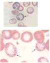
What is shown in this image?

A

Basophilic Stippling - characteristic of lead poisoning

  • Coarse dark blue inclusions in red cells, representing precipitated ribosomes
  • Seen in young red cells in conditions associated with RNA instability
46
Q

Describe the acute symptoms of lead toxicity:

A

•GI effects - crampy abdominal pain (lead colic), constipation

•Anemia—typically microcytic

•Neurologic effects – confusion, seizures, fatigue, headache, difficulty concentrating, sleep disturbance

  • Peripheral neuropathy (predominantly affecting motor nerves)
  • classic finding – wrist and foot drop
  • Muscle and joint pain
  • Decreased libido

•“Lead line” around teeth/gums

47
Q

Describe the chronic effects/symptoms of lead exposure:

A

•Hypertension (an association between lead levels in bone and blood and hypertension)

•Neuropsychiatric effects

  • Declines in neurocognitive function
  • Psychiatric symptoms – phobia, anxiety, depression, hostility
  • Distal sensory and motor neuropathy
  • EKG conduction delay
  • Decreased hearing acuity
  • Mortality – increased mortality with chronic exposure, even to low levels of lead
  • Reproductive effects:
  • Increased miscarriages and stillbirths with high maternal lead levels
  • Reduced birth weight and cognitive impairment in children born of mothers exposed to lead
  • Abnormal sperm morphology and decreased sperm concentration, total sperm count, and total motile sperm count.

•Lead nephropathy – pathology shows a chronic interstitial nephritis associated with high lead levels.

  • Associated with increased hypertension and gout
  • “Saturnine gout” (gout and depression/sadness)
  • May accelerate renal damage in patients with underlying renal injury
48
Q

How is lead toxicity diagnosed?

A

•Blood lead levels:

  • key clinical monitoring test for diagnosing lead toxicity
  • Make sure to use adult ranges for adults and childhood ranges for kids
  • Venous stick, rather than earlobe capillary stick (which can be contaminated by skin surface lead)

•Free erythrocyte protoprophyrin (FEP) and Zn protoporphyrin (ZPP) levels measure lead’s effect on heme production:

  • can be used as an indicator of lead exposure (and effect) over the preceding three-month period
  • If the FEP or ZPP are elevated, then need to do CBC, renal function testing.
49
Q

How should lead toxicity be managed?

A

Reduce lead exposure

Chelation therapy:

  • Used when there are symptoms of acute toxicity, or when levels need to be reduced quickly.
  • no randomized clinical trials that have proven the efficacy of chelation on clinical outcomes, particularly long-term outcomes, in lead poisoned adults
  • Chelation with any agent should not be undertaken unless exposure has been definitively curtailed, since its use in the presence of continuing exposure may result in enhanced absorption of lead and worsening, rather than amelioration of toxicity.
  • Two agents most commonly used are DMSA and Calcium EDTA (NOT disodium EDTA)