Pigments and tissue deposits, developmental anomalies, neoplasia part II, bones Flashcards

(331 cards)

1
Q

Matching! Which of these are pigments and which are tissue deposits?

A. Pigments

B. Tissue deposits

  1. Hematogenous pigments (hemoglobin, hemosiderin, bilirubin, porphyrins)
  2. Uric acid
  3. Calcification (dystrophic, metastatic, calcinosis cutis)
  4. Melanin
  5. Exogenous pigments
  6. Lipofuscin
  7. Amyloid
A

A. Pigments:

  • 1- hematogenous pigments (hemoglobin, hemosiderin, bilirubin, porphyrins)
  • 4- melanin
  • 5- exogenous pigments
  • 6- lipofuscin

B. Tissue deposits:

  • 2- uric acid
  • 3- calcification (dystrophic, metastatic, calcinosis cutis)
  • 7- amyloid
2
Q

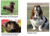
You are called out to a sheep farm with increased mortality and you notice several ewes are weak and have pale yellow mucous membranes. What is the term for yellow discoloration of a tissue?

  1. Gout
  2. Icterus
  3. Xanthosis
  4. Jaundice
A
  1. Icterus and 4. Jaundice are both increased bilirubin in tissues

grossly: yellow-green discoloration of tissue or fluid, most prominent in mucous membranes, adventicial surfaces (do not use fat to assess, especially in livestock because they store carotenoids there; sclera of eyes and intima of vessels a good place to look)

3
Q

What is causing the yellow color in jaundice/icterus?

  1. Hemosiderin
  2. Bilirubin
  3. Hemoglobin
  4. Haptoglobin
  5. Melanin
A

TEST QUESTION

  1. Bilirubin
    * (break down of RBC > macrophages break down hemoglobin into heme, globin, and iron > heme breaks down further into bilirubin)*
4
Q

Which enzymes break down heme (from hemoglobin) into bilirubin?

(pick 2)

  1. Biliverdin reductase
  2. Heme oxygenase
  3. Biliverdin oxygenase
  4. Heme reductase
A
  1. Biliverdin reductase and 2. Heme oxygenase

EXCEPT birds do not have biliverdin reductase, so the end product is biliverdin

5
Q

Once unconjugated bilirubin gets into circulation, hepatocytes will take it up and conjugate it with _______________ so it can be secreted into bile.

A

Diglucuronide

6
Q

What is too much bilirubin in the blood?

A

Hyperbilirubinemia

If it gets > 2mg/dL, you get jaundice (it can be conjugated or unconjugated, it will have the same color)

7
Q

What are the 3 ways that jaundice can occur?

A

TEST QUESTION

  1. Prehepatic hyperbilirubinemia: unconjugated bilirubin production exceeds hepatocellular uptake; caused by hemolysis (intravascular or extravascular) [WHAT DIAGNOSTIC TEST WOULD YOU USE? PCV]
  2. Hepatic hyperbilirubinemia: hepatocellular disfunction (decreased bilirubin uptake, decreased conjugation, decreased secretion in bile) = build up of conjugated and unconjugated bilirubin in blood; caused by hepatic insufficiency, hepatitis, hepatocellular degeneration; severe liver disease
  3. Posthepatic hyperbilirubinemia: reflux of conjuaged bilirubin into blood; caused by biliary obstruction (cholestasis) or rupture

KNOW THIS

8
Q

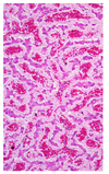
What will you see microscopically in a patient with jaundice?

A
  • Do not see pigment in jaundiced tissues!
  • Exception: severe cholestatic (obstructive bile flow) liver
  • Yellow-brown intracellular (hepatocytes, kupffer cells) or extracellular pigement (bile canalliculi)
9
Q

Decreased bilirubin uptake by hepatocytes is

  1. Prehepatic hyperbilirubinemia
  2. Hepatic hyperbilirubinemia
  3. Posthepatic hyperbilirubinemia
A
  1. Hepatic hyperbilirubinemia: hepatocellular disfunction (decreased bilirubin uptake, decreased conjugation, decreased secretion in bile); caused by hepatic insufficiency, hepatitis, hepatocellular degeneration
10
Q

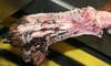
What mechanism of jaundice is to blame with this sheep with widespread jaundice, black kidneys, red-colored urine, and a normal liver?

  1. Prehepatic hyperbilirubinemia
  2. Hepatic hyperbilirubinemia
  3. Posthepatic hyperbilirubinemia
A
  1. Prehepatic hyperbilirubinemia

Urine is red from hemoglobinuria (red-brown coloration of kidney and urine, pink serum); microscopically, homogenous red-orange material in renal tubules

11
Q

How can you tell if hemoglobinuria is from RBCs, hemoglobin, or myoglobin?

A

Serum

Hemoglobin binds to haptoglobin to be carried in serum = pink serum; myoglobin does not = clear

12
Q

How does hemoglobinuria look microscopically?

A

Homogenous red-orange material in renal tubules

13
Q

In what type of hemolysis will you see hemoglobinuria?

  1. Intravascular hemolysis
  2. Extravascular hemolysis
A
  1. Intravascular hemolysis (RBC lysis in blood vessels > haptoglobin saturated > Hb free in blood > Hb freely filtered by kidney )

(extravascular hemolysis is what we usually see: abnormal RBCs taken out of circulation by phagocytes > Hb not free in blood to be filtered by kidney > no hemoglobinuria; but both can cause jaundice)

14
Q

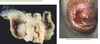
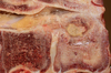
What is the etiology of a sheep with gun metal kidneys (black kidneys), widespread jaundice, normal liver, and red colored urine?

A

TEST QUESTION

Acute copper toxicosis

(sheep are insufficient in metallothionein for safe copper storage in the liver > chronic accumulation of copper in the liver > mild liver damage > acute copper release > oxidative RBC damage > intravascular hemolytic anemia > hemoglobinuria)

15
Q

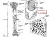
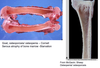
Will we see this change with intravascular hemolysis (intimal surface of a aorta from a horse)?

A

No, it is a post-mortem change; hemoglobin imbibition

16
Q

Which is the most severe?

  1. Prehepatic hyperbilirubinemia
  2. Hepatic hyperbilirubinemia
  3. Posthepatic hyperbilirubinemia
A
  1. Posthepatic hyperbilirubinemia

(no bilirubin leaving)

17
Q

Which has the slowest onset?

  1. Prehepatic hyperbilirubinemia
  2. Hepatic hyperbilirubinemia
  3. Posthepatic hyperbilirubinemia
A
  1. Hepatic hyperbilirubinemia
18
Q

Prehepatic, hepatic, or posthepatic hyperbilirubinemia? (dog, generalized jaundice, normal liver)

A

Prehepatic hyperbilirubinemia (IMHA, usually no intravascular hemolysis)

19
Q

Prehepatic, hepatic, or posthepatic hyperbilirubinemia? (puppy, generalized jaundice, smaller liver)

A

Hepatic hyperbilirubinemia (infectious canine hepatitis/adenovirus)

20
Q

Prehepatic, hepatic, or posthepatic hyperbilirubinemia? (fat cat, intimal surface of aorta is yellow, liver enlarged, pale/tan, greasy)

A

Hepatic hyperbilirubinemia (hepatic lipidosis)

21
Q

Prehepatic, hepatic, or posthepatic hyperbilirubinemia? (horse, green-brown liver)

A

Posthepatic hyperbilirubinemia (biliary calculus)

22
Q

What would you do with this sick foal to determine the cause of jaundice in this case? (list 4 things you can do clinically)

A

TEST QUESTION

  1. Urinalysis for intravascular hemolysis
  2. Serum chemistry to check bilirubin (liver enzymes)
  3. Liver imaging (x-ray, U/S)
  4. CBC (hematocrit or PCV for anemia; if there is no anemia = not prehepatic)
23
Q

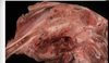
A sick foal was diagnosed with neonatal isoerythrolysis. Its mother (A/Q negative) had given birth to a foal with A/Q blood type with a A/Q positive stallion. The fetal cells passed to the mare’s blood during gestation and she became sensitized. She bred again to the A/Q stallion and the 2nd foal ingested colostrum packed with antibodies against its blood type and resulted in intravascular hemolysis. What other lesions would this foal have?

A

Red kidneys and hemoglobinuria

24
Q

What pigments are responsible for the color of this bruise?

A

Hemoglobin- red from oxygenized hemoglobin, blue from deoxygenized hemoglobin

Bilirubin- yellow/green

Hemosiderin- tan/brown

25
What happens to the iron that resulted from the break down of hemoglobin?
Macrophages bind to iron and is stored intracellularly as ferritin (bound to apoferritin)/ hemosiderin *_Gross_: must have a lot to impart gross brown color* *_Microscopic_: dark yellow-brown, coarse granular cytoplasmic pigment; **stains blue-black with prussian blue/Perl's stain*** ex. "heart failure cells" in the lungs due to chronic congestion
26
What causes hemosiderin accumulation?
Processes associated with RBC breakdown 1. _Local_: chronic congestion and hemorrhage 2. _Widespread_: hemolysis and inherited disorders of Fe storage
27
This pluck from a dog shows ____________ hemosiderosis on the lungs. 1. Mild 2. Moderate 3. Severe
3. Severe
28
What do the blue pigments from this Prussian blue stain represent?
**Iron**; Mynah bird liver, generalized hemosiderosis due to hemolysis
29
Erythropoietic porphyria is a developmental anomaly of calves, cats, and pigs in which they are deficient in \_\_\_\_\_\_\_\_\_\_\_\_\_\_\_\_\_\_\_\_\_\_\_\_\_\_, which causes defects in heme synthesis and causes porphyrin accumulation in bones and teeth.
Uroporphyrinogen III cosynthetase ## Footnote *(Erythropoietic porphyria: grossly will see pink-red discolored bones and teeth, "Pink Tooth", fluoresces with UV light)*
30
What pigment is black/brown tissue color grossly, and fine brown/black cytoplasmic granules when seen histologically.
Melanin
31
How can we get increased melanin in the epidermis?
Increased in tyrosinase, a copper binding enzyme, with UV light exposure, hormones (MSH), or inflammation
32
_Cutaneous_ hyperpigmentation (hypermelanosis) in dogs due to flea allergy dermatitis is a common place to see melanin. Where else can you find melanin?
* Sheep brain, _meningeal melanosis_ (intracellular, dark black granules confined to meninges); developmental anomaly * Sheep uterus, _endometrial melanosis_; developmental anomaly * Cow lungs, _pleural melanosis_; developmenal anomaly
33
Is this change the result of melanin accumulation?
No, it is a post-mortem change; pseudomelanin (hydrogen sulfide from bacteria)
34
What is the 'wear and tear' pigment that is derived from the breakdown of lipids commonly found in aged (neurons and muscle cells) and injured cells?
TEST QUESTION Lipofuscin *(they are un-degradable remnants of breakdown of organelles; composed of lipid complexes with protein; especially accumulates in post-mitotic cells)* _Gross_: usually nothing, tissue obtains a brownish color after large amount of accumulation _Histo_: golden brown, fine granular cytoplasmic pigment
35
What golden-brown pigment is seen in the intestines of a dog after a large amount of accumulation?
Lipofuscin ## Footnote *(bottom img: myocardiocytes)*
36
How can you differentiate hemosiderin from lipofuscin if both are yellow-brown cytoplasmic granules?
Prussian blue stain for iron! (hemosiderin)
37
What type of pigment is in this lesion (brain, transversely cut) from a young cat? 1. Endogenous 2. Exogenous
2. Exogenous ## Footnote *(black = melanin; inflammation or neoplasm, in this case it was inflammation, fungal hyphae = brown)*
38
What can you say about the pigment from this lesser omentum of a pony?
Not jaundice! It is **omental carotenoid pigmentation** (exogenous pigmentation); vitamin A accumulates in fatty tissues and causes yellow-orange discoloration
39
Is this pigment from a dog's lung endogenous or exogenous?
**Exogenous**; *pulmonary anthracosis* from inhaled carbon which depsoited in peribronchiolar macrophage aggregates; do not really see this grossly _Pneumoconiosis_- inhaled dust, anthracosis is a subtype of this *(it resembles hemosiderin, so the next step would be Perl's stain)*
40
Is the pigment from this sheep liver endogenous or exogenous?
**Exogenous**; chronic cholangitis (fibrotic walls), liver flukes digesting blood \> biliary parasitic hematin, looks like hemosiderin, but it is around parasites *(bottom img: macaque lung, chronic bronchiolitis and parasitic hematin & lung mites)*
41
During necropsy of a young dog, you notice white chalky plaques on the parietal pleura of the ribs and dry, white streaks on the gastric mucosa. The lungs failed to collapse and felt dry to the touch. Microscopically, it had basophilic intracellular and extracellular pleomorphic granular material that stained black with vonKossa stain. Is this a pigment or tissue deposit?
Tissue deposit \> calcification
42
What does calcification look like grossly and histologically?
TEST QUESTION _Grossly_: white, gritty granules/plaques; hard (can't cut through it) _Histologically_: basophilic amorphous granules of inconsistent size/shape; stains black with vonKoss
43
What are the 2 types of calcification?
1. Dystrophic 2. Metastatic
44
Which type of calcification is _local deposition_ of calcium in areas of injury, especially necrotic fat?
Dystrophic * Local deposition of calcium in **areas of injury** * Especially **necrotic fat**- calcium ions interact with fatty acids, producing insoluble calcium soaps ('saponification') * Also necrotic muscle, **granulomas, dead parasites**
45
Which type of calcification is _widespread deposition_ of calcium in otherwise normal tissues?
Metastatic * Widespread deposition of calcium in otherwise normal tissues * Caused by **hypercalcemia** * Favorite sites: vascular intima/adventicia, gastric mucosa, renal tubular epithelium, pulmonary interstitium, pleura, basement membranes
46
Which type of calcification is caused by too much calcium in circulation?
Metastatic
47
What is the best morphological diagnosis for this dog with white chalky plaques on the parietal pleura? 1. Metastatic calcification 2. Dystrophic calcification
TEST QUESTION (SAME IMAGE) 1. Metastatic calcification (widespread distribution) * (next, we want to know why the dog has hypercalcemia; from cholecalciferol rodenticide)*
48
What do the following do to the calcium levels in the blood? 1. Parathyroid hormone 2. Vitamin D 3. Calcitonin
1. Parathyroid hormone: increase in calcium 2. Vitamin D: increase in calcium 3. Calcitonin: decrease in calcium
49
What are the 2 causes of hypercalcemia?
1. **Excess PTH**: renal failure in small animals: hyperphosphatemia \> hypocalcemia \> PTH overcorrects \> hypercalcemia 2. **Excess vitamin D**: toxins
50
Does this pancreas and mesentery from a dog have dystrophic or metastatic calcification?
Dystrophic (necrotic fat); acute pancreatitis
51
Does this liver from a cow have dystrophic or metastatic calcification (gritty granuloma with necrotic center)?
Dystrophic ## Footnote *(bottom img: cow lung granuloma, tuberculosis)*
52
Does this heart from a cow have dystrophic or metastatic calcification?
Metastatic; a plant, *Cestrum diurnum*, toxicity (vitamin D analog \> leads to hypercalcemia)
53
Does this skin of a dog have dystrophic of metastatic calcification?
Not really metastatic or dystrophic **Calcinosis cutis:** * Dogs with hyperadrenocorticism * Pathogenesis not understood * Widespread mineralization of the dermal collagen and epidermal basement membrane *(characteristic of Cushing's disease)*
54
What do you call fibrils made of _stacked beta-pleated sheets_ that can be formed by lots of different protein monomers?
TEST QUESTION **Amyloid**; they are deposited and accumulates in extracellular spaces- compresses adjacent tissues causing atrophy
55
What does amyloid look like grossly?
TEST QUESTION _Grossly_: enlarged (only deposit that does this), firm organs with waxy appearance; stains blue violet when treated with iodine and sulfuric acid *(img: macaque liver, hepatic amyloidosis)*
56
What does amyloid look like histologically?
Histologically: amorphous homogenous eosinophilic extracellular material ("hyaline"); stains pink and has green birefringence with polarized light when stained with Congo Red ## Footnote *(img: dog liver, hepatic amyloidosis)*
57
What are the 4 types of amyloid?
1. **Amyloid light chain (AL)**: associated with monoclonal beta-lymphocyte proliferation; derived from IG light chain 2. **Amyloid A**: associated with chronic inflammatory conditions (breed predisposition; most common); made by liver 3. **Endocrine amyloid**: associated with diabetes 4. **A beta amyloid**: associated with Alzheimer disease
58
Is this subcutaneous mass from a young dog have dystrophic or metastatic calcification (discrete nodular mass, feels like bone)?
Dystrophic, **calcinosis circumscripta**
59
A dog that you treated for protein losing nephropathy with pitting edema died, and upon necropsy, you notice this kidney. What is the MDx?
Glomerular amyloidosis; consistent with the disease "reactive systemic amyloidosis"
60
What type of amyloid is deposited in "reactive systemic amyloidosis"? 1. Amyloid light chain 2. Endocrine amyloid 3. Amyloid of alzheimer's disease 4. Amyloid A
TEST QUESTION (FOCUS ON INFLAMMATION) **4. Amyloid A** (formed from _chronic inflammation_) Chronic inflammation \> liver produces SAA in response to IL-4 and IL-6 \> spontaneous conversion of SAA to AA \> formation of amyloid fibrils \*MOST COMMON FORM IN ANIMALS, HEREDITARY IN SHARPEIS AND ABYSSINIANS KIDNEY, LIVER, SPLEEN, LYMPH NODES\*
61
What is the disease in which uric acid accumulates in tissues?
Gout
62
Which species does not have **uricase**, in which uric acid is the end product?
Birds and reptiles; get gout from **decreased renal function,** dehydration
63
In mammals, what is uric acid converted into with uricase?
Urea is the end product
64
How do mammals get gout?
From diet, genetic disorders, chemotherapy
65
What does uric acid look like grossly?
Chalky, white foci on surface of visceral organs and serous membranes (liver, myocardium, spleen, pleura, air sacs, etc..."visceral gout"); may involve soft tissues around joints ("articular gout")
66
What does uric acid look like histologically?
Needle-like clear spaces (crystals dissolve out in processing); "Tophi"- granulomatous **inflammation** surrounding deposit (different from others deposits because it causes an inflammatory response) ## Footnote *(img: Macaw kidney)*
67
What deposit is on this snake lung and kidney with visceral gout?
Uric acid (differentiate from calcium deposits because this is "softer" and can scrape it off easily)
68
What deposit is on this swan heart with visceral gout?
TEST QUESTION (URIC ACID BUILD UP FROM RENAL DYSFUNCTION IN BIRDS) Uric acid
69
What could this pigment be? What is the MDx?
Melanin, MDx: vertebral melanosis
70
What's up with this hilar lymph node of a cow?
Severe anthracosis
71
T or F. Genetic lesions are defects of growth or development of a tissue or organ that are present, but not necessarily obvious, at birth.
TEST QUESTION False, do not confuse congenital with the terms genetic or inherited. *(congenital = born with the abnormality, genetic = can be born fine but the disease may manifest later)* CONGENITAL lesions are defects of growth or development of a tissue or organ that are present, but not necessarily obvious, at birth.
72
Which of these are developmental anomalies?
All are developmental anomalies (B is an anatomical anomaly, A and C are biochemic anomalies)
73
Matching! Which of the following are anatomic malformation and which are biochemical defects? A. Anatomic malformations B. Biochemical defects 1. Failure to fuse 2. Ectopic development 3. Involve an inability to synthesize adequate amounts of a particular enzyme or other protein 4. Abnormal development of a structure 5. Usually no grossly visible malformation 6. Haemophilia, lysosomal storage disease, dermatosporaxis, albinism 7. Failure to canalize/separate 8. Cysts 9. Failure of a structure to develop 10. Usually due to genetic mutation
A. Anatomic malfrmations: 1. Failure to fuse 2. Ectopic development 4. Abnormal development of a structure 7. Failure to canalize/separate 8. Cysts 9. Failure of a structure to develop B. Biochemical defects: 3. Involve an inability to synthesize adequate amounts of a particular enzyme or other protein 5. Usually no grossly visible malformation 6. Haemophilia, lysosomal storage disease, dermatosporaxis, albinism 10. Usually due to genetic mutation
74
Summer, NE NSW, many cows aborting late-term fetuses that look like this...what is morphologically abnormal about this aborted calf?
Limbs are fixed in flexion, MDx: arthrogryposis
75
Explain the pathogenesis of arthrogryposis.
Limbs do not move during development \> muscles don't develop correctly \> connective tissue matures \> limbs fixed in place; joints are usually fine
76
What is the MDx of this brain from a calf who was born like this during calving season (owner said it was dumb, depressed, some were blind)?
Hydranencephaly (cerebral hemispheres are missing, filled with cerebral spinal fluid)
77
Are both of these lesions abnormalities of the nervous system? Are these caused by the same thing?
Final disease Dx = **Akabane (Bunyavirus)** **All calves infected with akabane virus at the same time, but they are at different stages of gestation (some conceived earlier than others)** Infected 120-180 days- arthrogryposis (see first) Infected 80-100 days- hydranencephaly (see second)
78
Why do anatomical developmental anomalies occur?
They occur due to injury of cells during embryogenesis
79
The nature of the anatomical defect that results depends on what 2 things?
1. Timing of injury 2. Tissue injured
80
Injury during what period of gestation does developmental anomalies occur?
Embryo (organogensis)
81
What tissue would need to be injured to result in arthrogryposis?
Muscles and nervous tissues
82
This is a calf with severe nonsuppurative myelitis (spinal cord in early gestation), how can we determine the cause of the arthrogryposis and hydranencephaly? 1. Serology of dam 2. Serology of calves 3. Virus isolation 4. Histopathology
2. Serology of calves
83
What are some causes of developmental anomalies?
* **Genetic defect** * **_In utero_ infection- BVD, bluetongue, border disease, akabane, panleukopenia, classical swine fever,** * **_In utero_ exposure to teratogens- toxic plants, griseofulvin in cats** * _In utero_ nutritional deficiencies
84
What is the MDx and etiology of this brain from a cat?
MDx: cerebellar hypoplasia Etiology: Feline panleukopenia virus (parvovirus)
85
What is the MDx and etiology of this oral cavity from a cat?
TEST QUESTION MDx: palatoschisis ("cleft palate") Etiology: Griseofulvin used while pregnant (teratogen)
86
What is the MDx and etiology of this lamb?
MDx: synophthalmia (orbits and eyes fused) Etiology: *Veratrum californicum* day 14 gestation (toxic plant)
87
What is the MDx and etiology of the intestines from this foal?
Disease: Lethal white syndrome (no melanin, albino) MDx: colonic hypoplasia Etiology: inherited genetic trait
88
What is the MDx of this dog?
MDx: hydrocephalus (ventricles filled with fluid and surrounding tissues atrophy)
89
What are genetic etiologies of development anomalies?
* Inherited genes- often autosomal recessive * Spontaneous somatic genetic defect * Chromosomal anomalies * Breed dispositions *(chihuahuas with hydrocephalus)*
90
What are developmental anomalies of Persian cats? (FYI)
Polycystic kidney disorder (PKD), cardiomyopathy, cataracts, cleft palates and cleft lips, mega-esophagus, portosystemic shunt (liver), patent ductus arteriosus (PDA), entropion, amyloidosis, mucopolysaccharidosis
91
Which of the following is _not_ an anatomic malformation? 1. Failure to fuse 2. Abnormal development of a structure 3. Failure to canalize/separate 4. Ectopic development 5. Cysts 6. Usually due to a genetic mutation 7. Failure of a structure to develop
6. Usually due to a genetic mutation
92
What is the MDx, disease name, and type of anatomical defect of this pancreas from a dog?
MDx: pancreatic aplasia (no pancreas at all) Disease name: Exocrine pancreatic insufficiency Type of anatomical defect: failure to develop
93
What is the MDx, disease name, and type of anatomical defect of this pancreas from a dog?
MDx: pancreatic hypoplasia (just a little bit of pancreatic tissue) Disease name: Exocrine pancreatic insufficiency Type of anatomical defect: failure to develop
94
What is the MDx and type of anatomical defect of this calf and puppy?
MDx: spina bifida (neural tube defect, including skin) Disease name: Spina bifida Type of anatomical defect: failure to fuse
95
What is the MDx and type of anatomical defect of this brian from a calf?
MDx: meningoencephalocele *(meninges and brain protruding out into the subcutis around brain, head equivalent of spina bifida)* Type of anatomical defect: failure to fuse (bones didn't fuse)
96
What do you call a cleft in the dorsal tissues of the spine? 1. Schistosomus reflexus 2. Interventricular cardiac septal defect 3. Coloboma 4. Hypospadias 5. Cheiloschisis 6. Palatoschisis 7. Spina bifida
7. Spina bifida
97
What do you call a cleft in the hard palate (cleft palate)? 1. Schistosomus reflexus 2. Interventricular cardiac septal defect 3. Coloboma 4. Hypospadias 5. Cheiloschisis 6. Palatoschisis 7. Spina bifida
6. Palatoschisis
98
What do you call a hole communicating between the ventricles of the heart? 1. Schistosomus reflexus 2. Interventricular cardiac septal defect 3. Coloboma 4. Hypospadias 5. Cheiloschisis 6. Palatoschisis 7. Spina bifida
2. Interventricular cardiac septal defect
99
What do you call a cleft in the penis exposing the urethra? 1. Schistosomus reflexus 2. Interventricular cardiac septal defect 3. Coloboma 4. Hypospadias 5. Cheiloschisis 6. Palatoschisis 7. Spina bifida
4. Hypospadias
100
What do you call a cleft lip? 1. Schistosomus reflexus 2. Interventricular cardiac septal defect 3. Coloboma 4. Hypospadias 5. Cheiloschisis 6. Palatoschisis 7. Spina bifida
5. Cheiloschisis
101
What do you call a cleft in the internal structures of the eye? 1. Schistosomus reflexus 2. Interventricular cardiac septal defect 3. Coloboma 4. Hypospadias 5. Cheiloschisis 6. Palatoschisis 7. Spina bifida
3. Coloboma
102
Which of the following is _not_ a fusion defect? 1. Cheiloschisis 2. Palatoschisis 3. Pancreatic aplasia 4. Spina bifida 5. Schistosomus reflexus 6. Interventricular cardiac septal defect 7. Coloboma 8. Hypospadias
3. Pancreatic aplasia
103
What is the MDx and type of anatomical defect of this head from a calf?
MDx: maxillary brachygnathia and palatoschisis (cleft palate) Type of anatomical defect: failure to fuse
104
What is the MDx and type of anatomical defect in this calf?
MDx: schistosomas reflexus (ventrum not fused, so all viscera are eviscerated) Type of anatomical defect: failure to fuse *(sternal cleft, dorsal reflection of ribs, eventration of viscera, non-union of pelvic symphysis)*
105
What is the MDx and type of anatomical defect in this lamb?
MDx: atresia ani (failure to open) Type of anatomical defect: failure to canalize
106
What is the MDx, disease name, etiology, and type of anatomical defect of the kidneys from this cat?
MDx: renal cysts Disease: Polycystic kidney disease Etiology: inherited genetic defect Type of anatomical defect: cyst
107
What is the MDx, disease name, and type of anatomical defect of the kidneys from this cat?
MDx: pituitary cyst (Raphke's pouch cyst) [anterior pituitary does not form] Disease: Pituitary dwarfism Type of anatomical defect: cyst
108
What is the MDx, disease name, and type of anatomic defect?
MDx: proportionate dwarfism (both are from the same litter) Disease: Pituitary dwarfism Type of anatomical defect: cyst
109
What is the MDx and type of anatomical defect of the feet from this calf?
MDx: polydactyly Type of anatomical defect: abnormal development of a structure
110
What is the MDx and type of anatomical defect of the thoracic limbs from this sheep?
MDx: polymelia Type of anatomical defect: abnormal development of a structure *(don't confuse it for polydactyl, which is an error during embryo formation)*
111
What is the MDx, disease name, and type of anatomical defect of the coxofemoral joints from this dog?
MDx: coxofemoral malformation Disease name: Hip dysplasia Type of anatomical defect: abnormal development of a structure *(flat heads of both femurs, flat acetabular rims, one head luxated out of joint)*
112
What is the MDx and type of anatomical defect of heart from a calf?
MDx: ectopia cordis (heart outside of thoracic cavity) Type of anatomical defect: ectopic development
113
This calf with worsening neurological signs, otherwise looks okay. It was normal at birth. Does this rule out a _developmental_ anomaly?
No, because not all developmental anomalies are congenitally wrong.
114
What is the MDx, disease name and etiology of this brain from a calf (grossly, looks fine)?
MDx: vacuolar neuronal degeneration Disease name: Mannosidosis Etiology: genetic defect for mannosidase
115
Lysosomal Storage Disease is a _____________ deficiency of particular lysosomal enzymes.
Genetic deficiency
116
What does Lysosomal Storage Disease look like histologically?
TEST QUESTION (KNOW WHICH CELLS) Vacuoles filled with accumulated substrate of the deficient enzyme; post-mitotic (long-lived) cells most susceptible, accumulate more substrate: **neurons, skeletal and cardiac muscle**
117
What is the MDx, disease name, and etiology of the cerebellum from this raccoon?
MDx: vacuolar neuronal degeneration Disease name: Neiman Pick Disease Etiology: genetic defect for sphingomyelinase
118
What are some biochemical _developmental_ anomalies caused by single gene disfunctions?
* Albinism * Dwarfism * GM gangliosidiosis * Goitre * Malignant hyperthermia * Myotonia congenita * X-linked muscular dystrophy
119
What are the 2 incidences of cancer in veterinary medicine?
* **Experimentally-induced** (nude mice that lack T lymphocytes)- able to accept grafting, not reject; not only allografts but also xenografts (tissue from other species) * **Naturally occuring**- age-adjusted cancer incidence in dogs similar to humans (381 per 100,000); dogs present many advantages to study human neoplastic diseases, specially now that including the canine genome has been mapped ## Footnote *(in humans, 1 out of 5 die of cancer)*
120
Which of the following statements is _false_ about the molecular basis of cancer? 1. Genetic damage per se does not constitute mutation, mutation occurs during DNA replication; the alteration in DNA sequence caused by genetic damage needs to be imprinted in the genome 2. The genetic alterations that contribute to cancer development include inheritable changes (present in all cells within the organism) and somatic changes that accumulate in individual cells over time 3. Non-lethal genetic damage lies at the heart of carcinogenesis; such genetic damage (or mutation) may be acquired (environmental agents) or may be inherited in the germ line 4. All of the statements are true
4. All of the statements are true
121
T or F. Epigenetic changes and chromosomal alterations are observed in cancer phenotypes (tumor cells).
True; in addition to DNA mutations, epigenetic changes and chromosomal alterations are also observed in tumor cells
122
\_\_\_\_\_\_\_\_\_\_\_\_\_\_\_\_\_\_\_ refers to heritable changes in gene expression in somatic cells resulting from something _other than a change in the DNA sequence_ (most common ones are DNA methylation and histone modification).
Epigenetic changes (DNA methylation, imprinting, histone methylation, histone acetylation); results similar to DNA damage
123
# Fill in the blank. In tumor development 'initiation' is \_\_\_\_\_\_\_\_\_\_\_\_\_\_, 'promotion' is \_\_\_\_\_\_\_\_\_\_\_\_\_\_, and 'progression' is \_\_\_\_\_\_\_\_\_\_\_\_\_\_\_\_\_\_\_. 1. Nongenetic, reversible 2. Genetic, irreversible 3. Genetic/nongenetic, reversible/irreversible
# Fill in the blank. In tumor development 'initiation' is _2. Genetic, irreversible_, 'promotion' is _1. Nongenetic, reversible_, and 'progression' is _3. Genetic/nongenetic, reversible/irreversible._ ## Footnote *_(_tumor progression is a multi-step process at both the phenotypic and genotypic level)*
124
What are the 4 classes of normal regulatory genes that are the _main target of genetic damage_ and play a significant role in carcinogenesis?
1. Growth-promoting proto-oncogenes (DNA damage \> growth-promoting oncogenes) 2. Growth-inhibiting tumor suppressor genes 3. Genes that regulate programmed cell death (apoptosis) 4. Genes involved in DNA repair
125
This "guardian of the genome" gene is a growth-inhibiting tumor suppressor that acts as a "molecular policeman" when facing damage to the genome. Who am I?
TEST QUESTION P53 gene; *it will induce activation of P21 \> activation of CDK inhibitor enzymes that **stops mitosis** AND activation of GADD45 for **DNA repair**; it will also induce BAX, an **apoptosis** gene* *(img: role of P53 in maintaining the integrity of the genome)*
126
All of the following are characteristic of malignant phenotypes, _except_: 1. Sustained angiogenesis 2. Defects in DNA repair 3. Self-sufficiency in growth signals 4. Evasion of apoptosis 5. Insensitivity to growth-inhibitory signals 6. Ability to invade and metastasize 7. Apoptosis 8. Ability to escape from immunity and rejection 9. Limitless replicative potential
7. Apoptosis
127
The increase incidence of cancer in _______________ people and animals is the strongest argument for the existence of tumor immune surveillance.
**Immuno-suppressed people**; unfortunately tumor immune surveillance mechanisms are not as effective as they should be; the reason is that tumor cells have the capability to develop mechanisms to evade the immune system of the immunocompetent host.
128
Tumor antigens (tumor-specific and tumor-associated) can be used for: 1. Targets of effective immunosurveillance 2. Diagnosis 3. Monitoring 4. Immunotherapy 5. 1 and 4 only 6. All of the above
6. All of the above _Tumor specific_- expressed on the surface of tumor cells _Tumor-associated_- expressed on the surface of neoplastic cells AND on the surface of normal cells (more common)
129
What are the major immune defense mechanisms against tumors? 1. Tumor Necrosis Factor 2. NK cells 3. CD4+ helper T cells 4. CD8+ cytotoxic lymphocytes 5. Macrophages
TEST QUESTION 4. CD8+ cytotoxic lymphocytes **NK cells and macrophages** also play a role; **IFN-gamma** [a cytokine produced by T cells and NK cells] is a potent activator of macrophages; **antibodies** against tumor antigens are also part of the defense mechanisms of the host but there is little evidence that humoral immunity is effective against tumors *(img: tumor antigens recognized by CTLs)*
130
Which cells are involved in immune-surveillance against tumors?
CD4+ T-lymphocytes, B lymphocytes (antibodies produced), macrophages (chemokines produced), NK cells, CD8+ cytotoxic lymphocytes
131
What are the mechanisms in which tumors evade the immune system?
* Failure to produce tumor antigen *(T-lymphocytes cannot recognize it anymore)* * Mutations in MHC genes or genes needed for antigen processing *(no self-recognition)* * Production of immunosuppressive proteins *(TGF-beta)*
132
Matching! Which of the following are _macro-environmental (extrinsic) causes_ and which are _micro-environmental (intrinsic) causes_ of tumors? A. Macro-environmental (extrinsic) causes B. Micro-environmental (intrinsic) causes 1. UV light 2. Heritable genetic changes 3. Oncoviruses 4. Ionizing radiation 5. Reactive oxygen species 6. Chemical carcinogens
TEST QUESTION (WHAT IS AN ONCOVIRUS) A. _Macro-environmental (extrinsic) causes_ 1. UV light *(melanoma)* 3. Oncoviruses *(Feline leukemia virus, Bovine leukemia virus, Avian herpesvirus, **Marek's disease**)* 4. Ionizing radiation 6. Chemical carcinogens B. _Micro-environmental (intrinsic) causes_ 2. Heritable genetic changes *(mammary carcinomas in women)* 5. Byproducts of normal metabolism (reactive oxygen species)
133
T or F. Many of the familial cancer syndromes are due to mutation in dominant tumor suppressor genes.
False; many of the familial cancer syndromes are due to mutation in RECESSIVE tumor suppressor genes.
134
What are examples of chemical carcinogens?
* Bracken fern plant toxin in the environment: causes urinary bladder cancer in cattle grazing pastures containing the plant * Tobacco smoke contains potent carcinogens
135
Is Ocular squamous cell carcinoma (SCCa) in cattle caused by a macro-environmental (extrinsic) cause or a micro-environmental (intrinsic) cause?
Macro-environmental (extrinsic) cause, UV light; *"Cancer Eye" common in Hereford, dairy cattle, and those with unpigmented skin; histologically will see keratin circles/ pearls*
136
Is squamous cell carcinoma in the conjunctiva of this cow caused by a macro-environmental (extrinsic) cause or a micro-environmental (intrinsic) cause?
TEST QUESTION (SAME PICTURE) **Macro-environmental (extrinsic) cause**; _UV radiation_ causes dimerization and protein cross-links in DNA molecules; also, UV light induces formation of a carcinogen (cholesterol alpha oxide) from natural sterols in unpigmented skin (cattle and equine); very invasive, usually slow to metastasize via lymphatics Treatment: enucleate the eye or condemn the whole animal
137
Squamous cell carcinoma (SCCa) are common in what type of cats?
**White cats**, especially at the tip of the ears (UV light); it may be preceded by actinic (solar) keratosis (dysplastic changes that leads to the formation of carcinoma in situ, then invasive squamous cell carcinoma); invasive, locally aggressive
138
What are some oncogenic viruses of animals?
Retrovirus *(Avian sarcoma-leukosis = lymphocarcoma, feline leukemia and bovine leukemia = lymphomas, Feline Immunodeficiency virus = lymphomas)*, hepadnavirus, papovavirus, adenovirus, herpesvirus *(Marek's disease in chicken = lymphosarcoma)*, poxvirus
139
T or F. All tumors (even benign ones) may cause morbidity and mortality.
True (ex. benign tumors of the brain and pancreas, depends on the location)
140
What are effect of tumors on the host?
* **Local and hormonal effect** * **Paraneoplastic syndromes**: indirect and usually remote effects caused by tumor cell products rather than the primary tumor and its metastases; symptom complexes in cancer-bearing patients that can not readily be explained, either by the local or distant spread of the tumor, or the elaboration of hormones indigenous to the tissue of origin (ex. insulin is NOT a paraneoplastic syndrome)
141
What do you call the progressive loss of _body fat and lean body mass_, accompanied by profound weakness, anorexia and anemia?
TEST QUESTION **Cancer cachexia**; TNF, IL-1, IL-6, IFN-gamma, prostaglandins and proteolysis inducing factor are apparently involved in the pathogenesis of cancer cachexia; considered a paraneoplastic syndrome because it is associated with the production of certain molecules; lose taste so patients don't want to eat; one of the first signs of disease
142
\_\_\_\_\_\_\_\_\_\_\_\_\_\_\_\_ are important because they may represent the earliest manifestation of an occult neoplasm; in affected patients they may represent significant clinical problems and may even be lethal.
**Paraneoplastic syndrome**; *in humans, they occur in approximately 75% (10% if cachexia is not included) of patients with malignant disease*
143
All of the following are paraneoplastic syndromes in domestic animals, _except_: 1. Hypertrophic osteoarthropathy 2. Cachexia 3. Peripheral/sensory neuropathy ('tingling' in humans) 4. Hypoglycemia 5. Hypocalcemia of malignancy 6. Nodular dermatofibrosis (German Shepards) 7. Thrombotic disease
5. Hypocalcemia of malignancy; it should be HYPERcalcemia (common in dogs)
144
What is the most frequently observed paraneoplastic syndrome in dogs?
TEST QUESTION Hypercalcemia of malignancy; *due to the production of calcemic humoral substances (parathyroid hormone-related protein) by neoplastic cells from extra-osseous neoplasms*
145
T or F. Hypercalcemia due to osteolysis by skeletal metastases is a frequently observed paraneoplastic syndrome.
False, hypercalcemia due to osteolysis by skeletal metastases IS _NOT_ A PARANEOPLASTIC SYNDROME
146
Which of the following is not a clinical sign of hypercalcemia? 1. Vomiting 2. Diarrhea 3. Polyuria/polydipsia 4. Renal failure 5. Anorexia 6. Cardiac arrhythmia (rare) 7. Muscle weakness
2. Diarrhea
147
Even though neoplasia is the most common cause of persistent hypercalcemia, other causes should also be considered and ruled out, such as:
* Hyperparathyroidism * Renal failure * Hypoadrenocorticism * Hypervitaminosis D
148
In dogs, hypercalcemia of malignancy is primarily associated with apocrine gland carcinomas of the _________ (80-90% of cases) or with \_\_\_\_\_\_\_\_\_\_\_\_\_\_.
TEST QUESTION In dogs, hypercalcemia of malignancy is primarily associated with apocrine gland carcinomas of the _anal sacs_ (80-90% of cases) or with _lymphosarcomas_.
149
What is Marie's Disease in the dog?
**Hypertrophic Pulmonary osteopathy** (osteoarthropathy); this dog had a pulmonary tumor
150
Dogs (German shepards) affected with Nodular Dermatofibrosis (autosomal dominant) develop multiple benign cutaneous lesions almost always associated with \_\_\_\_\_\_\_\_\_\_\_\_\_\_\_\_\_\_\_\_\_.
TEST QUESTION Underlying bilateral renal disease: polycystic kidneys, renal cystadenomas or cystadenocarcinomas (most common)
151
What laboratory diagnostics would you use for tumors?
* **Histologic and cytologic examination**: clinical data is quite valuable for diagnosis, but have to have a good specimen * **Immunohistochemistry**: the availability of monoclonal anitbodies has greatly facilitated the identification of cell products or surface markers
152
What are the advantages of using immunohistochemistry in tumor diagnosis?
* **Categorization of undifferentiated malignant tumors**: use of antibodies against specific intermediate (cytoskeletal) filaments: Cytokeratins, Vimentin, Desmin * **Categorization of leukemias/lymphomas** * **Determination of site of origin of metastatic tumors** * **Determination of molecules that have prognostic or therapeutic signifcants**: e.g. determination of estrogen/progesterone receptors in breast cancer cells \> receptor positive breast cancers have a better prognosis/susceptible to anti-estrogen therapy (e.g. _Tamoxifen_ \> antagonist of the estrogen receptor in breast tissue)
153
_Grading_ tumors gives a semi-quantitative evaluation of the degree of differentiation of the tumor; it classifies cancer from ____ to ____ with increasing anaplasia.
**I to IV**; although histologic grading is useful, histologic appearance does not always correlate with biologic behavior
154
\_\_\_\_\_\_\_\_\_\_\_ is based on the size of the primary tumor, its extent of spread to regional lymph nodes, and the presence or absence of hematogenous metastases.
Staging
155
What is more useful in tumor evaluation, grading or staging?
Staging from a clinical point of view has proved to be more useful than grading
156
Explain the TNM system in staging.
**_T_**: primary tumor, with increasing size T1 \> T4; T0 is an "in situ" lesion **_N_**: regional lymph node involvement; N0 is no LN involvement; N1 to N3 would denote increase number and range of nodes **_M_**: blood-borne metastases; M0 is no blood-borne metastases, M1 or M2 indicates the presence of blood-borne metastases and some judgement as to their number
157
What type of carcinoma is not uncommon in horses and have large ulcerative and proliferative lesions? What clinical signs can be observed in affected horses?
**Gastric carcinoma** (squamous cell carcinoma), arising from the esophageal region of the gastric mucosa; bleeding through stomach, blood digested (black color), anemia
158
What is it called when gastric squamous cell carcinomas metastasize via tranceolomic spread (peritoneal spreading)?
**Peritoneal carcinomatosis**; multiple nodules in the mesentery and visceral peritoneum
159
T or F. Gastric carcinoma in dogs are of squamous cell origin.
False, only in horses. ## Footnote *(img: gastric carcinoma in a dog; thickening of wall of stomach, invasive carcinomatous cells have the ability to induce prominent desmoplasia (fibrous CT proliferation in stroma), that is the reason why some carcinomas may present a 'stony hard' (scirrhous) consistency; losing weight, V/D, melena, expansion of submucosa, bluish color = lymphocytes)*
160
What are the bluish foamy cytoplasms that infiltrate the muscle layer of the stomach in Mucinous gastric carcinoma (dog)?
Mucous-secreting neoplastic epithelial cells (goblet cells in tunica muscularis)
161
What special stain can be used in Mucinous gastric carcinoma (dog) to facilitate the visualization of invasive carcinoma cells that originate from the surface gastric epithelium?
**Alcian blue**; mucous contents stain dark blue; rows of malignant epithelial cells infiltrate the CT within the muscle layer of the stomach
162
What does the image show in a dog with Mucinous gastric carcinoma?
Tumor emboli within a lymphatic vessel (helpful in prognosis)
163
What does the circled area represent in the enlarged gastric lymph node from a dog with gastric carcinoma?
Metastatic nodule in the subcapsular surface *(hard to see so have to get multiple samples); paler, full of mucous secreting neoplastic cells; can further stain with Alcian blue or PAS (mucopolysaccharides present in the cytoplasm of the mucous secreting neoplastic cells stain blue or red)*
164
Is this tumor on the liver of a cat benign or malignant? What is the presumptive diagnosis?
**Malignant**; _hepatocellular carcinoma_ (could also be gall bladder or bile duct carcinoma), the liver appears diffusely infiltrated by the tumor; note peritoneal metastses *(bottom img: peritoneal implantation; transcoelomic spreading \> peritoneal carcinomatosis)*
165
What do you call this tumor that has metastasized to the pancreas?
Pancreatic carcinoma; **most tumors on the liver metastasize** ## Footnote *(bottom img: dog, metastasis from anal sac carcinoma; note the **umbilicated (crateriform) appearance of the lesion is often suggestive of carcinoma**)*
166
What do you call the peritoneal spreading from a pleomorphic pancreatic carcinoma from a cat?
Peritoneal carcinomatosis (multifocal to coalescing \> diffuse)
167
An 8-year old female cat came into your hospital because she was not eating properly. Upon examination, you notice oral lesions on the base of the tongue. What is your etiologic diagnosis?
TEST QUESTION Squamous cell carcinoma, a malignant tumor (locally aggressive) It can metastasize to the lymphatics and regional lymph nodes: retropharyngeal and submandibular. (not Feline Calicivirus because it would be smaller, ulcerated, and more cranially on the tongue)
168
A 9 year old female Rottweiler presented with a tumor on one of its left limb toes. The limb was swollen and had focal areas of ulceration in the back thigh (behind the stifle joint). The subinguinal and popliteal lymph nodes were brown/black in color upon sectioning. What is the etiological diagnosis?
TEST QUESTION (SAME PICTURE) Subungual melanoma (alopecia, nail is gone, edema hints at metastasis or an obstruction from a neoplasm; tumor destructed P3 and soft tissues, local lymphatics distended (lymphangiectasia) and filled with neoplastic cells; any melanoma in the nail bed or oral cavity is bad news, poor prognosis)
169
What are 2 common tumors in the distal limb of a dog?
* Subungual tumors- tumors in the nail bed * Squamous cell carcinoma of the nail bed
170
This is a tumor that has metastasized to the lungs. Why are some pigmented and some unpigmented?
The subclones from the original tumor can be melanomic or amelanomic
171
A 9 year old mixed breed neutered male (large breed) was diagnosed with a tumor in the left forelimb 1.5 months ago. He came back to the hospital because he has become more painful lately. The proliferative nodular mass is **hemorrhagic** with **necrosis** and infiltrated neighboring tissues. What is your diagnosis?
**Osteosarcomas** (common in large breed dogs) with pathologic comminuted fracture in the distal left radius and renal metastasis; _Predilection sites_ are close to the knee away from the elbow; distal femur, proximal tibia; metastasis to the _kidneys_ (white nodule on cortex) Bone weakened \> minor force _FRACTURED_ the bone (pathological fracture)
172
A dog with osteosarcoma has an enlarged axillary lymph node; does this confirm metastasis?
No, it can just be inflamed due to drainage Sinusoidal histiocytes, erythrophagocytosis and hemosiderosis (left axillary lymph node)
173
What is the etiologic diagnosis?
**Oral melanoma**, can be ulcerative (most common in dogs; around 90% of oral melanomas in dogs are malignant, _smaller breeds_ and _oral pigmentation_ are predisposing factors) *(bottom img: proliferative lesion on palate near molars, ulcerative, progressing in size)*
174
What is a common site that oral melanomas metastasize?
Lymph nodes and lungs (very quickly) Don't get it confused for hemangiosarcoma that also metastasizes to the lungs; difference is that hemangiosarcomas are dark red nodules
175
Besides dogs, what other domestic animals commonly get melanomas?
TEST QUESTION **Gray horses** (sometimes pigs); 80% of gray horses over 15 years old get it _Predilection sites_ are the base of tail, perineum, penis, and prepuce; it grows slowly and some metastasize without seeing clinical symptoms If it is benign: _melanocytoma_
176
If you suspect a melanoma in an older grey horse (nodules in the perineum), how would you check to see if it is benign or malignant?
Rectal exam to feel for enlarged intra-pelvic lymph nodes (perirectal LN), which would indicate metastasis *(cut surface would be very black)*
177
What is a common tumor seen in aged-female rabbits (not spayed)?
TEST QUESTION **Uterine carcinoma** (uterine adenocarcinoma, uterine endometrial carcinoma) that arises from the endometrial surface and protrudes into the lumen; it is the most common spontaneous neoplasm in domestic rabbits [focal enlargements of the right uterine horn, enlarged blood vessels in that area, areas of yellow discoloration upon segmenting (necrosis = malignant)] Sometimes it will infiltrate from the endometrial glands into the surrounding muscles or peritoneum (**peritoneal carcinomatosis** because it invaded the serosal layer) via transcoelomic infiltration
178
In addition to providing mechanical support and protecting organs from traumatic inury, bone plays an important role in ______ homeostasis.
Calcium
179
T or F. Bones are dynamic organs undergoing constant remodeling _throughout life_.
True
180
Where is the epiphysis, diaphysis and metaphysis of bones?
See image
181
What are the 2 types of bone?
Compact bone and spongy/cancellous bone
182
What part of the bone produces osteoprogenitor cells?
Periosteum and endosteum
183
T or F. Bone has the property of marked rigidity and strength whilst retaining some degree of elasticity.
True. Bone is a specialized connective tissue consiting of cells embedded within a gel-like substance that become mineralized.
184
Like other types of connective tissue, bone is composed of cells and organic extracellular matrix containing primarily glycoproteins and collagen fiber; this matrix is called \_\_\_\_\_\_\_\_\_\_\_\_\_\_\_\_.
Osteoid. Osteoid rapidly undergoes mineralization (by deposition of inorganic salts, mainly calcium hydroapatite crystals) to form bone.
185
Bone matrix also contains a variety of ________________ which probably play an important role in bone development, modeling and remodeling.
Growth factors
186
Which of the following is a type of cell that produces osteoid? 1. Osteoclasts 2. Osteocytes 3. Osteoblasts 4. Lining cells (inactive osteoblasts)
3. Osteoblasts; it becomes trapped within the osteoid and becomes osteocytes in lacunae
187
Which of the following are mature bone cells? 1. Osteoclasts 2. Osteocytes 3. Osteoblasts 4. Lining cells (inactive osteoblasts)
2. Osteocytes
188
Which of the following resorbs bone to maintain calcium levels in the body? 1. Osteoclasts 2. Osteocytes 3. Osteoblasts 4. Lining cells (inactive osteoblasts)
1. Osteoclasts
189
Which of the following are single squamous cells that get more plump as they become active? 1. Osteoclasts 2. Osteocytes 3. Osteoblasts 4. Lining cells (inactive osteoblasts)
4. Lining cells (inactive osteoblasts)
190
What does the pink/red area represent?
Osteoid
191
What are the large multinucleated cells (arrows)?
Osteoclasts
192
What are the 2 types of bone based on the degree of maturity?
* **Woven bone**- immature bone present during fetal development and in the early stages of bone repair; collagen fibers in woven bone are randomly arranged adopting a crisscross (woven) pattern microscopicallyg * **Lamellar bone**- mature bone; collagen fibers are arranged in a parallel pattern
193
During fetal life, what are the 2 ways that bone formation occurs?
Both ways involve replacement of connective tissue by bone * **Intramembranous ossification**- occurs within "membranes" of condensed primitive mesenchymal tissues \> flat bones of skull * **Endochondral ossification**- occurs in the majority of bones of the skeleton (limbs, vertebral column, pelvis, base of the skull); bone develops from a *cartilaginous model* (hyaline cartilage) that is subsequently *replaced by osseous tissue* present in the so called _ossification centers_
194
Where does endochondral ossification occur?
_Ossification centers of immature bones_ and in the _growth plates (epiphyseal plates) of developing bones_; once the growth plates are closed (mature animal), no further longitudinal growth (resulting in increase *length*) can occur Growth plate = longitudinal growth
195
What does the 'R', 'P' and 'H' represent (R is the top, P is below that and H below that [center of the image])?
Bottom (pink): beginning of metaphysis of bone (spongy bone), mineralized 'H': hypertrophy, chondrocytes become larger 'P': proliferative area 'R': replication Dorsal: area of resting cartilage because chondrocytes will be isolated there
196
Chondrodysplasia is a breed-associated, usually hereditary condition that occurs in \_\_\_\_\_\_\_\_\_, \_\_\_\_\_\_\_\_\_\_, and \_\_\_\_\_\_\_\_\_.
Dogs, cattle, and sheep
197
In chondropdysplasia, membranous ____________________ is normal but __________________ of cartilage is abnormal resulting in premature close of growth plates and decrease length of long bones (affects bones that form by endochondral ossification)
In chondropdysplasia, membranous **appositional growth** is normal but **interstitial growth** of cartilage is abnormal resulting in premature close of growth plates and decrease length of long bones (affects bones that form by endochondral ossification); results in short bones
198
What are other names for chondrodysplasia?
Chondrodystrophy, chondrodysplasia fetalis or disproportionate dwarfism
199
What breed of cattle is chondrodysplasia common in?
Beef breeds: Angus, Hereford In Dexter cattle, homozygote animals for the Dexter phenotype (Dexter 'bulldog' dwarfism) are severely deformed and are usually aborted before 7 months of gestation (lethal form of chrondrodysplasia); most severe form of chondrodysplasia
200
What will you see in a cow with Congenital lethal chondrodysplasia ('Dexter bulldog')?
Inherited condition; affected calves are often aborted and exhibit disproportionate dwarfism, short vertebral column, marked micromelia (shortening of the limbs), large head with short muzzle, protruding tongue (normal size) and large abdominal hernia
201
Chondrodystrophic dog breeds exhibit _localized chondrodysplasias_ involving specific bones; what are some breed examples?
* Dachshund- micromelic achondroplasia * Pug, bulldogs, boxers (brachicephalics) * Basset hound
202
This is a _localized skeletal dysplasia_ most common in *horses* and *large breed dogs* which results in dynamic or static compression of the cervical spinal cord by abnormal cervical vertebrae.
Cervico-vertebral stenotic myelopathy = Wobbler's syndrome * It can also be classified as a degenerative joint disease with affects the axial skeleton * Stenosis (narrowing) of vertebral canal \> compression of spinal cord * Primarily, the white matter is affected (proprioception)
203
In the ___________ form, spinal cord compression occurs when the neck is flexed.
In the **dynamic** form, spinal cord compression occurs when the neck is **flexed** ('ski-sloped lesion' can be seen in myelograms); in the _static_ form spinal cord compression occurs _no matter what position the neck is in_
204
What is Wobbler Syndrome?
Cervico-vertebral stenotic myelopathy *(img: severe narrowing of the vertebral canal \> ischemic injury to spinal cord \> axonal degeneration of white matter, static form)*
205
What is an inherited disease caused by osteoclast failure to reabsorb the primary spongiosa that results in an increased bone density and lack of medullary (bone marrow) spaces?
TEST QUESTION (SAME PICTURE) Osteopetrosis (petros = rock) a.k.a. Metaphyseal Dysplasia * Affected bones have an increased susceptibility to fracture
206
Since there is no space for the hematopoietic tissues of the bone marrow to grow in Osteopetrosis, affected animals often present \_\_\_\_\_\_\_\_\_\_\_\_\_\_\_\_.
TEST QUESTION Aplastic anemia (not enough RBC formation in bone marrow)
207
Osteopetrosis is reported in what species?
Mainly in dogs, sheep, cattle and horses
208
This is an autosomal recessive inherited condition in pigs where the limbs (primarily the forelimbs) appear swollen due to excessive deposition of radiating trabeculae on the periostal surface, and blockage of the local lymphatic circulation.
Congenital cortical hyperostosis of pigs * Most affected pigs are born dead or die within hours * This abnormality has been compared to Caffey's disease (infantile cortical hyperostosis) osberved in children and monkeys * Lame, hard to stand, edematous foreleg * Cause of death unknown
209
Which of the following refers to the absence of the proximal portion of a limb? 1. Amelia 2. Hemimelia 3. Polymelia 4. Phocomelia 5. Micromelia 6. Syndactylia 7. Polydactylia
4. Phocomelia
210
Which of the following refers to having supernumerary digits? 1. Amelia 2. Hemimelia 3. Polymelia 4. Phocomelia 5. Micromelia 6. Syndactylia 7. Polydactylia
7. Polydactylia
211
Which of the following refers to the absence of limbs? 1. Amelia 2. Hemimelia 3. Polymelia 4. Phocomelia 5. Micromelia 6. Syndactylia 7. Polydactylia
1. Amelia
212
Which of the following refers to the fusion of the digits? 1. Amelia 2. Hemimelia 3. Polymelia 4. Phocomelia 5. Micromelia 6. Syndactylia 7. Polydactylia
6. Syndactylia
213
Which of the following refers to abnormally small or short limbs? 1. Amelia 2. Hemimelia 3. Polymelia 4. Phocomelia 5. Micromelia 6. Syndactylia 7. Polydactylia
5. Micromelia
214
Which of the following refers to supernumerary limbs? 1. Amelia 2. Hemimelia 3. Polymelia 4. Phocomelia 5. Micromelia 6. Syndactylia 7. Polydactylia
3. Polymelia
215
Which of the following refers to the absence of the distal half of a limb? 1. Amelia 2. Hemimelia 3. Polymelia 4. Phocomelia 5. Micromelia 6. Syndactylia 7. Polydactylia
2. Hemimelia
216
Which of the following refers to the lateral deviation of the vertebral column? 1. Lordosis 2. Kyphosis 3. Scoliosis 4. Kyphoscoliosis
3. Scoliosis (img: hemivertebra = triangular)
217
Which of the following refers to the dorsal deviation of the vertebral column? 1. Lordosis 2. Kyphosis 3. Scoliosis 4. Kyphoscoliosis
2. Kyphosis (img: lumbar on left)
218
Which of the following refers to the ventral deviation of the vertebral column? 1. Lordosis 2. Kyphosis 3. Scoliosis 4. Kyphoscoliosis
1. Lordosis
219
Which of the following refers to the dorso-lateral deviation of the vertebral column? 1. Lordosis 2. Kyphosis 3. Scoliosis 4. Kyphoscoliosis
4. Kyphoscoliosis
220
Angular limb deformities are relatively common in ________________ and especially important in \_\_\_\_\_\_\_\_\_\_\_\_.
Angular limb deformities are relatively common in **young animals** and especially important in **horses**. They are present at birth or may develop later in life.
221
In many cases of Angular limb deformities, the origin of the deformity can be traced to an asymmetric lesion in a(n) \_\_\_\_\_\_\_\_\_\_\_\_\_\_\_\_\_.
Active growth plate *(etiology: asymmetric lesion in the active growth plate)*
222
Angular limb deformities are characterized by twisting of the bone of one or more limbs distal to the affected growth plate or joint; what is the name for the lateral (outward) deviation and the medial (inward) deviation?
* **Valgus deformity**- lateral (outward) deviation * **Varus deformity**- medial (inward) deviation
223
What are some causes of Angular limb deformities?
* Malposition in utero * Joint laxity * Hypothyroidism (congenital goiter) * Trauma (ischemia or reduced blood supply) * Malnutrition * Impaired endochondral ossification
224
What are osteodystrophies?
**Metabolic bone diseases**; results from disturbed bone growth, modeling or remodeling due to either *nutritional* or *hormonal* imbalance; it is characterized by failure of _production of bone matrix_, its _mineralization_ or its _maintenance_
225
What are nutritional etiologies for metabolic bone diseases (osteodystrophies)?
Vitamin C, vitamin D, Ca, P, protein deficiencies mostly (sometimes excess); in domestic animals, metabolic bone disease caused by nutritional deficiency is often caused by deficiency of more than one nutrient
226
What are hormonal etiologies for metabolic bone diseases (osteodystrophies)?
Parathyroid (PTH), thyroid (calcitonin), gonads (estrogen), adrenal problems (corticosteroids)
227
What are other etiologies for metabolic bone diseases (osteodystrophies) besides nutritional and hormonal?
* Disuse: physical inactivity * Toxic: lead and fluoride poisoning, hypervitaminosis A, etc.
228
T or F. Different forms of metabolic bone diseases (osteodystrophies) can coexist in the same individual.
True; can have more than one etiology
229
Metabolic bone diseases are traditionally classified as \_\_\_\_\_\_\_\_\_\_\_\_, \_\_\_\_\_\_\_\_\_\_\_\_, \_\_\_\_\_\_\_\_\_\_\_\_\_, and fibrous \_\_\_\_\_\_\_\_\_\_\_\_.
Metabolic bone diseases are traditionally classified as **osteoporosis**, **rickets**, **osteomalacia**, and **fibrous osteodystrophy**.
230
Osteoporosis is characterized by \_\_\_\_\_\_\_\_\_\_\_\_\_\_\_\_.
TEST QUESTION **Osteopenia**, a decrease in the amount of bone tissue; there is a decrease in the amount of bone but the bone that is present is normal; grossly there is a reduction in the thickness of the cortical bone and decrease in the number of trabeculae in the cancellous bone; vertebral bodies most affected (the older you get, the shorter you become) *(bottom img: left, medullary cavity is large; right, right, loss of bone and large medullary cavity)*
231
What are etiologies for nutrition-related osteoporosis?
**Starvation** (serous atrophy of fat in the bone marrow \> mobilization of fat), **Cu deficiency**, **vitamin C deficiency** *(img: serous atrophy of fat due to starvation; not very clear)*
232
Besides nutrition-related osteoporosis, there is osteoporosis from aging and osteoporosis from disuse; at what age in humans is the peak bone mass?
**30**, there after resorption outpaces bone formation (women more at risk); more at risk for _pathological fractures_
233
This is a metabolic bone disease of a complex etiology affecting _young growing animals_; the basic pathogenesis involves defective calcification of osteoid and defective endochondral ossification (defective mineralization of cartilage matrix).
TEST QUESTION **Rickets**; the etiology is multifactorial but typically involves _vitamin D_ or _phosphorous deficiency_, or both; exposure to sunlight (solar UV radiation is also important)
234
What are the gross lesions in an animal with Rickets?
TEST QUESTION * Irregular thickening of growth plates with tongues of uncalcified cartilage extending into the metaphysis * Widening of growth plates \> enlarged ends of long bones; enlargement of costochondral junctions (**"rachitic rosary"**); weight-bearing long bones may become bowed * Hemorrhages beneath the articular cartilage or in growth plates * Pathological fractures may occur * At necropsy ribs bend rather than snap
235
How did Rickets come about?
Smog-filled cities deprived children of sun-light (first "air pollution disease") during the Industrial Revolution
236
What is the best prevention and treatment of Rickets?
Sunlight!
237
The image shows enlargement of costochondral junctions due to Rickets; what is this called?
Rachitic rosary; osteodystrophic lines (growth-arrest) can be seen in animals with periods of starvation or malnutrition ## Footnote *(costochondral junction more prominent in vet med, so have to be careful not to misdiagnose)*
238
This is a disease of _grown (adult) animlas_ (closed growth plates). It has a *similar pathogenesis to rickets* in that there is a failure in the mineralization of osteoid primarily due to vitamin D or P deficiency.
Osteomalacia; unmineralized osteoid is resistant to osteoclastic resorption and accumulates in the bone (accumulation of ABNORMAL bone)
239
What will you see in an animal with osteomalacia?
Affected animals may exhibit bone pain (shifting lameness), pathologic fractures and deformities such as kyphosis, lordosis and scoliosis; collapse of articular surfaces may occur (instead of rounding of femoral head, it looks wrinkled)
240
This is a relatively common metabolic bone disease characterized by extensive bone resorption and replacement by fibrous connective tissue and poorly mineralized immature bone (woven bone).
Fibrous osteodystrophy = osteodystrophia fibrosa = osteitis fibrosa cystica
241
Fibrous osteodystrophy is the result of persistent elevations of plasma ________ levels.
PTH (hyperparathyroidism); decreased calcium, increased phosphorous
242
Fibrous osteodystrophy is more commonly seen in what species?
Horses, pigs, dogs and cats; also seen in reptiles and New World monkeys
243
Fibrous osteodystrophy due to _primary hyperparathyroidism_ is usually associated with functional _________________ in _dogs_.
Parathyroid adenomas (tumors); affected animals exhibit marked hypercalcemia and hypophosphatemia
244
Lesions associated with primary hyperparathyroidism may include:
FO, thyroid C-cell hyperplasia, _hypercalcemic nephropathy (nephrocalcinosis)_ and _metastatic mineralization in soft tissues_ (because there is excess calcium in blood)
245
Fibrous osteodystrophy due to secondary hyperparathyroidism can be ____________ or \_\_\_\_\_\_\_\_\_\_\_\_\_.
Nutritional or renal (secondary is most common)
246
Nutritional secondary hyperparathyroidism (fibrous osteodystrophy) is due to a dietary deficiency in \_\_\_\_\_\_\_\_, excess dietary \_\_\_\_\_\_\_\_, or deficiency in \_\_\_\_\_\_\_\_\_\_\_\_.
Nutritional secondary hyperparathyroidism is due to a **dietary deficiency of Ca**; **excess dietary P**, or **deficiency of vitamin D**; in most cases nutritional secondary hyperparathyroidism is the result of decreased calcium or increase phosphorous in the diet and, with the exception of horses, affects young growing animals *(bottom img: common in horses, also called "Big Head" or "Bran Disease"; bones of face primarily involved, swollen face and mandible more prominent; bran is rich in phosphorous \> hyperphosphatemia \> alters calcium-phosphorous ratio \> stimulates production PTH \> bone replaced with fibrous tissue and woven bone \> bone becomes softer than normal (but still firm) \> fibrous osteodystrophy; can happen to dogs with diet high in meat!)*
247
Renal secondary hyperparathyroidism has a complex etiology but ________ retention due to loss of glomerular function and the inadequate synthesis of __________________ by the kidney play a major role.
Renal secondary hyperparathyroidism has a complex etiology but **phosphorous** retention due to loss of glomerular function and the inadequate synthesis of **1, 25 dihydroxyvitamin D (1, 25 dihydroxy cholecalciferol, _calcitrol_)** by the kidney plays a major role. *P retention causes hyperphosphatemia \> imbalance of calcium and phosphorous \> l**ow calcium \> hyperplasia of parathyroid gland \> production of PTH \> lesion of fibrous osteodystrophy*
248
Renal osteodystrophy occurs more commonly in what species?
**Dogs**; bones (especially those of the head) become swollen and are firm rather than hard; _maxilla_ and _mandible_ are usually affected; the mandibles may become quite pliable (**"rubber jaw"**); teeth are often mobile and malpositioned within the swollen gums and alveolar bone
249
What disease is pictured (dog) where the teeth are "hidden" or fallen off?
**Fibrous osteodystrophy** due to secondary hyperparathyroidism (renal)
250
In _lead poisoning_, lead interferes with ____________ activity which causes subtle bone lesions leading to an increase in bone density (osteosclerosis) in the ______________ ("lead line").
TEST QUESTION In lead poisoning, lead interferes with **osteoclastic** activity which causes subtle bone lesions leading to an increase in bone density (osteosclerosis) in the **metaphysis** (_"lead line"_). *(Lead poisoning usually causes severe neurological signs)*
251
\_\_\_\_\_\_\_\_\_\_\_\_\_\_\_\_ toxicosis occurs in herbivores mainly in cattle and sheep. It affects the normal metabolism of bone and teeth primarily in growing animals.
**Fluoride toxicosis** occurs in herbivores mainly in cattle and sheep. It affects the normal metabolism of bone and teeth primarily in growing animals.
252
\_\_\_\_\_\_\_\_\_\_\_\_\_\_ and _____________ are markedly sensitive to excess fluoride and the result are soft dark-brown discolored teeth that wear down easily (defective enamel and dentine- odontodystrophy).
**Ameloblasts** and **odontoblasts** are markedly sensitive to excess fluoride and the result are soft dark-brown discolored teeth that wear down easily (defective enamel and dentin- _odontodystrophy_). Bones exhibit _periosteal hyperostosis_. Ameloblasts produces enamel and the odontoblasts produce dentin.
253
\_\_\_\_\_\_\_\_\_\_\_\_\_\_\_\_\_\_\_\_ is the most well known toxic osteodystrophy that is observed in cats that consume high quantities of bovine livers for a long period of time. This causes deforming _cervical spondylosis_.
TEST QUESTION **Hypervitaminosis A** is the most well known toxic osteodystrophy that is observed in cats that consume high quantities of bovine livers for a long period of time. This causes _deforming cervical spondylosis_. The disease is more common in Australia and Uruguay. Affected cats develop osteophyte formation around the joints of the cervical vertebrate, shoulder and elbow (also known as _cervical ankylosing spondylosis_ or _disseminating ankylosing osteoarthropathy_). Ankylosis- fixation of a joint (bone spurs/osteophytes)
254
What is the bone's reaction to injury? For example, ischemia from trauma or inflammatory and neoplastic bone diseases.
**Osteonecrosis** or **osteosis**. Necrotic bone appears paler Microscopically: cell death with loss of osteocytes from their lacunae
255
The outcome of osteonecrosis is influenced by the __________ and ____________ of necrosis and by the extent and strength of ______________________ and local repair mechanisms.
The outcome of osteonecrosis is influenced by the **size** and **extent** of necrosis and by the extent and strength of **collateral circulation** and local repair mechanisms.
256
What are the outcomes of osteonecrosis?
1. Complete _resorption_ and _replacement_ of necrotic bone may occur. 2. Formation of a _sequestrum_ may also occur, where a piece of necrotic bone is isolated from the remaining viable bone. * Attempts to wall off a **sequestrum** will result in the formation of a layer of granulation tissue and reactive bone known as an **involucrum**.
257
This type of bone fracture is when a normal bone is broken by excessive force. 1. Traumatic 2. Pathologic 3. Complete or incomplete 4. Closed (simple) or open (compound) 5. Comminuted 6. Avulsed 7. Microfracture/infarction
1. Traumatic
258
This type of bone fracture is caused by the pull of a ligament/muscle tendon at its insertion into bone. 1. Traumatic 2. Pathologic 3. Complete or incomplete 4. Closed (simple) or open (compound) 5. Comminuted 6. Avulsed 7. Microfracture/infarction
6. Avulsed
259
This type of bone fracture is when an abnormal bone is broken by minimal trauma or by normal weight bearing. 1. Traumatic 2. Pathologic 3. Complete or incomplete 4. Closed (simple) or open (compound) 5. Comminuted 6. Avulsed 7. Microfracture/infarction
2. Pathologic
260
This type of bone fracture is by fracturing of trabeculae without external deformation of cortical bone. 1. Traumatic 2. Pathologic 3. Complete or incomplete 4. Closed (simple) or open (compound) 5. Comminuted 6. Avulsed 7. Microfracture/infarction
7. Microfracture/infarction
261
In fracture repair, how long does it take for neovascularization (organizing blood clots) to occur?
Days It is important to _sterilize_ the area of a fracture for proper repair.
262
In fracture repair, how long does it take for woven bone to appear?
Weeks
263
In addition to an adequate blood supply, stability of ____________ is of prime importance in fracture repair.
TEST QUESTION (CALLOUS FORMATION) In addition to an adequate blood supply, stability of **bone fragments** is of prime importance in fracture repair. We don't want the repair to result in connective tissue or cartilage. *(img: callous is big, rib is always moving because of breathing)*
264
What are complications associated with bone fractures?
* Bone necrosis and formation of a sequestrum * Nonunion fracture \> pseudoarthrosis (false joint) formation * Osteomyelitis (compound fractures; open fractures open to the environment; inflammation that makes healing more difficult) * Cachexia (wild animals) *(img: femoral fracture healed improperly \> false joint formation)*
265
This type of fracture is where the skin is still closed.
Closed (simple) fracture
266
This type of fracture is where a part of the bone breaks through the skin.
TEST QUESTION Open (compound) fracture
267
This type of fracture is one that results in multiple fragments.
TEST QUESTION Comminuted fracture
268
T or F. Bone fractures result in areas of hematomas.
True
269
A 6-month old heifer had a history of ataxia and progressive paresis of the hind limbs. Upon necropsy, the vertebral column was observed. What is the morphologic diagnosis?
_Morphologic diagnosis_: chronic suppurative osteomyelitis and suppurative meningomyelitis *(Compression of the spinal cord, vertebral abscess (yellowish discoloration), inflammation and necrosis, pale vertebral body T13)*
270
What is the diagnosis?
Severe congenital chondrodysplasia fatalis ("Dexter" bulldog) *Will be aborted at 7 months of age*
271
What is your diagnosis of the femoral head from a young growing pig on the left?
Articular collapse, degeneration of the cartilage and underlying bone; **osteochondrosis**/ metabolic bone disease
272
What is your diagnosis of this femoral condyle from a young, growing pig?
Osteochondrosis/ degeneration of the cartilage, can also affect the subchondral bone
273
What is your diagnosis of this bone from a fast growing pig?
TEST QUESTION Osteochondritis dessicans of the femoral condyle; multifactorial disease and nutritional disease; formation of cartilagenous "flaps" \> breaks off \> joint mice that floats in synovial joints; villous hyperplasia of synovial membrane; nodules of the cartilage on the affected condyle indicate unsuccessful regenerative attemps
274
What is your diagnosis of this elbow joint from a pig?
Osteochondrosis/ osteochondritis dessicans, formation of cartilagenous flaps, exposure of subchondral bone, villous hyperplasia of synovial membrane
275
What should the articular cartilage look like normally?
It should have an EVEN thickness
276
What is a common sequel to a tail bite lesion in a pig?
TEST QUESTION Vertebral osteomyelitis, can result in a pathological fracture (compressive myelopathy)
277
What is your diagnosis of this horse head (brain included)?
Fracture in the sphenoid bone due to trauma (fall backwards)
278
What is the morphological diagnosis of the distal humerus from a pig?
Chronic suppurative osteomyelitis (very close to the growth plate because the blood vessels in the bone are close to that area!) Cavitating lesions filled with pale-tan pasty exudate are present adjacent to the growth plate
279
A feedlot heifer had a chronic history of lameness on the forelimb. What is your morphologic diagnosis from this distal forelimb?
Focal osteomyelitis affecting P2 and P2 or digital suppurative osteomyelitis; sequestrum in the middle (isolated piece of bone) with involucrum around it
280
A heifer had a history of severe lameness. What is the morphologic diagnosis of this distal forelimb?
Chronic (gray discoloration = fibrosis) osteomyelitis or chronic sole abscess
281
What is the morphological diagnosis? (You also see abandoned bone proliferation)
Healed fracture (may have been osteomyelitis); couldn't have been just 1 week
282
Is this from a young or an old animal? What is the morphologic diagnosis?
Young animal (due to epiphysis); chronic osteomyelitis/ humeral fracture (open comminuted fracture)
283
What is the secondary condition this cow had with subluxation of the hip joint and rupture of the round ligament?
Chronic osteoarthritis (degenerative joint disease) *(bottom img: loss of articular cartilage with exposure of the subchondral bone; in addition periarticular fibrosis and villous hypertrophy/hyperplasia were also seen)*
284
Is this lesion from a heifer acute or chronic (metacarpus)? What type of fracture is it?
Acute (a lot of hemorrhage seen); closed, comminuted fracture of a long bone
285
What type of fracture is this?
Pathological fracture from a young growing pig; metabolic bone disease
286
What is the morphologic diagnosis?
Bovine actinomycosis ## Footnote *(bottom img: macerated specimens; observe the symmetric enlargement of the mandible; affected bone exhibits cavitations because of osteolysis and periosteal hyperostosis (bone proliferation) due to remodeling; these changes are evident in the bleached specimen)*
287
What is your diagnosis (dog)?
Osteosarcoma
288
The trauma from aseptic inflammation in osteomyelitis/osteitis may cause ________________ with formation of exostosis (osteophytes).
Osteoperiostitis ## Footnote *(the infection may be local or systemic)*
289
Osteomyelitis is most common in ________________ as the result of bacteremia or septicemia.
Young farm animals * *Omphalophlembitis* is a common source of osteomyelitis in _neonates_ (umbilical cord) * In _piglets_ the infection often localizes in *vertebral bodies* * Infection also tends to localize in **metaphysis (growth plates) of long bones** due to the microanatomy of vessels * There may be suppurative lesions in the skin in chronic conditions (fistulus tracts) *(top img: left, physitis has extended to the periosteum and metacarpal phalangeal joint of a foal [can notice a color change with rim of inflammation surrounding the area of osteomyelitis]; right, embolic osteomyelitis in a foal, not the area of bone necrosis)*
290
What is the morphological diagnosis for this cow with "lumpy jaw?
MDx: chronic pyogranulomatous osteomyelitis (neutrophils and macrophages) * Fistulus tracts that reached the skin from the bone (focal ulcerations) * Bone replaced by fibrous CT
291
What is the etiological diagnosis for this cow with "lumpy jaw"?
TEST QUESTION Etx: bovine actinomycosis (involvement of the left maxilla \> osteolysis; mandible more common) * Etiology: Actinomyces bovis is a **gram positive** bacteria (stains bluish) that affects primarily bone * Feed material impacted into gingival crevice \> tooth abscess * **Sulfure granules** (dried granules of exudate) produced (Nocardia and Actinomyces produce these); microscopically: large bacterial colonies surrounded by eosinophilic filamentous material (**Splendore Hoeppli phenomenon: position of Ag-Ab complexes**) * Cavitations in mandible \> bone remodeling by woven bone
292
What is the morphological and etiological diagnosis of this cat with "Lumpy Jaw"?
MDx: chronic pyogranulomatous osteomyelitis Etx: Nocardia spp. (identified by culture and PCR); very similar to Bovine Actinomycosis *(bottom img: top is affected bone, bottom in normal bone)*
293
This bone disease has been sporadically reported in _humans_ ("Marie's Disease") and _domestic animals (especially dogs)_. It is usually observed in individuals with an **intra-thoracic space-occupying mass** (tumor, abscess) and *does not have a clear etiology*.
Hypertrophic pulmonary osteoarthropathy or osteopathy * It is characterized by **painful swelling of limbs** caused by **periosteal bone proliferation** (periostitis, hyperostosis) in long bones * Bone changes can regress if the space-occupying lesion in the thoracic cavity is removed * Occasionally observed in _young dogs_ with *rhabdomyosarcomas of the bladder* and _mares_ with *ovarian tumors*
294
This bone disease is a proliferative disorder confined to the bones of the skull, especially the _mandible, occipital and temporal bones_. It is most commonly found in _West Highland White Terriers_ where a genetic etiology is suspected (still an unclear etiology).
Canine craniomandibular osteopathy ("Lion Jaw") * Usually recognized at 4-7 months of age * Affected dogs may exhibit **discomfort when chewing or inability to open the mouth to eat**
295
T or F. Primary bone tumors (bone neoplasia) are more common than secondary tumors (metastatic tumors).
True * Primarily observed in dogs and to a lesser extent cats * Tumors of _bone_ and _cartilage_ cell-lines are most common * In _dogs_, most tumors of bone are *malignant* * In _horses, cattle and oher domestic animals_ *benign* bone tumors are more common
296
All of the following are primary bone neoplasms, except: 1. Chondroma 2. Chondrosarcoma 3. Osteochondritis Dissecans 4. Osteoma 5. Osteosarcoma 6. Ossifying fibroma 7. Multilobular tumor of bone
3. Osteochondritis Dissecans * _Chondroma_ * _Chondrosarcoma_ * _Osteoma_- tumor of bone, mostly bengin * _Osteosarcoma_ * _Ossifying fibroma_ (bengin)- commonly seen in young horses; located in the cranial/proximal end of the mandible * _Multilobular tumor of bone_- tumor in the bones of the skull in dogs; locally aggressive; grows slowly, but the location causes fatalities *(img: benign osteoma of the maxilla in a sheep; larve of O. ostertagi)*
297
This is the most common primary bone tumor in dogs and cats, more commonly in male dogs around 7.5 years old (dogs less than 2 years old affected too).
Osteosarcoma * More common in males * Predilection site: close to the knee (hind limbs) and away from the elbow (fore limbs) * Some create new bone, some cause lysis of bone * Metastasis hard to detect (lungs)
298
The highest incidence of osteosarcoma is in what breed of dogs?
Large breeds: St. Bernard, Great Dane, Irish Setter, Boxer, Doberman, Rottweiler and Labrador Retrievers *(img: osteosarcoma in a 2 year-old Poodle, distal femur)*
299
This bone neoplasm accounts for approximately 10% of primary bone tumors in _dogs_ and is rare in other domestic animals. In all species, it _involves flat bones_ (ribs, nasal turbinates and pelvis) more often than long bones.
Chondrosarcoma * (top img: chondrosarcoma in the humerus of a Mastiff, \*exception)* * (bottom img: left, locally aggressive chondrosarcoma in the frontal lobe; right, chondrosarcoma in the ribs of a cat, cartilaginous appearance in some areas, transparent)*
300
\_\_\_\_\_\_\_\_\_\_\_\_\_\_\_ are formed when 2 or more bones are united by fibrous, elastic or cartilaginous tissues or by a combination of these tissues.
Articulations or joints
301
What are the 3 types of joints?
1. Fibrous joints (synarthroses) 2. Cartilaginous joints (amphiarthroses) 3. Synovial joints (true joints or diarthroses)
302
Which types of joint unites 2 bone ends covered by hyaline articular cartilage and an articular capsule surrounds a cavity filled with synovial fluid? 1. Fibrous joints (synarthroses) 2. Cartilaginous joints (amphiarthroses) 3. Synovial joints (true joints or diarthroses)
3. Synovial joints (true joints or diarthroses)
303
Which type of joint is described by bones united by _hyaline cartilage_ (e.g. costochondral joints) or _fibrocartilage_ (pelvic and mandibular symphyses, intervertebral joints)? 1. Fibrous joints (synarthroses) 2. Cartilaginous joints (amphiarthroses) 3. Synovial joints (true joints or diarthroses)
2. Cartilaginous joints (amphiarthroses)
304
Which type of joint is described by bones united by _fibrous tissue_? 1. Fibrous joints (synarthroses) 2. Cartilaginous joints (amphiarthroses) 3. Synovial joints (true joints or diarthroses)
1. Fibrous joints (synarthroses) * E.g. suture: cranial bones * Syndesmosis: tibial-fibular joint * Gomphoses: teeth and alveolar bone
305
The most common joint lesions in domestic animals involve what kind of joints? 1. Fibrous joints (synarthroses) 2. Cartilaginous joints (amphiarthroses) 3. Synovial joints (true joints or diarthroses)
3. Synovial joints (true joints or diarthroses)
306
Injury to articular cartilage results in \_\_\_\_\_\_\_\_\_\_\_\_\_\_, \_\_\_\_\_\_\_\_\_\_\_\_\_\_\_\_, or ________________ formation.
TEST QUESTION (FIBRILLATION) Injury to articular cartilage results in _fibrillation (fraying)_, _eburnation (ulceration)_ or _"joint mice"_ formation * Fibrillatin (fraying): loss of ECM (proteoglycans \> less collagen) and cartilage * More severe injury \> ebumation, where the whole articular cartilage is gone and the subchondral bone is exposed (shiny ivory appearance from chronic trauma) *(img: epiphyseal bone = subchondral bone)*
307
\_\_\_\_\_\_\_\_\_ and _________ formation, and ____________________ of the synovial membrane may be sequels of _chronic joint injury (chronic arthritis)_.
_Osteophyte_ (bone spurs) and _pannus_ formation, and _villous hypertrophy/hyperplasia_ of the synovial membrane (should be smooth) * (img: villous hypertrophy/hyperplasia in a dog with hip dysplasia)* * (bottom img: osteophyte formation in between the periosteum and articular cartilage)*
308
Pannus formation is another possible sequel to chronic joint injury; what is is it and what can it lead to?
Pannus is a **fibrovascular (granulation) and histiocytic tissue** that develops within the **synovial membrane** **at its junction with the periosteum and cartilage margins** (transitional zone) and can spread over the articular surfaces as a **velvety membrane** Inflammatory cells within the pannus along with collagenases from synovial fibroblasts may further damage the articular cartilage and **_lead to_** **_ankylosis (fixation) of the joint_.**
309
What is the generic term used to describe a joint with severe chronic injury?
End-stage joint * Affected joints exhibit variable degrees of damage to the articular cartilage, deformation, osteophyte and pannus formation, capsular fibrosis, synovial villous hypertrophy/hyperplasia and occasional ankylosis (fixation, immobility)
310
\_\_\_\_\_\_\_\_\_\_\_\_\_\_\_\_\_\_\_\_ is a developmental joint disease characterized by abnormal growth and maturation of the articular cartilage.
Osteochondrosis (dyschondroplasia) * Severe degenerative joint disease is a common sequel
311
What is the most common cause of lameness in _domestic animals, especially swine, horses, poultry and large breed dogs?_
Osteochondrosis (dyschondroplasia) * Pathogenesis is multifactorial but poorly understood (nutrients) * Whatever the underlying causes ischemic damage to the growing cartilage seems to play a significant role * In rapidly growing pigs, the incidence may reach close to 100%
312
This is a specific form of osteochondrosis characterized by the _separation of a piece (flap) of articular cartilage_ from the _subchondral bone_.
Osteochondrosis/ Osteochondritis Dissecans (OCD) * Broken off piece of flap commonly referred to as "joint mice" (floats around in the synovial cavity)
313
In dogs, Osteochondrosis/ Osteochondritis Dissecans (OCD) is more common in:
TEST QUESTION (WHO IT AFFECTS) Young fast-growing males of **large/ giant breeds** and affect primarily the shoulder and elbow joints * Lesions in the elbow joint form part of the "elbow dysplasia syndrome" which also includes **ununited anconeal process and fragmented (or ununited) medial coronoid process of the ulna** * Important orthopedic conditions in young dogs * Also an important disease of horse (common cause of lameness in young animals) where lesions are widespread (especially at the stifle, hock and fetlock) * (top img: cavitations between the _articular cartilage_ and _subchondral bone_; flap starting to form)* * (bottom img: uneven thickness of the articular cartilage)*
314
What condition can be seen from the distal femur of a horse?
OCD lesions, distal femur, lateral ridge (not abnormal growth of the articular cartilage and subchondral bone resulting in deformation of the trocheal groove and ridges)
315
What condition can be seen in the femoral head of a pig?
Trick question, this is normal! ## Footnote *(bottom img: osteochondrosis of the femoral head; articular collapse)*
316
What condition can be seen from this bone of a pig?
Osteochondrosis (severe); can affect up to 100% of growing pigs ## Footnote *(img: villous hypertrophy, cavitation resulting in flaps, etc.)*
317
This is a developmental joint disease that is a _very important orthopedic disease in large breed dogs_ (occasionally seen in other species). It is characterized by a lack of conformity between the femoral head and the acetabulum.
Hip dysplasia * Lack of conformity \> subluxation \> DJD * Joint laxity (instability) is an early finding * German Shepards have a genetic susceptibility for this *(bottom img: articular cartilage practically gone; shiny subchondral bone with a marbly appearance \> ebumation)*
318
What causes hip dysplasia?
Polygenic mode of inheritance has been postulated in dogs but environmental factors as nutrition and rapid growth plays a significant role
319
What condition can be seen in this aged German Shepard?
DJD of the hip joint (hip dysplasia)
320
This degenerative joint disease occurs in ALL dog breeds as part of the aging process. It is one of the most common causes of paresis in dogs.
TEST QUESTION (PICTURE OF A DACHSUND) Intervertebral disk disease, IDD * There are differences between IDD in chondrodystrophic and non-chondrodystrophic breeds regarding the age of onset and the nature of the degenerative changes * 1. _Non-chondrodystrophic breeds_ (associated with aging) \> ID protrusion: Hansens type II IDD (degeneration of the outer layer of the intervertebral disk \> annulus fibrosus protrudes) * 2. _Chondrodystrophic breeds_ (Dachsund) \> ID extrusion, more severe: Hansens type 1 IDD (degeneration of the annulus fibrosis causes sudden rupture \> nucleus pulposus into the spinal canal); neurological signs (paresis and paraylsis)
321
What condition can be seen in this vertebrae of a dog?
Intervertebral Disk Disease in non-chondrodystrophic breeds (focal compression of the spinal cord \> neurological signs)
322
This is a common degenerative disease of the vertebral column characterized by the formation of osteophytes at the ventral and lateral margins of vertebral bodies adjacent to vertebral spaces. The initial lesion appears to be degenerative changes in the ventral annulus fibrosus.
TEST QUESTION Spondylosis [spondylosis deformans, ankylosing spondylosis(itis)] * Common in _**old bulls**, pigs (sows and boars) and dogs_ * Lesions are seen in almost every bull past the middle age and is especially common in _those used for artificial insemination_ (chronic trauma) * Usuallly an incidental finding but may cause mild to severe clinical signs (posterior weakness, ataxia or even paralysis) * (top img: ankylosing spondylosis of an aged bull, macerated and bleached bone specimen)* * (bottom img: ventral and lateral instability \> osteophyte proliferation between vertebrates \> fixed)*
323
This 31 year-old gelding in thin body condtion (395 kg) was losing weight. In addition to swayback, this horse had severe degenerative joint disease (lame). Poor tail tone and mild proprioceptive deficits in the hind lmbs were noticed on examination. Because of progressive deterioration, its owners elected euthanasia. What was the most likely disease?
Intervertebral Disk Disease (IDD); sub-luxation of L3-L4; ankylosing spondylosis related to age
324
Matching! A. Primary DJD (synovial joints) B. Secondary DJD (synovial joints) 1. Associated with an underlying abnormality in the joint or its supporting structures which leads to premature degeneration of the articular cartilage (e.g. hip dysplasia in dog, trauma, inflammation) 2. No apparent predisposing cause; generally observed in older animals (age-related DJD)
B. Secondary DJD: 1. Associated with an underlying abnormality in the joint or its supporting structures which leads to premature degeneration of the articular cartilage (e.g. hip dysplasia in dog, trauma, inflammation) A. Primary DJD: No apparent predisposing cause; generally observed in older animals (age-related DJD) *(they will both look the same)*
325
What is the secondary degenerative disease commonly seen in horses used in the rodeo or Polo affecting the inter-phalangeal joints?
TEST QUESTION Ringbone * Rodeo events or Polo \> chronic trauma * The result of abrupt stops, turns and twists * Bone spurs formed
326
Inflammatory Join Disease is most common in farm animals (especially young), where it is a common sequel to:
Neonatal bacteremia, suppurative osteomyelitis at the growth plates that travel to the synovial membranes, omphelophlebitis, localized puncture wound
327
Can you name some common bacteria isolated from septic arthritis (FYI)?
* Trueperella (Arcanobacterium) pyogenes: cattle and swine * Erysipelothrix rhusiopathiae: swine, sheep, birds * E. coli: claves, piglets; all * Streptococcus suis: pigs 2-10 weeks old * Haemophilus suis and parasuis: 5-12 week old pigs * Mycoplasma hyorrhinis: pigs 3-10 weeks old * Mycoplasma hyosynoviae: pigs more than 10 weeks old * Histophilus somni: cattle * Mycoplasma bovis: cattle * Borrelia burgdorferi (Lyme disease), humans and dogs
328
\_\_\_\_\_\_\_\_\_\_\_\_\_\_\_\_\_\_\_ is a slowly progressive disease of goats caused by a retrovirus. The virus also produces a respiratory and neurologic syndrome. Joint lesions are characterized by a lymphocytic hyperplastic synovitis with the formation of carpal hygromas.
Caprine Arthritis-Encephalitis (CAE)
329
What are chronic lesions (seen in Caprine Arthritis-Encephalitis) that appear as flattened cystic fluid-filled subcutaneous distensions over the dorsal carpus.
Hygromas * Usually there is no communication with the carpal joint or tendon sheath * A high prevalence of hygromas and lameness in a goat herd is a characteristic feature of CAE
330
\_\_\_\_\_\_\_\_\_\_\_\_\_\_\_\_\_\_\_ occurs most often in _dogs and cats_ and affect multiple joints (polyarthritis). It has an immune-mediated etiology (rheumatoid-like arthritis).
Non-infectious arthritis * The inflammation is in response to persistence of antigen in the synovial membrane of affected joints. likely the sequel of previous infection, or to the deposition in the synovium of immune complexes derived from inflammatory lesions everywhere * Erosive and non-erosive forms are described (due to protease formation)
331
Malignant neoplasms of joints are uncommon in dogs (rare in other species) and arise from the _synovial membrane_. What are the 2 types?
1. **Synovial cell sarcoma**: malignant tumors arising from synovial _fibrocyte_ origin 2. **Histiocyte sarcoma**: malignant tumors arising from cells of _histiocytic_ phenotype, possible dendritis (Langerhans) cells present in the synovium *Osteosarcomas and chondrosarcomas spare the joints, so you would suspect these malignant tumors if they are at the joints.*