Intro, post-mortem morphologies, anomalies, etiologies, cell adaptations and injury, inflammation Flashcards

(353 cards)

1
Q

Types of pathology studies

A

structural, biochemical, functional in response to injurious agents and deprivations

2
Q

General pathology

A

common reactions of cells and tissues to injurious stimuli

(e.g. acute inflammation in response to bacterial infections)

3
Q

Systemic pathology

A

alterations and underlying mechanisms in organ specific diseases

(e.g. liver failure)

4
Q

Disease

A

abnormal body process with or without characteristic signs; begins at the molecular and cellular level

5
Q

Rudolph Virchow

A

father of modern pathology

6
Q

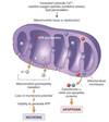
Reversible cell injury

A

injury during the decline of cell function

7
Q

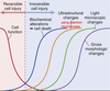
Irreversible cell injury

A

injury during incline of biochemical alterations (cell death), ultrastructural changes, light microscopic changes, and gross morphologic changes

8
Q

4 aspects of a disease process that form the core pathology

A

etiology, pathogenesis, molecular and morphologic changes, and clinical manifestations

9
Q

Etiology (Etx)

A

the cause (causative agent, etiologic agent) or origin of a disease or disorder; the study or theory of the factors that cause disease and the method of their introduction to the host

(ex. Canine parvovirus type 2)

10
Q

What are the 2 major classes of etiology?

A

genetic/intrinsic (e.g. inherited mutations, disease-associated gene variants, polymorphism) and acquired (extrinsic, infectious, nutritional, chemical, physical)

11
Q

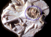
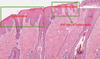
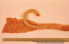
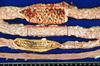
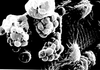
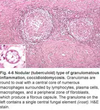
Tissue from a raven; what is the etiology?

A

Poxvirus

12
Q

Pathogenesis

A

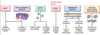
the mechanisms of disease development; sequence of events from INITIAL stimulus to the ULTIMATE expression of the disease in the response of cells or tissues to the etiology; always end it with the morphological diagnosis

(ex. oropharyngeal infection > viremia > infection and death of rapidly dividing cells from crypts > necrohemorrhagic enteritis)
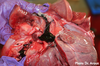
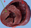
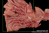
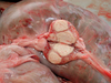
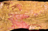
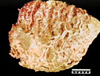
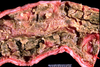
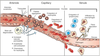
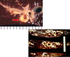
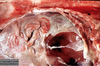
13
Q

Pathogenesis (img)

A

Grain overload, rumenal acidosis, mucosal damage, opportunistic fungal infection, vasculitis, ischemia and mucosal ulceration

14
Q

Molecular and morphological changes

A

refers to biochemical and structural alterations induced in the cells and organs of the body; the changes may be characteristic (pathognomic) of a disease or diagnostic of an etiologic process

15
Q

Clinical manifestations

A

results of genetic, biochemical and structural changes in cells and tissues; manifests as functional abnormalities such as signs (animals) and symptoms (humans, what the patient feels and tells you); injury to the cells and to the extracellular matrix finally leads to tissue and organ injury

16
Q

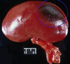
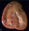
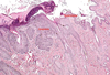
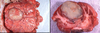
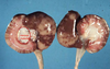
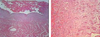
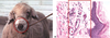
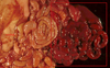
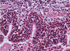
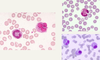
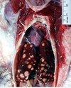
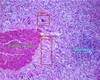
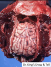
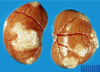
What are the molecular and morphologic changes of the image? ( MDx)

Clinical history and signs: 3 week old puppy, anorexia, dypsnea, abd pain upon palpation, normal rectal temperature

A

Multifocal, acute renal necrosis and hemorrhage or necrohemorrhagic nephritis

17
Q

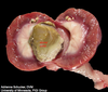
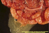
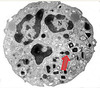
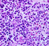
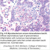
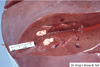
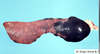
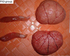
What is the etiology?

Clinical history and signs: 3 week old puppy, anorexia, dypsnea, abd pain upon palpation, normal rectal temperature

A

Canine herpesvirus-1 (CHV-1)

  • red spots are from the virus targeting endothelium
18
Q

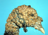
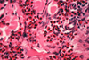
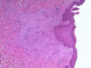
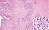
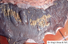
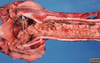
What is the pathogenesis?

Clinical history and signs: 3 week old puppy, anorexia, dypsnea, abd pain upon palpation, normal rectal temperature

A

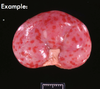
Transmission CHV-1 to pup at birth >incubation period of up to 1 week > virus replicates at temperature lower than 37C (98.6F) > endothelial cell tropism > multifocal necrosis in numerous organs

19
Q

Diagnosis

A

concise statement or conclusion concerning the nature, cause, or name of a disease

20
Q

Types of diagnosis

A

clinical diagnoses, differential diagnoses (DDx), morphologic diagnoses (MDx), etiologic diagnoses (Edx, agent and organ), clinical pathologic diagnosis

21
Q

Clinical diagnosis

A

based on case history (Hx), clinical signs, physical examination; may provide differential diagnosis

22
Q

Differential diagnosis (DDx)

A

list of diseases that could account for the evidence or lesions of the case; DAMNITV scheme; clinical or morphological

(ex. clinical: anticoagulant rodenticides, canine distemper, hemorrhagic gastroenteritis due to Clostridium spp.)

23
Q

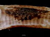
Morphologic diagnosis (MDx)

A

based on the predominant lesion(s) in the tissue(s); macroscopic, microscopic; summary of the lesion, but generally does not describe what is causing the lesion

  1. location (organ tissue)
  2. distribution (focal, multifocal, locally extensive, diffuse)
  3. severity (mild, moderate, severe)
  4. duration/time (acute, chronic)
  5. nature (degenerative, inflammatory, neoplastic) of the lesion; if inflammatory remember to classify the type of exudate

(ex. [small intestine], segmental necrohemorrhagic enteritis)

24
Q

Main types of pathological processes

A

degeneration/necrosis, inflammation & repair, tissue deposits/pigmentations, circulatory disorders, disorders of growth (adaptation vs. neoplasia vs. malformations)

25
Etiologic diagnosis (EDx)
a more definitive dx and names the specific _cause_(s) of the disease; restricted to 2 words only (causative agent and site of lesion) ## Footnote *(ex. parvoviral enteritis)*
26
What is the MDx?
Pulmonary artery, diffuse chronic arteritis with intralesional nematodes (Dirofilaria immitis)
27
What is the EDx?
Verminous/parasitic arteritis or Dirofilarial arteritis
28
What is the EDx?
Bacterial dermatitis or Streptococcal dermatitis
29
Clinical pathologic diagnosis
based on the changes observed in the chemistry of fluids and the hematology
30
Prognosis
it combines pro (before) and gnosis (knowing); refers to the course of a disease or the **outcome**; diagnosis enables the clinician to predict the prognosis; "good", "intermediate", or "poor"
31
Pathognomonic
(test question) characteristic or indicative of a specific disease *(ex. canine herpesvirus-1 nephritis)*
32
Description of gross lesions is based on:
* localization (where is the change?) * distribution (focal, multifocal, diffuse) * size (measure, weight or estimate what % of the organ is affected) * shape (ovoid, flat, nodule, well delimited, encapsulated) * color * consistency (soft, firm, hard; lung with pneumonia is dense compared to normal elastic) * texture (fluid, pasty, fluctuant, smooth/rough) * outline (surfaces: excavated- something missing, prominent- something added) * number/extent * content * odor
33
What is the MDx and EDx of this cat?
(test question) MDx: multifocal to coalescing, granulomatous nephritis EDx: mutated feline coronavirus
34
Autolysis
(test question) self-digestion or degradation of cells and tissues by the hydrolytic enzymes normally present in tissues (postmortem change); occurs after somatic death; those tissues with greater concentration of proteolytic enzymes will undergo autolysis first (GI tract, pancreas, gall bladder, liver, kidney)
35
Somatic death
due to total diffuse hypoxia; cells degenerate as described for hypoxic cell injury
36
Putrefaction/decomposition
process by which post mortem bacteria break down tissues; gives color, texture changes, gas production, odors
37
Death of the whole body
death due to discontinued cardiac activity and respiration
38
Morphologic appearance of postmortem changes vary depending on:
cause of death, environmental and body temperature, microbial flora
39
Cool environmental temperature and refrigeration delays autolysis, delaying putrefaction, except:
ruminants forestomach, equine cecum and ascending colon
40
T or F: ingesta will continue to undergo bacterial fermentation (producing gas and heat) even after death?
True
41
Early postmortem changes
1. **Livor mortis** (lividity, hypostasis): if an animal dies on the left side, more blood will travel to that side 2. **Rigor mortis** (rigidity): depends on the size of the animal, starts from the head down to the tail, depends on the outside temperature (slower if colder) 3. **Algor mortis** (cooling): depends on the outside AND body temperature 1. Corneal clouding 2. Tache noire: black discoloration of the whites of eyes 3. Drying and dark discoloration of lips, tongue, scrotum
42
Rigor mortis
refers to the contraction of the muscles after death; begins at 1-6 hours post death, persistent 1-2 days; high heat and high activity before death accelerate the onset of rigor mortis NO RIGOR MORTIS IN CARCASS WITH ANTRHAX because edema factors inhibits muscle contraction stimulating cAMP
43
Algor mortis
cooling of the body post mortem, depends on the temperature of the body (and environment) at the time of death
44
How do you obtain the temperature from a carcass?
hole in the skull, site where the temperature in brain decreased in a relatively straight line to ambient temperature about 18 hours after death; other sites many variants (wool, fat); changes of temperature curve
45
Livor mortis
(test question) aka hypostatic congestion; occurs about 1 hour post-mortem; gravity will pull blood post death; variation in color of tissues like skin, lung, heart, kidneys, liver; in some areas the tissues will be more red and in other areas pale due to that the blood was kept away
46
Describe post-mortem clotting
(test question) shiny in appearance, never attached to vessel wall; occurs several hours post death in the heart and vessels; influenced by ante mortem changes in blood (ex. Warfarin poison, hereditary coagulapathies can delay or cause failure of blood to clot)
47
Chicken clot appearance (postmortem clotting)
due to separation of RBCs to the bottom and clotted serum at top (red and yellow); differentiate this from pre-mortem clotting!!
48
What type of clots are pictured (pre-mortem, post-mortem, or chicken clot)?
Top: normal post-mortem clots Bottom: "Chicken" clots
49
What type of clot is this (pre-mortem or post-mortem)?
Pre-mortem clot (mural thrombi and thromboemboli): attached to vessel walls (arterial type), loosely attached to vessel walls (venous thrombi, may resemble post mortem clots), dry and duller, laminated, friable
50
What type of clot is this (pre-mortem or post-mortem)?
Post mortem clots: unattached to vessel walls, shiny and wet, perfect cast of vessel lamina, elastic
51
What is hemoglobin imbibition?
post-mortem red staining of tissue, especially the heart, arteries and veins; Hg is released by lysed RBCs and penetrates the vessel wall and extends into the adjacent tissues; however, Hg staining can occur in acute intravascular hemolysis (pre-mortem)
52
Bile imbibition
when bile in the gallbladder starts to penetrate the wall and stains the adjacent tissues yellowish to greenish brown post-mortem; tissues stained are those in contact with the gall bladder (liver, intestines, diaphragm); takes a while to occur
53
Is bloating a pre-mortem or post-mortem morphological change?
(test question) results from **post-mortem** bacterial gas formation in the lumen of the GI tract; herbivores bloat faster; differentiate from ruminant tympany (pre-mortem) *Comparison: pre-mortem bloat will cause a line of color change in the distal third of the esophagus*
54
What causes corneal opacity/clouding (morphologic appearance of postmortem change)?
due to dehydration of cornea/due to cold temperatures of the carcass; most different from cataracts; differentiate from Blue Eye disease (canine Coronavirus)
55
What is pseudomelanosis?
refers to greenish-black discoloration of tissues post-mortem; decomposition of blood by bacterial action forming **hydrogen sulfide with iron**; occurs **soon after death** (like in the **gut**), also will be common to see in those tissues in contact with the gut (kidney, liver, spleen, even the gut wall itself)
56
Marcus Terentius Varro (1st century BC, ancient Rome)
warned his contemporaries to avoid swamps and marshland because of the risk of diseases
57
Miasma theory (medieval times Europe, India, China)
diseases such as Cholera, Chlamydia, or the Black Death were caused by a miasma, a noxious form of "bad air" also known as "night air"
58
Robert Koch
formed Koch's postulates; identified the specific causative agents of **tuberculosis**, **cholera**, and **anthrax**; gave experimental support for the concept of infectious disease
59
Example of infectious diseases \> one etiological agent \> one disease
Cryptococcosis ## Footnote *(img: thalamus, cerebellum and mesecephalon; "cavitational" lesions caused by Cryptococcus neoformans; multifocal distribution in both gray and white matter)*
60
Example of single gene disorder \> one inherited disease
Spider lamb chonrodysplasia ## Footnote *(img: multiple, disorganized ossification centers resulting in variation of the size, shape and orientation of the vertebrate)*
61
Example of one etiological agent \> multiple diseases
(test question) Hepatosis dietetica (nutritional hepatic necrosis) from vitamin E (antioxidant) and/or Selenium deficiency *(img: diffuse, hemorrhagic)* Also, encephalomalacia in chicken, Rickets in puppies, malaisia in adult dogs from vitamin E deficiency (also A and D)
62
Example of multifactorial \> multiple factors, etiologies, external triggers (risk factors, promoters) \> one disease
(test question) **most common method**; gastric ulcer, pulmonary edema, autoimmune diseases, Bovine Respiratory Disease (BRD) complex, pneumonias of pigs, bovine enzootic pneumonia
63
What are some causes of gastric ulcer (pars esophagea) in pigs? (multiple factors \> one disease)
(test question) in growing pigs; stratified squamous epithelium surrounding the cardia (pars esophagea); causes include ingestion of finely ground grain or pelleted feed, possibly vitamin E deficiency, fermentation of sugars in the feed, stress of confinement rearing
64
Animals dying of sepsis, toxemia, aspiration of gastric contents, pancreatitis \> fulminating pulmonary edema because of: (what is the pathogenesis?) (multiple factors \> one disease)
hyperreactive macrophages -\> directly or indirectly generate too many cytokines -\> some cytokines prime neutrophils stationed in the lung capillaries -\> release of enzymes and free radicals -\> diffuse endothelial and alveolar damage -\> pulmonary edema; analogous to **ARDS/shock lung** (acute/adult respiratory distress syndrome) in humans
65
What is a factor that causes Systemic Lupus Erythematosus (autoimmune diseases)? (multiple factors \> one disease)
Type III hypersensitivity
66
What are some factors that cause pneumonia of pigs? (multiple factors \> one disease)
_Host_ (age, genetic makeup, immune status), _infectious agents_ (virus, bacteria, mycoplasma), _environmental determinants_ (humidity, temperature, ammonia concentrations, ventilation), _management practices_ (crowding, mixing of animals, air quality, nutrition, stress)
67
What are the main types of etiologies?
genetic, microbial, immune mediated, toxic, trauma, idiopathic, nutritional, metabolic
68
What are the building blocks for _general_ pathology?
**adaptation, degeneration and cell death, tissue deposits and pigmentation, inflammation and repair, circulatory disorders, genetic disorders, diseases of immunity, neoplasia, microbial infections**
69
What type of pathological process is this?
adaptation, degeneration and cell death: squamous metaplasia of the esophagus (parrot) * (Description: avitaminosis A, no lubrication, glandular cells replaced by keratin)* * Another example: prostatic hyperplasia, benign but the large size can cause a lot of damage (dog)*
70
What type of pathological process is this?
inflammation and repair: embolic nephritis (horse kidney) *(Description: Actinobacillus equuli; growth in the cortex [glomerulus] usually occurs first, red around the pus, bacteria lodged in the glomerulus)*
71
What type of pathological process is this?
(top normal, bottom abnormal) tissue deposits and pigmentations ## Footnote *(Description: defect in heme synthesis caused by a deficiency in uroporphyrinogen III cosynthetase; decoloration of bones, can see discoloration in teeth as well, accumulation of bilirubin, can see in UV light)*
72
What type of pathological process is this?
circulatory disorder: chronic passive congestion ("nutmeg liver") of a cow, right sided heart failure, ## Footnote *(Description: ingestion of hepatotoxin (Wedelia glauca); venous blood not reaching the heart, hepatocytes do not get oxygen which causes tissue death, replaced by connective tissue and lipid inclusions)*
73
What type of pathological process is this?
(test question) genetic disorder: globoid cell leukodystrophy (a type of lysosomal storage disease, dog) * (Description: defect in an enzyme (galactosylceraminidase); special stain used for myelin (blue), periphery not stained because not enough myelin, impairment of function of lysosomes)* * Another example: male pseudohermaphrodite, external female genitalia and testicles*
74
What type of pathological process is this?
disease of immunity: bovine nasal epithelium, acute allergic rhinitis with secondary plant foreign body ## Footnote *(Description: type I hypersensitivity reaction (plant allergen); mast cells and eosinophils accumulate)*
75
What type of pathological process is this?
neoplasia: bovine abomasal lymphoma, bovine leukemia virus
76
What type of pathological process is this?
microbial infection: epithelial plaques, papular stomatitis (inflammation of mouth and lips), hard palate mucosa (calf), ## Footnote *(Description: parapoxvirus [causes Orf in sheep and goats]; plaques are yellow and multifocal)*
77
DAMN IT V
acronym for a clinical investigation plan, based on probable pathophysiologic causes of the disease present * **degenerative**, developmental (degenerative is a mechanism) * allergic, **autoimmune** * **metabolic**, mechanical * **nutritional, neoplastic** * **inflammatory**, **infectious**, **iatrogenic**, immune-mediated, ischemic, **idiopathic** * **toxic, traumatic**, **vascular** (vascular is a mechanism)
78
Idiopathic
unknown cause or spontaneous origin ## Footnote *(ex. epilepsy, canine polyarteritis, pulmonary fibrosis, laryngeal hemiplegia, renal amyloidosis, hypertrophic cardiomyopathy)*
79
Cell adaptation
occurs when the cell homeostasis is distorted by stresses or pathologic stimuli; in this stage, cells preserve viability and function; it is a reversible change; principal responses of adaptation are atrophy, hypertorphy, hyperplasia, metaplasia, dysplasia
80
Homeostasis
tendency to stability in the normal body states of the organism; it is the ability to maintain internal equilibrium by adjusting its physiological processes
81
Atrophy
(test question) decrease in size and/or number of the cells and their metabolic activity _after normal growth has been reached_ (cells are not dead); decrease in protein synthesis and increase in protein degradation in cells; _causes_: decreased workload (immobilized animal), denervation, decreased blood supply or oxygen, inadequate nutrition (generalized atrophy because body is trying to adapt), loss of endocrine stimulation, aging; a type of adaptive cell change
82
Examples of atrophy
_muscle disuse_ in a limb that is in a cast, _sedentary atrophy_, atrophy of adrenal cortex by _reduction of ACTH stimulation_ (steroid therapy), atrophy in tissues adjacent to a tumor due to _pressure and compromised blood supply_, _physiologic atrophy_ (non-lactating mammary gland) * ex. _serous atrophy of fat_ (loss of nutrients, vitamin E deficiency), no fat on the heart, white lines seen on the surface (dilated lymphatic vessels), yellow, gel-like bone marrow* * ex. _hydrocephalus with compression atrophy_ (cat), fluid accumulation in the brain, defect with draining*
83
Hypertrophy
increased **size** of cells and their **functions** (synthesis of more organelles and structural proteins, bigger cells), more common in cells with little replication (stable or permanent cells: cardiomyocytes, neurons); a type of adaptive cell change
84
Examples of hypertrophy
(test question) hypertrophic pregnant uterus, weightlifter (physiologic hypertrophy), x-linked muscular hypertrophy (genetic disease in cats), cardiac hypertrophy from hypertension or aortic valve disease (pathologic hypertrophy, common in animals with longer life spans, transcription factors produced from increased work load such as mechanical stretch, agonists, growth factors)
85
Concentric vs. eccentric hypertrophy
eccentric hypertrophy is thinner because it is compensating (left concentric, right eccentric)
86
Hypertrophic cardiomyopathy (HCM) is common in what breed of cats?
common in Maine coon cats (more prone to have this dz earlier than other species), mutation in MYBPC3 gene (inherited autosomal dominant)
87
Hyperplasia
increase in the **number** of cells of an organ, cells capable of replication, may occur concomitantly with hypertrophy; a type of adaptive cell change
88
Examples of hyperplasia
* _hormonal_ (breasts during pregnancy) * _compensatory_ (hepatectomy, also in kidneys) * pathologic most commonly caused by excessive hormonal or growth factor stimulation (epidermal thickening from repeated irritation, respiratory mucosa in viral infections), * _fibrous_ hyperplasia (formerly part of fibrous or fibromatous epulis, gingival hypertrophy, canine) SCC epidermal hyperplasia proceeds to dysplasia, carcinoma in situ and invasive squamous cell carcinoma (_carcinoma in situ,_ when cells have already gone neoplastic transformation but not invading basal membrane yet) (*ex. cat's ears from sun exposure, horses eyelids/oral cavity)*
89
Dysplasia
refers to abnormal development (mostly of epithelial cells); term mostly used in neoplastic processes, near synonym is _carcinoma in situ_; cells lose polarity
90
Metaplasia
change in _phenotype_ of a differentiated cell, response to _chronic irritation_ (cell withstand stress), may result in _decreased functions or increased propensity_ for malignant transformation (neoplasia), _reversible_ if cause is removed, most often in _epithelial cells_ (urinary bladder, distal esophagus canine)
91
Examples of metaplasia
chronic irritation in lungs, vit-A deficiency, estrogen toxicity (mainly in urinary bladder/genital tract), mammary tumors
92
What is the MDx (atrophy, hyperplasia, hypertrophy, metaplasia, or dysplasia)?
feline kidney, nephrolith (xanthinuria) with hydronephrosis, cortical and medullary **atrophy** and medullary fibrosis, diffuse
93
Is this atrophy, hyperplasia, hypertrophy, metaplasia, or dysplasia?
cat: squamous cell carcinoma, **dysplasia**
94
What is the MDx and etiology (atrophy, hyperplasia, hypertrophy, metaplasia, or dysplasia)?
snake stomach, diffuse marked chronic gastric **hypertrophy** from Cryptosporidium serpentis
95
What is the MDx (atrophy, hyperplasia, hypertrophy, metaplasia, or dysplasia)?
feline stomach gastric lymphoid **hyperplasia**, multifocal (white nodules)
96
What is the MDx (atrophy, hyperplasia, hypertrophy, metaplasia, or dysplasia)?
canine liver, hepatocellular carcinoma with nodular **hyperplasia**
97
What are the 2 MDx (atrophy, hyperplasia, hypertrophy, metaplasia, or dysplasia)?
canine kidney, MDx1: hydronephrosis with secondary severe diffuse cortical **atrophy** MDx2: ureter, hydroureter
98
Normal cell components
cell membranes, cytosol, mitochondria, nucleus, nucleolus, ER, Golgi complex, lysosomes, cytoskeleton, extracellular matrix
99
How is DNA of a normal cell organized?
antiparallel, 5' to 3' and 3' to 5'; purine (adenine, guanine) is bound to a pyrimidine (cytosine, thymine) by hydrogen bonds (A:T and G:C); the helix occurs naturally because of the bonds in the phosphate backbone; organized by histones into nucleosome bead that are coiled and packed into chromosomes
100
How is chromatin of a normal cell organized?
DNA is organized around histones into nucleosomes; nucleosomes are wound into a helix to form chromatin; chromatin wound again into supercoiled chromosomes
101
What is the function of the nucleus?
(test question) storage of genetic material/location of the genome; DNA complex to protein = **chromatin**: **euchromatin** (uncoiled, transcriptionally _active_) and **heterochromatin** (coiled, transcriptionally _inactive_)
102
What is a nucleolus?
(test question) organelle of the nucleus, composed of RNA, protein, chromatin, functions in the synthesis of rRNA, **prominence is a subjective measure of a cell's synthetic activity**
103
Normal cellular functions are compartmentalized within membrane-bound organelles which allow for:
isolation of potentially harmful substances (*e.g. degradative enzymes, reactive metabolites*) and the creation of unique intracellular environments (*e.g. low pH, high Ca++ concentrations*)
104
What is the function of the rough endoplasmic reticulum?
site of protein synthesis (esp. EC); contains ribosomes
105
What is the function of the smooth endoplasmic reticulum?
locus of enzymes that metabolize steroids, drugs, lipids and glycogen; lacks ribosomes
106
What is the function of the Golgi apparatus?
synthesis of complex proteins and the production of secretory vesicles and lysosomes
107
What is the function of endosomal vesicles?
shuttles internalized material within cell and directs newly synthesized material to cell surface or cell organelle
108
What is the function of lysosomes?
digests macromolecules (waste management)
109
What is the function of proteasomes?
selectively degrades denatured proteins and release peptides (waste management)
110
What is the function of peroxisomes?
breakdown fatty acids and generates hydrogen peroxide (waste management)
111
Autophagy vs. heterophagy
autophagy is to "eat oneself" while heterophagy is the digestion within a cell of a substance taken in by phagocytosis
112
Cell polarity
refers to the **spatial differences** in shape, structure, and function of cells ## Footnote *(ex.: epithelial cells: apical surface and basilar surface are exposed to different environments and have different functions)*
113
What are the functions of the cytoskeleton?
(test question) contents and position of cell organelles are regulated by the cytoskeleton; responsible for **cell movement**, maintains **cell shape** and intracellular **organization**, can **move organelles** and proteins within the cell
114
What are the components of the cytoskeleton?
* actin microfilaments (cell structure and movement) * intermediate filaments (support) * microtubules (protein movement and signaling)
115
What are the 2 main functions of cell membranes?
selective barrier and structural base for enzymes and receptors
116
What are the functions of membrane proteins?
transport channel, enzyme, cell surface receptor, cell surface markers, cell adhesion, attachment of cytoskeleton
117
Mitochondria characteristics:
evolved from ancestral prokaryotes engulfed by eukaryotes, **contain their own DNA, maternal inheritance**
118
What are the functions of mitochondria?
(test question) site of aerobic metabolism and regulator of apoptosis (leakage of cytochrome C \> activation of caspases)
119
Which part of the nucleus is not being actively transcribed (heterochromatin, euchromatin, histones, or nucleolus)?
(test question) heterochromatin
120
What is cell injury?
damage or pathologic alterations in molecules and/or structure that can occur in cells and extracellular components
121
What are some extrinsic causes of cell injury (etiology)?
physical trauma, viruses, toxins
122
What are intrinsic causes of cell injury (etiology)?
genetic mutations (enough to become neoplastic, apoptosis)
123
What are both extrinsic and intrinsic causes of cell injury?
workload imbalance, nutrional abnormalities, immune dysfunction
124
When does cell injury occur?
when the cell cannot maintain a steady state
125
What are some causes of cell injury?
**oxygen deficiency**, physical agents, **infectious agents**, nutritional deficiencies and imbalances, genetic derangement, workload imbalance, chemicals, drugs, toxins, **immunologic dysfunction**, aging
126
What is oxidative phosphorylation?
when O2 is required for cellular respiration
127
What is hypoxia?
(test question) partial reduction in O2 delivery to a tissue, inadequate oxygenation of blood, reduced transport of O2 in blood, reduction in blood supply (ischemia), blockage of cell respiratory enzymes *(e.g. cyanide)*
128
What is anoxia?
no oxygen delivery to a tissue, necrosis
129
What are physical agents that cause cell injury?
trauma, temperature extremes, electrical injury, ionizing radiation (UV light, microwaves)
130
What are infectious agents that cause cell injury?
* viruses * obligate intracellular parasites; use host cell enzyme systems * cell survival depends on method viruses leave the cell * bacteria * toxins, overwhelming and uncontrolled replication * fungal (mycosis) * progressive, chronic inflammatory disease * protozoan * replicate in specific host cells -\> cell destruction * metazoan parasites * inflammation, tissue distortion, utilization of host nutrients
131
What are immune dysfunctions that cause cell injury?
immune system fails to respond to infectious agents (test question) * **congenital defects: SCIDS (Arabian foals**, lymphocytes not fully developed) * **acquired defects** * may be transient (but not always) * results from damage to lymphoid tissue (steroids) * viral infections, chemicals, **drugs** * **autoimmune diseases** * **hypersensitivity reactions** (overzealous mast cells, eosinophils)
132
Manifestation of cell injury is variable depending on:
type of agent, extent of injury, duration of injury, cell type
133
6 mechanisms of cell injury:
1. **decreased ATP** 2. **mitochondria damage** 3. **entry of Ca++** 4. **increased reactive oxygen species** 5. **membrane damage** 6. **protein misfolding/DNA damage** * all mechanisms have multiple effects on cells; cells have systems in place to mediate and repair damage; severe damage leads to cell apoptosis or necrosis
134
Explain the mechanism of cell injury: "depletion of ATP"
(test question) * **ATP is required for almost all _synthetic_ and _degradative_ processes within the cell** * associated with **hypoxic injury** and **toxic injury** (e.g. cyanide stops the electron transport chain) * **fundamental cause of necrotic cell death** * **depletion of 5-10% = BAD** * **​**​NA+ K+ ATPase pump failure * cell **swelling**, ER swelling, plasma membrane damage * altered cell metabolism * anaerobic glycolysis: increased lactic acid -\> decreased pH -\> loss of enzyme function * ribosome detachment * decreased protein synthesis * culminates in **irreversible** mitochondrial and lysosomal membrane damage -\> **cell necrosis (cell swelling)**
135
Explain the mechanism of cell injury: "mitochondrial damage"
(test question) 3 major consequences: * formation of the **mitochondrial permeability transition pore** (MPTP, forms in outer membrane of mitochondria) * production of **reactive oxygen species** * activation of **apoptotic pathways**
136
Explain the mechanism of cell injury: "loss of calcium homeostasis"
(test question) accumulation Ca++ * **opening of MPTP -\> decreased ATP** * **enzyme activation** (phospholipases, proteases, endonucleases, ATPases) * activation of **caspases -\> induction of apoptosis**
137
Explain the mechanism of cell injury: "accumulation of reactive oxygen-derived free-radicals"
(test question) mechanism: * unstable configuration * interacts with adjacent molecules: proteins, lipids, carbohydrates, nucleic acids, etc. * initiates autocatalytic reactions
138
Mechanism of cell injury: "inflammation"
1. recognition and attachment: microbe bind to phagocyte receptors 2. engulfment: phagocyte membrane zips up around microbe 3. killing and degradation
139
What are pathologic effects of free radicals?
(test question) * lipid peroxidation in membranes * MOA: forms peroxides -\> autocatalytic reaction (propagation) * extensive membrane damage * oxidative modification of proteins * MOA: oxidation of amino acid side changes, formation of protein cross-linkages (disulfide bonds), oxidation of protein backbone * damage active sites, change conformation, enhance degradation * lesions in DNA * MOA: single or double stranded breaks, cross-linking of DNA strands, formation of adducts * cell aging, malignant transformation
140
What are the ways to remove free radicals
* **spontaneous decay** O2\*(superoxide anion) + H2O -\> O2 + H2O2 * **antioxidants** (donors of electron): vitamin E, vitamin A, glutathione * blocks initiation, inactivate (scavenge) * **storage and transport proteins**: transferrin, ferritin, ceruloplasmin * bind reactive metals (Fe, Cu) * **enzymes**: catalase, superoxide dismutase, glutathione peroxidase * break down H2O2 and O2\* * located near sites where oxidants are formed * ex. superoxide dismutase (SOD) **in mitochondria (endogenous)** 2O2\* + 2H + SOD -\> H2O2 + O2
141
What are reactive oxygen species (ROS)?
(test question) a type of oxygen-derived free radical; degraded and removed by cell defense systems; production by: * during mitochondrial respiration * absorption of radiant energy * inflammation * enzymatic metabolism of drugs or toxins * transition metals (Fe, Cu)
142
What is oxidative stress?
when **reactive oxygen species production exceeds antioxidant capacity** (cell injury, cancer, aging, degenerative disease; system becomes overwhelmed) *classic example: **inflammation** (neutrophils and macrophages; mediators for destroying microbes, dead tissue, etc.)*
143
Explain the mechanism of cell injury: "membrane damage"
(test question) * reactive oxygen species * decreased phospholipid synthesis * increased phospholipid breakdown * cytoskeletal abnormalities * activation of proteases -\> damaged cytoskeleton * cells stretch and rupture
144
What are the consequences of membrane damage?
* mitochondrial membrane damage * opening of the MPTP -\> decreased ATP * release of pro-apoptotic proteins * plasma membrane damage * loss of osmotic balance -\> influx of fluids and ions (Ca++) * loss of cell contents and metabolites * injury to lysosomal membranes * leakage of enzymes into the cytoplasm (RNases, DNases, proteases, phosphatases, glucosidases) * enzymatic digestion of RNA, DNA, proteins, etc.
145
What is the clinical pathology correlation to membrane damage?
* chemistry panel * analytes used to determine hepatocellular injury: * alanine aminotransferase (ALT) * located in the cytoplasm of hepatocytes * convertes alanine into pyruvate * pyruvate is used for gluconeogenesis or the Kreb's cycle * when hepatocytes undergo cellular injury, ALT is released * cell necrosis (ruptures and releases ALT) * membrane blebs that contain ALT
146
Explain the mechanism of cell injury: "damage to proteins and DNA"
cells have repair mechanisms when overwhelmed \> initiates **apoptosis**
147
What is inflammation?
reaction of **vascularized** **living tissues** to injury; involves changes in vascular bed, blood, connective tissue; main job is intended to eliminate irritant and repair damaged tissue; initiated by exogenous and endogenous stimuli; leading to the accumulation of fluid (plasma proteins) and WBCs in **extravascular tissues**
148
Signs of inflammation
**redness, heat, swelling, pain, loss of function** *(img: tissue from a pigeon, etiology: mosquito bite)*
149
What are the roles of inflammation?
* dilute, contain and isolate injury * destroy invading microorganisms and/or inactivate toxins * achieve healing and repair
150
What are the outcomes of inflammation?
* ideal conditions- _return to normal_ * elimination of the source of injury, resolution of inflammatory process, restoration of normal tissue architecture and physiologic functions * intense inflammatory response- _attempt to separate injured tissue_ (ex. fluid tries to dilute toxic -\> lymphatics) * attempt to isolate inflammatory process, formation of a wall (such as a capsule in an abscess) * failure to eliminate insult- _sequel_ * persistence of inflammatory cells (acute -\> chronic) * scar formation (ex. chronic dermatitis in a cow, thickening of skin)
151
Inflammation generalities
* occurs only in living tissue * is a response and as such it requires an initiating stimulus (etiology; used in forensics) * it can be more harmful than the initiating stimulus (ex. anaphylactic shock) * is fairly stereotypical/predictable irrespective of the initiating stimulus (chemotactic factors) * involves multiple participants * it is critically tied to the blood (plasma, circulating cells, blood vessels, cellular and extracellular components of connective tissue) * it is highly redundant with many promoters and regulators (because we have to be cautious of failure of other components)
152
When does inflammation stop?
when the stimulus is eliminated (sometimes there is an overreaction even when the stimulus is eliminated) * mediators are broken down and dissipated * WBCs have short life span in tissues * anti-inflammatory mechanisms are activated
153
When does repair begin (inflammation)?
* repair begins during inflammation and it is completed when injurious stimuli have been neutralized * damaged tissues are replaced * commonly by combination of both: * regeneration of native parenchymal cells * filling of the defect with fibrous tissue (scarring)
154
What is exudation?
escape of fluid, proteins and blood cells from the vascular system into the interstitium or body cavities; exudation implies **alteration of the normal permeability of local blood vessels**
155
What is exudate?
inflammatory extravascular fluid that has a high protein concentration, much cellular debris, and a specific gravity above 1.020 (more than transudate)
156
What is transudate?
essentially an ultrafiltrate of blood plasma and results from hydrostatic imbalances across the vascular endothelium; a transudate is a fluid with a low protein content and a specific gravity of less than 1.020
157
What is an edema?
denotes an excess of fluid in the interstitial tissue or serous cavities, it can be an exudate (inflammatory edema) or a transudate (changes in hydrostatic pressure) ## Footnote *(img: inflammatory edema in a case of acute hemorrhagic abomasitis, EDx: Clostridial abomasitis)*
158
What is pus?
an inflammatory exudate rich in leucocytes (primarily viable and degenerated neutrophils) and parenchymal cell debris; damage surrounding tissue
159
How do you classify inflammation?
* extent, duration, distribution, exudate, anatomic modifiers, organ
160
Explain the extent of inflammation
how bad is the inflammation? * minimal * mild: absent to minimal tissue damage, few inflammatory cells, slight vascular involvement * moderate: some tissue damage, inflammatory cells evident, moderate edema and evidence of hemorrhage * severe: substantial tissue damage, inflammatory cells abundant, massive edema and hemorrhage may be seen *(img: severe)*
161
Explain the duration of inflammation: peracute
"how long did the inflammation persist?" usually caused by a potent stimulus (anaphylactic shock, bee sting), usually the animal has no time to response, less common than acute disease processes _time_: 0-4 hours _vascular involvement_: hyperemia, slight edema, hemorrhage _inflammatory cells_: few inflammatory cells, few leucocytes _clinical signs_: shock, sudden death
162
What type of inflammation is pictured? (peracute, subacute, acute, chronic)
peracute inflammation; infectious canine hepatitis left: congestion and edema of abdominal viscera right: intranuclear inclusions within glomeruli
163
What type of inflammation is pictured? (peracute, subacute, acute, chronic)
acute inflammation; migration of neutrophils, migrate to wall/edge "paving" of endothelium
164
Lymphadenitis
reactive inflammation of lymph node(s) occurs in acute, subacute and chronic inflammation *(img: chronic inflammation of mesenteric lymph nodes in a llama, due to tuberculosis)*
165
Lymphangitis
inflammation of lymphatic vessel(s) ## Footnote *(img: thickening of serosal lymphatic vessels due to Johne's disease, goat small intestine)*
166
What type of inflammation is pictured? (peracute, subacute, acute, chronic)
subacute stomatitis (inflammation of the mouth) in a bovine
167
What type of inflammation is pictured? (peracute, subacute, acute, chronic)
chronic inflammation of the mandible
168
What type of inflammation is pictured? (peracute, subacute, acute, chronic)
chronic colitis, multifocal, ulcerative (feline)
169
What type of inflammation is pictured? (peracute, subacute, acute, chronic)
chronic lymphangitis, Johne's disease in a goat
170
Explain the duration of inflammation: acute
_clinical signs_: warm, red, swollen, painful, loss of function _time_: 4-6 hours _vascular involvement_: active hyperemia, edema (due to endothelial damage-lymphatics and small blood vessels), occasional fibrin thrombi within vessels _inflammatory cells_: leucocyte infiltration is variable (dependent upon TIME), in general neutrophils predominate, but sometime mononuclear cells (lymphocytes and plasma cells) can also be present; no macrophages _lymphatics_: lymphatic vessels have a role in moving away the exudate; the transporation of the exudate (inflammatory cells and necrotic debris) can lead to acute regional **lymphadenitis**
171
Explain the duration of inflammation: subacute
a gradual change, between acute and chronic _time_: can vary from a few days to a few weeks _vascular involvement_: there is a decline in the magnitude of vascular changes, compared to acute inflammation (less hemorrhage, hyperemia and edema) _inflammatory cells_: "**mixed**" or "**pleocellular**" inflammatory infiltrate; stilly may be primarily neutrophilic but usually is also associated with an infiltration by **lymphocytes**, macrophages and **plasma cells**; **_fibrosis and neovascularization (angiogenesis) are not features of subacute inflammation_** _lymphatics_: increased lymphatic drainage, repair of endothelial damage
172
Explain the duration of inflammation: chronic
(test question) often the result of a persistent inflammatory stimulus in which the host has failed to completely eliminate the causative agent; inflammatory response usually is accompanied by an **immune response**; **macrophages present**; it may follow an acute inflammatory phase or it may develop as an insidious, low-grade, subclinical process without history of a prior acute episode _time_: variable (weeks to months), _vascular involvement_: proliferation of capillaries and small blood vessels (**angiogenesis/neovascularization**) resulting in hemorrhage and congestion _host involvement_: parenchymal regeneration or repair by **fibrosis/scarring** _inflammatory cells_: primarily mononuclear inflammatory cells (lymphocytes, **macrophages** for **phagocytosis** and **tissue debridement** in a chronic lesion, plasma cells, fibroblasts) _lymphatics_: lymphatic involvement variable _clinical signs_: **many of the changes represented in chronic inflammation are also seen in areas of repair**
173
What is the definition of focal inflammation?
single abnormality or inflamed area within a tissue
174
What is the size of a focal inflammation?
varies from 1 mm to several cm in diameter
175
What type of inflammation is pictured? (focal, multifocal, diffuse, or locally extensive)
(test question) focal inflammation
176
What type of inflammation is pictured? (focal, multifocal, diffuse, or locally extensive)
focal lesion in the brain of a deer; cut surface reveals presence of cloudy, yellowish material (interpreted as pus)
177
What type of inflammation is pictured? (focal, multifocal, diffuse, or locally extensive)
(test question) focal keratitis (inflammation) in a bovine
178
Define a multifocal inflammation
arising from or pertaining to many foci
179
What is the size of a multifocal to coalescent inflammation?
variable; each focus of inflammation is separated from other inflamed foci by an intervening zone of relatively normal tissue
180
What type of inflammation is pictured? (focal, multifocal, diffuse, or locally extensive)
multifocal to coalescent hepatitis in a peacock
181
What is a locally extensive inflammation?
(test question) involves a considerable zone of tissue within an inflamed organ; possible origins are severe local reactions that spread into adjacent normal tissue and coalescence of foci in a multifocal reaction
182
What type of inflammation is pictured? (focal, multifocal, diffuse, or locally extensive)
locally extensive; cranioventral aspect of the lungs are involved (dark red) while the dorsal portions are spared
183
What type of inflammation is pictured? (focal, multifocal, diffuse, or locally extensive)
locally extensive pneumonia in a pig
184
What is a diffuse inflammation?
although variations in severity of the inflammation may occur, the entire tissue is involved (ex. interstitial pneumonia); diffuse lesions are often viral or toxic in etiology
185
What type of inflammation is pictured? (focal, multifocal, diffuse, or locally extensive)
diffuse; mucosal surface of the stomach, enterotoxemia
186
Explain the classification of inflammation: exudate
the inflammatory process can be classified according to the predominant type of inflammatory cells and exudate * suppurative (pus)- lesions are often bacterial in origin * fibrinous * serous (watery) * serofibrinous * fibrino-purulent * purulent * granulomatous
187
What is suppurative/purulent exudation?
consist of contains pus; associated with the formation of pus
188
What is pus?
a liquid inflammation product composed of accumulated dead cells (tissue and inflammatory cells), variable numbers of viable leucocytes (mostly neutrophils), and fluids added by the inflammatory edema-forming process
189
What type of inflammation is pictured? (exudate)
suppurative bronchopneumonia in a dog, locally extensive; need histology to make diagnosis left: grey discoloration in cranioventral areas of the lung right: neutrophils within alveolar spaces
190
What type of inflammation is pictured? (exudate)
severe suppurative pyelonephritis in a dog (ascending)
191
What type of inflammation is pictured? (exudate)
severe suppurative inflammation, pyometra in a dog
192
Define suppuration
the process by which pus (suppurative exudation) is formed; the use of the terms suppuration implies that neutrophils and proteolytic enzymes are present, and that necrosis of host tissue cells has occurred
193
Define abscess
a circumscribed (partially walled-off) collection of pus; therefore, an abscess is a localized form of suppurative inflammation gross appearance is yellow-white to gray-white and varies from watery to viscous depending on fluid content
194
What type of inflammation is pictured? (exudate)
focal abscess (suppurative) of the liver
195
What type of inflammation is pictured? (exudate)
chronic (neutrophils taking time) suppurative osteomyelitis (affecting bone and bone marrow) \*osteitis is just affecting bone
196
What is the pathogenesis of fibrinous exudation?
severe injury to *endothelium and basement membranes* results in leakage of plasma proteins including fibrinogen, which polymerizes **perivascularly** as fibrin
197
What are the contents in fibrinous exudation?
fibrin
198
What type of inflammation is pictured? (exudate)
fibrinous exudation of a joint space; pale yellow = fibrin
199
What is the gross appearance of fibrinous inflammation?
yellow-white, or pale tan, stringy, shaggy meshwork (or fibrillar material) which gives a rough irregular appearance to the tissue surfaces; casts of this friable material may form in the lumen of tubular organs; goes away when it gets into the peritoneum
200
How long does it take for a fibrinous inflammation to occur?
acute process, it can form in seconds
201
What type of inflammation is pictured? (exudate)
fibrinous enteritis, lumen of intestine
202
What type of inflammation is pictured? (exudate)
fibrinous exudate, severe diffuse enteritis, bovine intestine mucosal surface; circled portion is a part of the fibrin lifted up \*Fuentealba likes this image\*
203
What type of inflammation is pictured? (exudate)
fibrinous cholecystitis (inflammation of the gall bladder) right: fibrin cast
204
What is fibrinopurulent exudate?
an inflammatory process in which neutrophils and fibrin are abundant; fibrin mixes with pus (can tell histologically)
205
What type of inflammation is pictured? (exudate)
fibrinopurulent pericarditis in a foal ## Footnote *example: Hardware disease in ruminants can cause this*
206
What type of inflammation is pictured? (exudate)
diffuse severe fibrinosuppurative pericarditis, porcine
207
What is the difference between fibrinous exudation with fibrosis?
* presence of a fibrinous exudate involves an **acute** process, fibrosis is a **chronic** process * fibrin on tissue surfaces can be easily broken down (friable); microscopically, fibrin is composed of thread-like eosinophilic meshwork that sometimes forms masses of solid amorphous material * fibrin provides the support for the eventual ingrowth of fibroblasts and neocapillaries; the transformation of the fibrinous exudate into well-vascularized connective tissue (a chronic process known as **organization** of the exudate) * fibrin is often removed by enzymatic fibrinolysis or by phagocytosis by macrophages before organization can occur
208
What type of inflammation is pictured? (exudate)
fibrinous pleuritis in a horse left: pleural surface covered by friable material right: homogenous, eosinophilic material, bacterial colonies (arrows) are basophilic with HE stain
209
Which is fibrinous and which is fibrous?
left fibrinous, right fibrous; if both fibrin and fibrous connective tissue are present, the inflammatory process should be interpreted as chronic
210
Which is a fibrinous inflammation?
left is fibrin over lung, comes off easily (right is fibrous connective tissue adhesions)
211
Fibrinous or fibrous? (heart)
(test question) fibrinous pericarditis (severe), epicardial surface is covered by fibrin; caused by infectious agent
212
Fibrinous or fibrous? (liver cross section)
chronic fibrosing (fibrous) cholangitis, thickening of bile ducts
213
What is serous exudation?
inflammatory process in which the exudate occurs in tissues in the absence of a prominent cellular response; may be a dominant pattern of exudation for a wide variety of mild injuries (ex. traumatic blisters, sunburn)
214
What are the contents of serous exudation?
outpouring of fluid relatively rich in protein, and derived from blood and locally injured cells
215
What type of inflammation is pictured? (exudate)
serous inflammation right: separation in the dermis
216
What is the gross appearance of serous exudate?
yellow, straw-like color, fluid commonly seen in very early stages of many kinds of inflammatory responses; ulceration will follow the rupture of a vesicle
217
What type of inflammation is pictured? (exudate)
serous exudate
218
How long does it take for a granulomatous inflammation to occur?
always chronic!
219
What is a granulomatous inflammation?
granulomatous refers to an inflammatory response characterized by the presence of lymphocytes, macrophages and plasma cells with the predominant cell being the **macrophage**
220
What type of inflammation is pictured? (exudate)
granulomatous, multifocal granulomatous hepatitis
221
What is a granuloma?
(test question) macrophages are clustered in a characteristic elliptical formation around the causative etiologic agent, or around a central necrotic area, or simply as organized nodules; large cells with abundant cytoplasm, referred to as **epithelioid cells** and **multinucleanted giant cells** are also commonly present in this type of inflammatory response
222
What type of inflammation is pictured? (exudate)
diffuse granulomatous inflammation, bovine intestine; **cerebroid** appearance of affected intestine (brain-like)
223
What is the etiology of a granulomatous inflammation?
usually some non-digestible organism or particle which serves as a chronic inflammatory stimulus, delayed-type hypersensitvity is often required * organisms: mycobacterium, actinomyces, blastomyces, coccidioides, etc * noninfectious agents: **mineral oil** (in cats, goes into lungs), complex polysaccharides, **foreign bodies**
224
What are other types of exudate (modifiers) that can be used in morphologic diagnoses besides serous, purulent, and granulomatous?
necrotizing, hemorrhagic, mucoid
225
Based on the histological characteristics, what are other types of exudate (modifiers) that can be used in a morphologic diagnosis?
eosinophilic, lymphocytic, lymphoplasmacytic and non-suppurative
226
What type of inflammation is pictured? (exudate modifier)
hemorrhagic and proliferative (when parenchymal cells are proliferating) enteritis, porcine
227
What is a necrotizing inflammation?
necrosis is the main feature and exudation is minimal; in general, the process is interpreted as inflammatory (rather than purely ischemic) if an infectious etiology is suspected
228
What type of inflammation is pictured? (exudate modifier)
necrotizing enteritis
229
What is a hemorrhagic inflammation?
hemorrhage is the main feature of this type of inflammation; the presence of an etiologic agent will indicate that the process is inflammatory rather than a primary circulatory disturbance
230
What type of inflammation is pictured? (exudate modifier)
hemorrhagic inflammation, caprine intestine (Clostridium infection)
231
What type of inflammation is pictured? (acute or chronic)
both! **segmental** hemorrhagic enteritis
232
What type of inflammation is pictured? (exudate modifier)
segmental hemorrhagic enteritis *(from Parvovirus infection*)
233
What type of inflammation is pictured? (exudate modifier)
diffuse severe hemorrhagic cystitis (*left: urinary bladder serosal layer; right: mucosa, tissue should look pale pink*)
234
What type of inflammation is pictured? (exudate modifier)
mucoid enteritis, rabbit
235
What is a mucopurulent or catarrhal inflammation?
(test question) the inflammatory exudate is composed of **mucus and pus** (neutrophils and cell debris)
236
What type of inflammation is pictured? (exudate)
(test question) mucopurulent or catarrhal, bovine catarrhal rhinitis
237
What type of inflammation is pictured? (exudate)
catarrhal enteritis
238
What are some adding modifiers to the classification of inflammation?
* anatomical: interstitial * multifocal to coalescent * segmental * bilateral/symmetrical * fibrino-hemorrhagic * fibrinonecrotizing
239
What type of inflammation is pictured? (adding modifier)
**multifocal to coalescent**; severe multifocal to coalescent dermatitis etiology: turkey pox virus
240
What type of inflammation is pictured? (adding modifier)
**bilateral;** bilateral locally extensive pneumonia
241
What type of inflammation is pictured? (adding modifier)
**fibrino-necrotizing** enteritis, pig ileum ## Footnote *(if it was just fibrin, it would peel off easily; in fibrino-necrotizing, it is stuck onto the mucosa [dead])*
242
What type of inflammation is pictured? (adding modifier)
**fibrino-necrotizing** enteritis ## Footnote *(blue bacterial colonies on the bottom right)*
243
What is a fibrino-necrotizing inflammation (adding modifier)?
further examination will show sloughed epithelium admixed with fibrin= means necrosis and supporting a diagnosis of fibrinonecrotizing enteritis
244
What type of inflammation is pictured? (anatomical location/organ)
lungs, diffuse severe fibrinous pleuritis
245
What are the 2 subdivisions of inflammatory cells in exudate?
**Polymorphonuclear leukocytes** (granulocytes: neutrophils, eosinophils, basophils and mast cell) and **mononuclear cells** (lymphocytes and plasma cells, monocytes and macrophages, platelets)
246
What 2 types of leukocytes are NOT normal inhabitants of the circulating blood?
plasma cells and mast cells
247
Neutrophil characteristics
* crucial to the entire inflammatory process * constitute the **first line of cellular defence** (usually the first to gather at sites of acute inflammation) * they develop in the bone marrow and the maturation process takes about 2 weeks * high motility (rapid amoeboid movement) * response to many chemotactic compounds (through endothelial cells) * phagocytic and bactericidal activites (major defence against **bacteria**)
248
What is the function of neutrophils?
to eliminate (ingest, neutralize destroy) microorganisms, tumor cells, and foreign material by phagocytosis; secretion of pro-inflammatory substances kills microorganisms by: * producing oxygen free radicals (kills everything surrounding it, even tissue) * hydrogen peroxide * lysosomal enzymes **mediates tissue injury** via release of oygen free radicals and lysosomal enzymes; they **regulate inflammatory response** via releasing chemical mediators such as leukotrienes and platelet activating factor
249
Neutrophil morphology
* 10-12 micrometers in diameter (a little bit bigger than RBCs) with multilobed nucleus * contains abundant cytoplasmic granules * 2 classes of granules: * **azurophil granules** (primary granules)- large, oval and electron dense FYI * **specific granules** (secondary granules)- smaller, less dense and more numerous FYI * neutrophils of rabbits, guinea pigs, rats, reptiles, fish and birds have eosinophilic granules called heterophils (pink)
250
What type of inflammatory cell is pictured? (leukocyte)
neutrophils, fluid and fibrin (pink meshwork) within the alveolar spaces (which should be empty)
251
What type of inflammatory cell is pictured? (leukocyte)
neutrophils within a bronchi lumen, going under degeneration so it looks different from neutrophils in blood smear
252
What type of inflammatory cell is pictured? (leukocyte)
neutrophils in blood smear
253
What type of inflammatory cell is pictured? (leukocyte)
neutrophils in tissues (suppurative exudate), not going to look polymorphonuclear at tissues
254
What types of inflammatory cells (leukocytes) are seen degranulating against the surface of a nematode?
equine eosinophils
255
What type of inflammation is pictured? (exudate)
suppurative endometritis, cow (neutrophils), diffuse
256
When can you find an abundance of eosinophils?
(test question) sites of inflammation in diseases of immunologic, parasitic or allergic origin (ex. mosquito bites, rhinitis)
257
What is the function of eosinophils?
they have unique functions as effector cells for **killing helminths** and their propensity for both _causing_ and _assisting_ in the regulation of tissue damage in **hypersensitivity** (immediate-type); phagocytic but less active phagocytes than neutrophils; also kills helminth parasites by antibody-dependent cell-mediated cytotoxicity (ADCC); they are attracted to sites of helminth invasion in sensitized hosts by chemotactic factors; adheres to helminths and degranulates against them (major basic protein)
258
What is the morphology of eosinophils?
similar to neutrophils, but are slightly larger; affinity of their coarse cytoplasmic granules for the acid dye eosin; lysosomal granules of the eosinophil contains a wide variety of catalytic enzymes similar to those in neutrophils, except that they lack lysozyme and phagocytin FYI; small granules, primary granules, large secondary (specific) granules such as: * major basic protein * eosinophilic cationic protein * eosinophil-derived neurotoxin * eosinophil peroxidase
259
What type of inflammatory cell is pictured? (leukocyte)
severe eosinophilic bronchitis
260
What is the red arrow pointing to in the eosinophil?
primary granule; tests done to see if there is an imbalance of primary or secondary granules
261
What type of inflammatory cell is pictured? (leukocyte)
eosinophils in tissues, eosinophilic dermatitis in a horse (eosinophilic granules red in tissues)
262
What is a major basic protein? (large secondary granule of eosinophils)
strongly toxic to parasites as well as other kinds of cells; even low concentrations of eosinophil major basic protein can mimic the destructive effects of intact eosinophils on the helminths; additionally, major basic protein can cause histamine release from mast cells and basophils, as well as neutralize heparin
263
What is an eosinophilic cation protein? (large secondary granule of eosinophils)
contributes to parasite killing and also shortens coagulation time and alters fibrinolysis
264
What type of inflammatory cells are pictured? (leukocytes)
eosinophils in blood smear
265
What are the key cellular mediators of both the immediate antibody response and the delayed cellular hypersensitivity response?
lymphocytes and plasma cells
266
What are some examples of eosinophilic infiltration?
* allergies such as contact dermatitis * habronema infection in horses * eosinophils are also prominent in mast cell tumors, particularly in dogs
267
What is the morphology of lymphocytes?
(test question) heterogeneous in both size and morphology, but are generally smaller than neutrophils; densely staining nucleus and a scant amount of cytoplasm; in addition to the division of lymphocytes into T and B cells, there is functional heterogeneity among different subclasses (express different cell surface proteins that act as "markers", **CD4 helper T cells and CD8/cytotoxic suppressor T cells** -\> important in treatment of tumor cells)
268
What type of inflammatory cells are pictured?
lymphocytes, plasma cells and eosinophils (red)
269
Where do macrophages come from?
they are derived form circulating blood monocytes of **bone marrow** origin (reacts to etiologic agent, fungal, etc.); a smaller portion may come from immature resident mononuclear phagocytes in the tissue (also bone marrow origin) (reacts to toxic agent, ex. liver cells)
270
Explain the process from blood monocytes to tissue macrophages.
monocytes do not have a large reserve pool in the bone marrow, but remain longer (up to 72 hours) in the circulation compared to neutrophils; blood monocytes are functional cells but they require activation under the influence of various chemical mediators before they can achieve their maximal functional competence as macrophages; once monocytes migrate into tissue they are referred to as macrophages; macrophages are sluggishly motile but are responsive to chemotactic influences (from lymphocytes), have a long life span (30-60 days), may proliferate at sites of inflammation (maintain mitotic capability in tissues)
271
What is the role of macrophages in chronic inflammation?
in addition to phagocytosis, when macrophages are activated they increase in cell size, increase levels of lysosomal enzymes, increase metabolism, and have a greater ability to phagocytose and kill ingested microbes
272
Explain the biology of macrophages
macrophages are part of the mononuclear system which consists of closely related cells of bone marrow origin (including blood monocytes and tissue macrohpages); tissue/resident macrophages are diffusely scattered in connective tissue or clustered in organs such as the liver (Kupffer cells ), spleen and lymph nodes (sinus histiocytes), and lungs (alveolar macrophages)
273
What are the 4 functions of macrophages?
* **phagocytosis**: major scavengers in the inflammatory response * **modulation** of inflammatory and repair processes * **regulation** of immune response (important effector cells in certain delayed-type hypersensitivity responses) * **production** of IL-1
274
What is the morphology of macrophages?
larger (15-20 micrometers) than neutrophils, have a prominent (usually central) nuclei which may be folded or bean-shaped; macrophages are bigger than monocytes and have a variable number of azurophilic granules and remnants of ingested material
275
What type of inflammatory cell is pictured? (lamina propria of the bovine jejunum)
activated macrophages, chronic, granulomatous; crypts of Leiberkuhn spaced out because of macrophage infiltration
276
What are the 4 events of acute inflammation?
* **stimuli** for onset of acute inflammation * **vascular changes** * **cellular events** (extravasation, chemotaxis, phagocytosis, release of leukocyte products) * **termination** of acute inflammatory response (go back to normal or become chronic)
277
What are possible stimuli (etiology) for acute inflammation?
* infectious agents (bacteria, viruses, fungi, parasites, microbial toxins) * trauma (foreign bodies) * necrotic tissue/cells (body walls it off because it is now NOT recognized as "self") * immune reactions
278
What are released from the effects of inflammatory stimuli?
chemical mediators (vasoactive amines, plasma proteases: complement, lipid mediators such as arachidonic acid, platelet activating factor, cytokines, chemokines, nitric oxide) FYI, will be very helpful to know
279
During acute inflammation, why are there vascular changes?
to maximize movement of cells and plasma proteins from within circulation to site of injury (tissues); increased vascular flow (hyperemia) and caliber of blood vessels, increased vascular permeability (capillaries and post-capillaries)
280
During acute inflammation, what is the purpose of extravasation (cellular event)?
to deliver white blood cells to the site of injury; * injury, endothelium separates, transudate (just fluid, low proteins) passes through, WBCs (first neutrophils) gets closer to the endothelial wall and escapes to go to the site of inflammation, RBCs will be seen because of these gaps (not necessarily hemorrhage), lymphocytes move later on, chemotactic compounds released, blood flow becomes more sluggish, margination, rolling, activation to adhere, migration
281
What are the steps in extravasation?
* margination (tethering) * rolling * activation and adhesion * transmigration
282
What is the disease due to leukocyte adhesion failure?
(test question) Leukocyte adhesion deficiency (LAD), important in **cattle**
283
What causes Leukocyte adhesion deficiency?
type I mutation in beta-1 integrins CD18; will see **neutrophilia** with impaired transmigration because neutrophils are unable to adhere and go through
284
What are the clinical signs and treatment in Leukocyte adhesion deficiency (LAD)?
gingivitis, tooth loss, ulcers in oral and enteric mucosa, cutaneous ulcers, pneumonia; mainly in surface areas; no treatment
285
What is chemotaxis? (cellular event in acute inflammation)
the process where WBCs emigrate in tissues toward the site of injury; occurs right after extravasation; granulocytes, monocytes and some lymphocytes respond to chemotactic stimuli at different speeds
286
What are chemotactic agents?
attracts inflammatory cells to the site of infection * endogenous: chemical mediators (component of complement system or lipoxygenase pathways) * exogenous: infectious agents
287
Explain phagocytosis in acute inflammation (cellular event)
involves the accumulation of WBCs at the site of injury followed by release of enzymes by neutrophils and macrophages to eliminate injurious agent; involves 3 interconnected processes: 1. **recognition** and **attachment** of the particle to be ingested 2. **engulfment** with subsequent formation of phagocytic vacuole 3. **killing** or degradation of ingested material
288
Explain the release of leukocyte products and leukocyte-induced tissue injury in acute inflammation (cellular event)
see image for answer neutrophils release lysosomal enzymes, phagocytosis of neutrophils, goes through apoptotic cell death, ingested by macrophages -\> resolution on inflammatory process
289
Explain the termination of the acute inflammatory response
* degradation of mediators of inflammation * stop signals are produced once stimulus is gone * _stop signals_: * switch from pro-inflammatory leukotrienes to anti-inflammatory lipotoxins from arachidonic acid * liberation of anti-inflammatory cytokines such as TGF-beta from macrophages and other cell * neural impulses resulting in inhibition of TNF production in macrophages
290
Where can you usually find mast cells?
connective tissue and sub-mucosa
291
What are the 4 outcomes of acute inflammation?
1. **complete resolution** (mild injuries) 2. **healing by scarring** (after substantial tissue destruction, or when the inflammation occurs in tissues that do not regenerate, or when there is abundant _fibrin_ exudation) 3. **abscess formation** (occurs particularly in infections with pyogenic organisms) 4. **progression to chronic inflammation**
292
Complete resolution of acute inflammation
in ideal conditions, all inflammatory reactions, once they have succeeded in neutralizing the injurious stimulus, should end with restoration of the site of acute inflammation to normal
293
What is resolution?
involves neutralization of the chemical mediators, with subsequent return of normal vascular permeability, cessation of leucocytic infiltration and finally removal of edema fluid, leucocytes, foreign agents and necrotic debris
294
What outcome of acute inflammation is pictured?
healing by formation of scar tissue
295
What outcome of acute inflammation is pictured?
abscess formation
296
What outcome of acute inflammation is pictured?
abscess (pus) formation
297
What is chronic inflammation?
(test question) a type of inflammation resulting from injurious persistent stimuli (often weeks or months), which leads to a predominantly _**proliferative**, **rather than exudative, reaction**_; in most tissues, **fibrosis** is the hallmark of chronic inflammatioin
298
What are the 3 clinical _origins_ of chronic inflammation?
1. it may follow acute inflammation, either because of the persistence of the inciting stimulus, or because of some interference in the normal process of healing; * ex. the persistence of pneumonia (acute inflammation) and the organisms of their products leads to tissue destruction, a smouldering inflammation, and a chronic lung abscess 2. Repeated bouts of acute inflammation may also be responsible, with the patient showing successive attacks of fever, pain and swelling 3. chronic inflammation may begin insidiously as a low-grade smouldering response that does not follow classic symptomatic acute inflammation * ex. persistent infection by intracellular microorganisms which have low toxicity but evoke an immunologic reaction [delayed hypersensitivity], prolonged exposure to non-degradable but potentially toxic substances [asbestosis], immune reactions against the individual own tissue [autoimmune disease]; neutrophils can't do much
299
What inflammatory cells are characteristic of chronic inflammation?
* **mononuclear cells** (primarily macrophages), **lymphocytes**, and **plasma cells** * proliferation of **fibroblasts** and in many instances, small blood vessels (angiogenesis, neovascularization)
300
What happens to tissue during chronic inflammation?
increased connective tissue (fibrosis) and tissue destruction
301
What is a granulomatous inflammation?
a specific type of chronic inflammation characterized by accumulation of modified macrophages **"epithelioid cells"** and initiated by a variety of **infectious and noninfectious agents**; formation of a granuloma; _diffuse; common in tubular structures_
302
What is a granuloma?
small, 0.5 - 2mm, organized collections of modified macrophages called **epithelioid macrophages**, usually surrounded by a rim of **lymphocytes**; presence of Langhan's **(multinucleated) giant** or foreign body-type cells and presence of fibrous connective tissue
303
What is the cause of a granulomatous inflammation?
stimuli resistant to phagocyte killing and degradation; etiologies include: * inert particles (silica, asbestos) * lipids resistant to metabolism (mineral oil) * bacteria resistant to lysosomal degradation (Mycobacterium) * systemic fungal agents (Histoplasma, Blasomyces, Coccidioides) * Foreign bodies (wood splinters, suture material, hair shafts)
304
What type of inflammation is pictured with Mycobacterium avium sp. as the etiology? (exudate)
granulomatous inflammation ## Footnote *(Mycobacterium usually goes along with granulomas)*
305
What type of inflammation is pictured with Mycobacterium avium sp. as the etiology? (exudate)
granulomatous inflammation
306
What type of inflammation is pictured with Mycotic Airsacculitis as the etiology? (exudate)
granulomatous inflammation (multifocal granulomas in the air sac) ## Footnote *(high mortality in birds)*
307
What type of inflammation is pictured? (exudate)
_fibrinous_ peritonitis
308
What type of inflammation is pictured? (exudate)
fibrinous pneumonia
309
What is fibrous connective (fibrosis) tissue formed by?
fibroblasts and collagen (chronic)
310
What cells are involved in a granulomatous inflammation?
* **epithelioid cells** * **mutlinucleated giant cells** * **lymphocytes (T)** (img: caseous necrosis, multinucleated giant cells and clusters of lymphocytes)
311
What causes activation of macrophages?
activation signals such as lymphokines (gamma-interferon) secreted by sensitized **T lymphocytes**, bacterial endotoxins, and contact with fibronectin-coated surfaces and a variety of chemicals, some of which are generated during acute inflammation
312
What are epithelioid cells?
specialized macrophages, large, 15-30 microns in diameter, pale-staining macrophages and a shape resembling epithelial cells, abundant lightly eosinophilic, plump cytoplasm and eccentrically located round to oval nucleus, rich in endoplasmic reticulum, Golgi apparatus, vesicles and vacuoles, specialized extracellular secretion, less phagocytic activity than non-specialized macrophages ## Footnote *(purple circles = glands of Lieberkuhn, surrounding it are epithelioid cell proliferation)*
313
What are multinucleated giant cells?
(test question) formed by coalescence and fusion of epithelioid cells (macrophages, this fusion is induced by cytokines; 40-50 micrometers in diameter with over 50 nuclei; nuclei are sometimes arranged around the periphery (creating a horseshoe pattern: Langhan's giant cells); functions similar to that of epithelioid cells
314
What are the functions of T-lymphocytes (during a granulomatous inflammation)?
* to produce lymphokines and interferon * to attract and activate macrophages * to induce formation of multinucleated giant cells
315
What are 2 possible gross patterns seen in chronic inflammation?
1. _diffuse_ thickening of affected tissue (Johne's disease) 2. _solid, firm, nodular_ lesion (Blastomyces dermatitidis) which may compress adjacent tissue these lesions may contain organized granulomas with necrotic or suppurative centers (*fungal organisms commonly cause granulomatous inflammation*)
316
What are the histologic features of chronic inflammation?
* dense accumulations of macrophages, epithelioid cells, giant cells and lymphocytes * neutrophils and plasma cells are often present
317
What is a pyogranulomatous inflammation?
if significant numbers of neutrophils are present in the center of a granulomatous reaction; usually around blood vessels ## Footnote *(ex. FIP)*
318
What are the 2 types of granulomas?
* **simple granuloma**- organized accumulation of macrophages and epithelioid cells, often rimmed by lymphocytes * **complex granuloma**- granuloma with a central area of necrosis * necrosis may lead to calcification/mineralization * necrosis may be due to: * release of oxygen free radicals * release of lysosomal enzymes * ischemia * *in some neoplasms, will see mineralization so have to make sure to differentiate between the two*
319
What is L, MGC, and CN in this hepatic granuloma?
L = lymphocytes MGC = multinucleated giant cells CN = caseous necrosis
320
What type of granulomatous inflammatory cell is pictured in the red box?
multinucleated giant cells
321
What are sequelae of chronic inflammation or granulomatous inflammation?
* destruction of stimuli \> resolution of inflammation \> **repair of tissue** * persistence of stimuli \> progression of inflammatory reaction \> **continuation of disease** ## Footnote *(img: fibrosis secondary to granulomatous inflammation in blue)*
322
What is the post-mortem MDx and Etx of this horse?
MDx: post-mortem artifactual nasal froth Etx: common artifact of dying Lesion or Dx name: nasal froth (when lungs collapse and pushes everything out of the orifices; occurs frequently in horses who just exercised)
323
What is the post-mortem MDx of this cat?
MDx: pleural cavity, euthanasia solution artifact (from intracardiac administration)
324
What is the post-mortem MDx and EDx of this cow from the slaughter house?
(test question) MDx: liver, artifact reverse flow of brain EDx: increased air pressure resulting during slaughter description: pale, white soft brain tissue in the hepatic vein
325
What is the post-mortem MDx from this Caribou calf?
(test question) MDx: carcass, post-mortem caused carcass damage Description: margins not infiltrated with blood suggests that this finding is only an artifact (animal predation) caused after death
326
What is the post-mortem MDx of this pig?
MDx: liver, bile ducts subacute purulent (suppurative) cholangitis Description: black outlines: pseudomelanosis is considered a post-mortem change due to the bacterial action on the blood producing disulfides
327
What is the post-mortem MDx from this horse colon that has been washed out?
MDx: colon, acute fibrinonecrotizing colitis Description: the grey appearing mucosa of surrounding bowel is primary of autolytic change and artifactual
328
What is the post-mortem MDx of this dog?
MDx: meninges, unilateral darkened (hypostatic congestion) Description: more blood on the right side of the meninges
329
What is the post-mortem MDx of this pig?
MDx: kidney, terminal congestion with intestinal loop pressure artifacts of no blood Description: pressure of the intestines imprinted onto the kidney
330
What is the post-mortem MDx of this cow?
MDx: liver, normal (hemorrhage), artifact during slaughtering process Description: broken capillaries
331
What is the post-mortem MDx of this dog?
MDx: spleen, unequal expulsion of blood Description: result of some scattered areas of smooth muscle contraction preventing blood escape at death or soon after it is considered a post-mortem artifact
332
What is the post-mortem MDx of this cow?
MDx: eye, corneal clouding/opacity
333
What is the post-mortem MDx of these fish?
MDx: bodies, skin, artifacts Description: artifact color change with death and recognized by pale splotches in the dark areas of normal skin
334
What is the post-mortem MDx and Etx of this cow?
MDx: larynx, trachea, severe, diffuse fibrinonecrotic laryngitis and tracheitis Etx: Bovine herpesvirus-1
335
What is the post-mortem MDx and 3 possible etiologies of this pig?
MDx: abdominal serosal surfaces, severe, acute, diffuse, fibrinous peritonitis Etiology: Haemophilus parasuis, Streptococcus suis, Mycoplasma hyohinis, E. coli
336
What is the post-mortem pathogenesis of this cat tongue?
renal insufficiency \> elevated levels of circulating BUN \> breakdown of oral bacteria produces ammonia \> caustic burns \> ulcerative glossitis (can also happen in the lungs, but all starts at the kidneys)
337
What is the post-mortem MDx and Etx of this cat?
(test question) MDx: kidney, multifocal to coalescing, pyogranulomatous nephritis Etx: Feline coronavirus (mutated)
338
What is the post-mortem pathogenesis and 2 possible sequelae of this cat?
pathogenesis: renal secondary hyperparathyroidism: chronic renal disease \> decrased GFR \> inadequate phosphorous secretion \> hyperphosphatemia \> excess phosphorous binds with Ca++ in serum \> decreased ionized calcium \> increased parathyroid hormone secretion \> increased bone resorption resultng in parathyroid gland hyperplasia possible sequelae: fibrous osteodystrophy, soft tissue mineralization
339
What is the post-mortem MDx and etiology of this dog?
MDx: kidney, severe, acute, multifocal to coalescing petechial, cortical hemorrhages Etx: canine herpesvirus
340
What is the post-mortem MDx, Etx, disease name, and special stain of this sheep?
MDx: intestine, diffuse granulomatous enteritis Etx: Mycobacterium avium subspecies paratuberculosis Disease name: Johne's disease Special stain: Ziehl-Neelsen acid fast
341
What is the post-mortem etiologic Dx and etiology of this horse?
Etx: verminous endarteritis (with aneurysm and thrombosis) Etiology: Strongylus vulgaris 4th stage larva
342
What is the post-mortem MDx and associated lesion of this cat?
MDx: aorta, internal iliac arteries; occlusive fibrinous thromboembolus (saddle thrombus) associated lesions: hypertrophic cardiomyopathy, renal/other tissue infarcts
343
What is the post-mortem MDx and etiology of this cat?
(test question) MDx: cerebellum, diffuse congenital hypoplasia Etiology: in utero **feline panleukopenia** virus infection (feline parvovirus)
344
Neoplasms
new, abnormal growth (usually related to a cancer); name of the neoplasm and the organ in it is located (ex. renal carcinoma; certain conditions may be summed up in a single word (palatoschisis, cyclopia)
345
What is a free radical?
chemicals with a **single unpaired electron** in an outer orbit; when free radicals are low, cells maintain a steady-state
346
During injury to the lining of a blood vessel, what type of fibers are exposed to begin the clotting process?
collagen fibers
347
After injury to the lining of blood vessels, what causes vessels to contract and form plugs in formation of a fibrin clot?
platelets
348
During the clotting process of blood vessels, what seals the wound until the vessel wall heals?
(test question) fibrin
349
During the clotting process of blood vessels, what polymerizes fibrinogen to fibrin?
thrombin
350
Is this fibrinous or fibrous?
fibrous connective tissue
351
What cells are in a thrombus?
platelets, WBCs, fibrin
352
What is the difference between neoplasia and hyperplasia?
neoplasia is localized and hyperplasia is generalized
353
What are the first stage causes (primary agents) and second stage causes (opportunistic bacteria) that result in Bovine enzootic pneumonia? (multiple etiologies)
(test question) First stage: **PI-3**, BRSV, Mycoplasma, Ureaplasma, Chlamydophila Second stage: Pasteurella multicoda, Arcanobacterium (Actinomyces) pyogenes, Histophilus somni, Mannheimia haemolytica, E. coli