Pictures! Flashcards

Mast cell activation in hypersensitivity I- bovine anaphylaxis

Granule granulomas Hypersensitivity III

Multi-focal morphology

Hemorrhagic exudate from profound tissue damage and vascular permeability
Acute, diffuse hemorrhagic enteritis from Lawsonia

Granulation tissue zone- necrotic debris and fibrin
Circled: macrophage

Granulomatous inflammation-
epitheliod macrophages and multinucleated giant cells

Fibrinoid necrosis

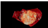
Chronic fibrinopurulent pericarditis and granulation tissue

Fibrinopurulent - fibrin and neutrophils
Puppy with perforating roundworm infection
Sheets of fibrin forming on hepatic surface, neutrophils and blood leaking from vessels
Red, inflamed hyperemic intestines
Acute, diffuse fibrinopurulent peritonitis

Coccidioides immitis spherule

Hemorrhagic exudate from hemorrhagic enteritis
Common in Parvovirus

Simple granuloma- compact and organized exudate collection

Fibrinous pleuritis with fibrous adhesions (blue arrow)

Chronic immune complex glomerulonephritis

Focal

Necrotic exudate
Multifocal necrotizing abomasitis
Change in abomasal acidity allowing fungal growth - Fungal hyphae cause necrosis and hemorrhage

Fibrinous exudate
ABundant fibrin, hyperemic vessels

Fibrinous exudate

Epithelioid cells from macrophages
(called such because they form sheets that look like epithelium)

Suppurative (purulent) exudate of rabbit uterus
Neutrophil dominated
Bacteria- pasturella multocida infection

Shigella gingivitis - inflammatory reaction - swelling, heat

Schizogany occuring- leads to proliferative response of mucosa

Complex granuloma- Have central areas of necrosis or calcification

Granule granulomas Hypersensitivity III