Pharengeal Apparatus Flashcards

1
Q

Structurs that give rise to the head & neck develop form the pharengeal apparatus. This consists of what 4 types of structures?

A

Pharyngeal arches

Pharyngeal pouches

Pharyngeal grooves

Pharyngeal membranes

2
Q

Pharyngeal Arches are fromed from what types of tissues?

A

somitomeric mesoderm and neural crest cells

3
Q

Pharyngeal Arches:

the mesoderm tissues differentiates into which structures?

A

muscles and arteries (aortic arches)

4
Q

Pharyngeal Arches:

the neural crest cells differentiate into which structures?

A

bone and connective tissue

5
Q

Each pharengeal arch is associate with what?

A

a cranial nerve

6
Q

By what week are 4 pharengeal arches visible externally?

Why are the 5th and 6th arches not visible?

A
  • by week 4
  • arches 5 + 6 are rudimentary; not visible externally
7
Q

Pharengeal Pouches are invaginations of what type of tissue?

A

endoderm

(lining the foregut)

8
Q

Pharengeal Grooves are invaginations of what type of tissue?

A

ectoderm

(located between each arch)

9
Q

Pharengeal Memebranes consits of which types of tissues?

A

ectoderm

mesoderm

endoderm

nerual crest cells

(located between each arch)

10
Q

The 1st pharyngeal arch is also called _______?

It gives rise to what 2 prominences?

A

“mandibular arch”

- maxillary prominence

- mandibular prominence

11
Q

The maxillary prominence (1st arch) gives rise to what structures?

A
  • maxilla
  • zygomatic bone
  • a portion of the vomer
12
Q

The mandibular prominence (1st arch) gives rise to what structures?

A
  • mandible
  • squamous temporal bone
13
Q

The second pharyngeal arch is also called _________?

It contributes to the formation of the hyoid bone, with contributions from which other arches?

A

” hyoid arch”

  • parts of the 3rd + 4th arches help form the hyoid bone
14
Q

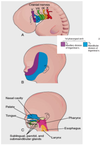
What is the stomodeum?

A

(primordial mouth)

  • appears as a slight depression of the surface ectoderm
15
Q

Where is the function of the oropharengeal membrance?

What types of tissue does it consist of?

A
  • to separate the stomodeum from the cavity of the primordial pharynx
  • ectoderm externally + endoderm internally
16
Q

Each pharengeay arch contains what structures?

A
  • artery
  • vein
  • cartilage
  • muscular component
  • sensory + motor nerves
17
Q

where does the arteries of the pharengeal arches arise from?

Where does it carry blood to?

A
  • arises from ** truncus arteriosus** of the primordial heart
  • goes to the** aorta**
18
Q

What does the cartilage of the pharyngeal arches form?

A

the skeleton of the arches

19
Q

What does the muscular component of the pharyngeal arches become?

A

differentiates in to muscles fo the head + neck

20
Q

What is the cartilage at the dorsal end of the 1st pharyngeal arch called?

What does it develop into?

A

Meckel cartilage

  • develops the malleus + incus of the middle ear
21
Q

The middle part of the Meckel cartilage regresses. What is fromed by its remaining perichondrium?

A
  • anterior ligament of malleus
  • sphenomandibular ligament
22
Q

When does the cartilage of the 1st pharyngeal arch disappear?

What process occurs?

A
  • disappears as the mandible develops around it
  • intramembraneous ossification
23
Q

What is the cartilage of the 2nd pharyngeal arch called?

What does it develop into?

A

Reichert cartilage

gives rise to:

  • stapes of the middle ear
  • styloid process of the temporal bone
  • stylohyoid ligament
  • lesser cornu of the hyoid bone
24
Q

What does the cartilage of the 3rd pharyngeal arch develop into?

A

ossifies to form:

  • greater cornu of the hyoid bone
  • inferior part of the hyoid bone
25
The **1st pharyngeal arch** is supplied by what nerve? What other structures does this nerve innervate?
**CN V**- **trigeminal nerve** _Also innervates the:_ - face - teeth - mucous membranes of the nasal cavities - palate - mouth and tongue | (principle sensory nerve of head + neck)
26
The **2nd pharyngeal arch** is supplied by what nerve?
**CN VII - Facial nerve**
27
The **3rd pharyngeal arch** is supplied by what nerve?
**CN IX - Glossopharyngeal nerve**
28
The **4th pharyngeal arch** is supplied by what nerve? This nerve supplies what other arch?
**CN X - Vagus nerve** **(superior laryengeal branch + recurrent laryngeal branch)** - also supplies the **6th arch**
29
**First (Mandibular) Arch:** 1. gives rise to what muscles? 2. gives rise to what skeletal structures? 3. gives rise to what ligaments?
**1. MUSCLES** - muscles of mastication - mylohyoid + anterior belly of digastric - Tensor Tympani - Tensor veli palatini **2. SKELETAL** - malleus - incus **3. LIGAMENTS** - anterior ligament of malleus - sphenomandibular ligament
30
**Second (Hyoid) Arch:** 1. gives rise to what muscles? 2. gives rise to what skeletal structures? 3. gives rise to what ligaments?
**1. MUSCLES** - muscles of facial expression - stapedius - stylohyoid - posterior belly of digastric **2. SKELETAL** - stapes - styloid process - lesser cornu of hyiod bone - upper part of body of hyoid bone **3. LIGAMENTS** - stylohyoid ligament
31
**Third Arch:** 1. gives rise to what muscles? 2. gives rise to what skeletal structures?
**1. MUSCLES** - stylopharyngeus **2. SKELETAL** - greater cornu of hyoid bone - lower part of body of hyoid bone
32
**Fourth Arch:** 1. gives rise to what muscles? 2. gives rise to what skeletal structures? 3. gives rise to what ligaments?
**1. MUSCLES** - cricothyroid - levator veli palatini - constrictors of pharynx - intrinsic muscles of larynx - striated muscles of esophagus **2. SKELETAL** - thyroid cartilage - cricoid cartilage - arytenoid cartilage - corniculate cartilage - cuneiform cartilage
33
The First Pharyngeal Pouch expands into along which recess?
tubotympanic recess
34
The distal part of the 1st pharengeal pouch is in contact with the first pharyngeal groove. This gives origin to what membrane?
tympanic membrane
35
The cavity of the **tubotympanic recess** becomes the ___________ cavity and __________ antrum.
- tympanic cavity - mastoid antrum
36
The cavity of the **tubotympanic recess** gives to the \_\_\_\_\_\_\_\_\_\_\_\_\_\_\_\_\_ tube.
tubotympanic tube
37
The **thymus** is derived from what type of cells?
epithelium
38
The epithelium cells of the thymus come form what tissue in which pharyngeal arch?
- endoderm - 3rd pharyngeal arch
39
Thymus growth is not complete at birth. How does its growth differ from childhood to adulthood?
- very large in children - starts decreasing towards adulthood
40
What cells are derived from hematopoietic stem cells in the thymus
Lymphocytes
41
Is the pancreas still functional in the adult? If so, what does it secrete? If not, don't worry 'bout it...
thymic hormones primes thymocytes (T- cell precursors)
42
The 2nd pharyngeal pouch gives rise to what?
palatine tonsil
43
The connection between the Fourth Pharyngeal Pouch and what structure, degenerates during development? ## Footnote
the pharynx
44
During week 6, each dorsal part of the 4th pharyngeal pouch forms a ...?
**superior parathyroid gland**
45
The parathyroid glands derived from the **third pouches** goes with what structure? These glands are more **inferior/superior** than parathyroid glands derived from the fourth pouches?
- thymus **- inferior** to glands from 4th pouch
46
**Parathyroid glands:** Epithelium from the which pouches proliferates and forms small nodules? (forming a capillary network)
**third** and **fourth **
47
Which **type of cells** grow in the small nodules formed by the proliferating 3rd + 4th pouches? What overall structure will this form?
**mesenchyme** - capillary network
48
What is regulated by the parathyroind in the developing fetus?
fetal calcium metabolism
49
What is the first endocrine gland to develop in the embryo?
thyroid gland
50
When does the thymus gland begin to develop? What type of tissue does it arise from? (name & location)
**24 days** after fertilization **endoderm** of the pharynx
51
thyroglossal duct
connection between the tongue and the thyroid
52
The thyroid becomes a solid mass of cells and divides into **right and left lobes.** What are these lobes connected by?
the **isthmus of the thyroid gland**
53
A part of the thyroglossal duct persists as the ...?
foramen cecum
54
a cyst in the tongue or in the anterior part of the neck (usually under the hyoid bone) is most commonly fromed from what?
remnants of thymus tissue | (asymptomatic unless infected)
55
A thyroglossal duct cyst located at the base of the tongue is called...?
a **lingual cyst**
56
Branchial Fistula
abnormal canal - opens internally into **tonsilar sinus** - opens externally into **side of the neck**
57
Branchial fistulas are generally found where?
along a**nterior border of sternocleidomastoid muscle**
58
Branchial Cysts are remnants of what?
second pharyngeal groove
59
What causes branchial cysts to become enlarged?
**accumulation of fluid** and **cellular debris** (painless)
60
Branchial Vestiges
pharyngeal cartilages fail to disappear - may appear under the skin in the side of the neck
61
First Arch Syndrome is caused by what?
insufficient migration of neural crest cells into the first arch
62
What are the 2 types of Frist Arch Syndrome?
- Treacher Collins Syndrome - Pierre Robin Syndrome
63
What are some anomolies seen in First Arch Syndrome?
_Anomalies of the:_ eyes ears mandible palate
64
Treacher Collins Syndrome
Mandibulofacial dysostosis ***autosomal dominant*** gene **_clinical signs:_** - Malar hypolasia - downslanting palpebral fissures - defects of the lower eyelids - deformed external ears
65
Pierre Robin Syndrome
***Autosomal recessive*** _Clinical signs:_ - hypoplasia of the mandible (micrognathia) - bilateral cleft palate - defects of the ear and eye
66
DiGorge Syndrome
Pharyngeal pouches 3 + 4 **fail to differentiate** into thymus + parathyroid glands _Clinical signs:_ - facial anomalies = first arch syndrome - cardiovascular anomalies (abnormal neural crest cell migration) - immunodeficiency (absence of thymus gland) - hypocalcemia (absence of parathyroid glands)
67
DiGorge Syndrome is characterized by what condition?
**congenital hypoparathyroidism:** - increased susceptibility to infections - anomalies of mouth (fish mouth) - low set notched ears - nasal clefts - thyroid hypoplasia - cardiac abnormalities
68
Ectopic Parathyroid Glands are found where?
near or within thyroid gland or thymus
69
Ectopic location of which parathyroid gland is more frequent? Where can they be found?
**inferior parathyroid gland** near carotid artery or in thorax
70
What is the most common presentation of an Ectopic Thyroid Gland?
ectopic **lingual thyroid tissue**
71
Why is it clinically important to differentiate an ectopic thyroid gland from a thyroglossal duct cyst (or accessory thyroid tissue)?
to **prevent surgical removal of the thyroid gland**
72
**Congenital Hypothyroidism** (or Cretinism) causes what?
causes impaired skeletal growth + mental retardation
73
What causes congenital hypothyroidism?
* thyroid deficiency during the early fetal period due to:* - severe **lack of dietary iodine** - thyroid agenesis - **mutations** involving the **biosynthesis of thyroid hormone**
74
Congenital hypothyroidism is characterized by what signs/symptoms?
dry, rough skin wide-set eyes periorbital puffiness flat, broad nose large protuberant tongue
75
In the developed world, nearly all cases of congenital hypothyroidism are detected. ## Footnote **When/how is this detected?** **Leves of what substances are measured?**
detected = **newborn screening program** (2nd or 3rd day of life) measured = **Thyroid Stimulating Hormone (TSH) or thyroxine (T4)**
76
What are the 2 parts of the tongue during early stages of development?
**oral part =** anterior 2/3 of tongue **pharyngeal part =** posterior 1/3 of tongue
77
The 2 parts of the tongue are seperated by what?
**terminal sulcus** | (v-shaped depression)
78
The **tongue buds** are formed by the which parts of the tongue?
**median tongue bud =** oral part **2 distal tongue buds =** floor of pharynx associated w. 1st pharyngeal arch
79
The distal tongue buds overgrow the median tongue bud and fuse in the midline, forming what?
the **median sulcus**
80
What 4 types of **papillae** characterize the oral part of the tongue?
filiform papillae (no taste buds) fungiform papillae (taste buds) foliate papillae (taste buds) circumvallate papillae (taste buds)
81
_Name the nerves that do the following:_ 1. carry **generl sensations** from the mucosa 2. carry **taste sensations** form the mucosa 3. **innervated** the developing **tongue muscles**
1. Trigeminal nerve (CN V) - *lingual branch* 2. Facial nerve (CN VIII) 3. Hypoglossal nerve (CN XII)
82
What part of the tongue forms from the **copula** and **hypopharyngeal eminence?** These develop in the floor of the pharynx associated with what pharyngeal arches?
- pharyngeal part - 2, 3 + 4
83
The hypopharyngeal eminence overgrows the copula, eliminating contribution of what pharyngeal arch in the formation of the definitive adult tongue?
**pharyngeal arch 2**
84
Fissuring of the tongue, macroglossia, microglossia and hypertrophy of the lingual papillae, and some characteristics of Down Syndrome are all example of what?
Congenital Anomalies of the Tongue
85
Congenital lingual cysts and fistulas may produce discomfort or dysphagia. **What does this term mean?** What causes these fistulas?
- ** difficulty in swallowing** - persistence of the thyroglossal duct
86
Ankyloglossia
**Lingual frenulum** is short + extends to the tip of the tongue * \*lingual frenulum (connects inferior surface of tongue to floor of mouth)* - interferes in tongue protrusion - makes breast feeding difficult
87
Glossoschisis | (Bifid or cleft Tongue)
Incomplete fusion of lateral lingual swellings = a deep midline groove (very uncommon)
88
Facial primordia appear around what week of development? What structure/area do they form around?
Week 4 - the stomodeum
89
What are the 5 facial primordia?
- One frontonasal prominence - Two Maxillary prominences - Two mandibular prominences
90
What are the bilateral ectodermal thickenings that develop on the ventrolateral aspects of the frontonasal prominence?
nasal placodes
91
The nasal placodes invaginate into the mesoderm to form the nasal pits, producing what?
a ridge that forms the **medial nasal prominence** and **lateral nasal prominence**
92
The nasolacrimal groove forms between what 2 prominences? This forms the ___________ and \_\_\_\_\_\_\_\_\_\_\_\_.
**maxillary prominence + lateral nasal prominence** - nasolacrimal duct + lacrimal sac
93
What are the 2 stages of palate development?
1. The development of the primary palate 2. The development of a secondary palate
94
Primary Palate
two medial nasal prominences fuse together at the midline = intermaxillary segment (caused by medial growth of the maxillary prominences)
95
The intermaxillary segment forms the ... (3)?
philtrum of the lip four incisor teeth primary palate
96
Secondary Palate
outgrowths of the maxillary prominences called the **palatine shelves** - project downward on either side of tongue; later attain a horizontal position + fuse along the **palatine raphe** to form **secondary palate**
97
Where do the primary and secondary palates fuse to form the definitive palate?
at the **incisive foramen**
98
Bone forms in which parts of the palate?
primary palate + anterior secondary palate (not in posterior secondary palate = **soft palae + uvula**)
99
What develops from the medial nasal prominences and fuses with the definitive palate?
nasal septum
100
What is the most common craniofacial anomaly? What are the 3 types of this?
**cleft lip + cleft palate** **- Anterior cleft anomalies** (palatine shelves fail to fuse with the primary palate) **- Posterior cleft palate** (palatine shelves fail to fuse with each other and with the nasal septum) **- Anteroposterior cleft palate** (combination of both defects)
101
Unilateral cleft lip
_Results from:_ 1. Failure of the maxillary prominence to fuse with the medial nasal prominence. 2. Failure of the underlying mesoderm and neural crest to expand, resulting in a persistent labial groove. * (most common congenital malformation of head + neck)*
102
Bilateral Cleft Lip
_Results from:_ Failure of the mesenchymal masses in both maxillary prominences to meet + unite with the merged medial nasal prominences **complete bilateral cleft** of lip + alveolar part of maxilla = medial palatal process hangs free and projects anteriorly - defects affect **orbicularis oris muscle** (closes mouth + purses lip)
103
Complete Cleft Palate
- maximum degree of clefting - cleft extends through soft palate and to the incisive fosa (IF)
104
At what stage of development can facial anomalies be detected? What is used to detect them?
- by the beginning of the **second trimester** **-** sonograph + other imaging techniques