Peripheral Vascular Disease Flashcards
What is an aneurysm? Is it congenital or acquired? what are the 2 types?
- congenital or acquired
- localized dilation of a blood vessel (aorta) or heart
- types:
- True: involves all 3 layers of wall
- false (pseudoaneurysm): wall defect leading to extravascular hematoma
What are the 3 factors affecting collagen structure or function
- Inadequate or abnormal sythesis of collagen
- Marfan syndrome
- Ehlers Danlos
- Excessive connective tissue degradation
- Occurs with increased matrix metalloproteinase (MMP) or decreased tissue inhibitors of metalloproteinase (TMP) (in the setting of inflammation (atherosclerosis) polymorphisms of MMP and or TMP genes may predispose to aneurysm function)
What is Marfan? What is EHler Danlos syndrome?
- Marfan
- defective synthesis of fibrilin
- fibrilin is “Scaffoliding” for deposition of elastic tissue
- results in cystic medial necrosis of aorta
- leads to aneruysm and arotic dissection
- Ehlers Danlos
- one variant has defective synthesis of Type II collagen
- leads to aneurysm formation
How does smoking influence connective tissue degradation?
smoking influences the balance between MMPs and TMPs
What is the pathogenesis of Aneurysms?
- loss of smooth muscle cells
- thickening of intima (due to atherosclerosis)
- leads to ischemia of inner media
- systemic hypertension
- narrows vasa vasorum, leading to ischemia of outer media
- morphologic result is cyctic medial degradation
- thickening of intima (due to atherosclerosis)
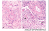
***image shows destruction of the elastic tissue that occurs with an anerysm compared to normal elastc tissue

what are the 2 most common causes of aneurysms? where o these anuerysms occur?
Atherosclerosis-abdominal aorta
Hypertension- ascending aorta
what are some uncommon causes of aneurysm?
- congenital defects
- berry aneurysm: bifurcation of cerebral arteries, subarachnoid hemorrhage
- infections (bacterial, fungi)
- mycotic aneurysm: septic emboli, direct extension, direct infection ny circulating organisms
- syphyllis
- trauma-AV fistula aneurysm
- Vasculitis (inflammation of vessels leads to weakening ot the wall
- IgG4 disease
- Genetic defect in collagen (Marfans and Ehler’s Danlos)
*
What is the most common location of an aneurysm?
Abdominal aortic anerysm (AAA) below the renal arteries and above the bifurcation
arch of the aorta thoracic aorta and iliac arteries also occur but are less common
what is the pathogenesis of an aortic aneurysm(who is it common in and what is the most common cause)?
Describe the morphology (up to X cm etc)
Pathogenesis: more common in men and smokers, most common cause-atherosclerosis
morphology: up to 15 cm diameter.thinning and destruction of media,mural thrombus, saccular or fusiform (one sided or both) variants!
- variants: inflammatory or unkown etiology, mycotic-secondary infection (salmonella gastroenteritis) of an atherosclerotic wall

what is the clinical course of AAA? who is it most common in? how does it present? what are the complications? how do we treat?
more common in men over 50
present as pulsatile abdominal mass
complications
- rupture into peritoneum or retroperitneum
- vascular obstruction-renal, mesenteric, spinal arteries
- embolism of atheroma or mural thrombus to kidenys or lower extremities
aggressive management for large (>5cm) aneurysms, risk of rupture is directly proportional to size
How do we treat?
aggressive management for large (>5cm) aneurysms, risk of rupture is directly proportional to size
open reppair or stent graft inserted through right and left femoral arteries

What are the causes of a thoracic aneurysm? what are the clinical features?
- Causes
- HTN, Marfans syndrome, Tertiary syphyllis
- Clinical Features:
- Mediastinal encroachment
- tracheal compression
- esophageal compression
- bone erosion
- cough due to irritation of the recurrent laryngeal nerve
- cardiac symptoms
- heart failure due to aortic valve insufficiency
- aortic rupture
- Mediastinal encroachment
What is aortic dissection? What is the pathogenesis/etiology?
- blood between and along the laminar planes of media
- causes a blood filled channel that easily ruptures
- pathogenesis/etiology:
- HTN (40-60 yo) >90% of cases
- connective tissue abnormalitiy (younger ages) in association with Marfans syndrom or Ehlers Danlos syndrome
- ascending aorta most commonly involved
- rare causes: post procedural-arterial cannulation; pregnancy
What is the morphology of aortic dissection like? Where does the dissection usualyl occur? inwhat part of the wall?
- initmal tear 10 cm above the arotic valve
- dissetion plane between middle and outer 2/3 of wall in media
- dissection usually extends anterograde
- usually ruptures “out”
- may reenter and form a double barreled lumen
WHat 2 things are associated with an aortic dissection in hypertensive patients?
vasa vasorum: hyaline arteriossclerosis
media: loss of smooth muscle cells
pre-existing pathology of cystic medial degeneration in patients with connective tissue disorder (Marfan syndorme)
How does aortic dissection happen?
Intimal tear begins the process, but trigger for tear is not usually known

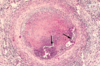
what is shown by these pictures?

dissection!

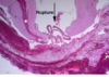
what is this a picture of?

dissection
How does an aortic dissection occur in Marfan Syndrome?
cyctic medial necrosis causes connnective tissue weakness Pools of blue mucinous ground substance disrupt elastic fibers

Describe the clinical course of aortic dissection?
- depenent on level of aorta involved
- sharp pain of anterior chest, uneven pulses and widened meadiastinum
- types A and B
- classical symptoms
- sudden onset of tearing or stabbing pain in anterior chest radiating to the back
what are some possible complications of aortic dissection?
hemopericardium occurs when blood dissects through media proximally
may also rupture into pleural or peritoneal cavities
What is Vasculitis? what 3 things are thought to be involved in the pathogenesis?
inflammation of vessel walls; may affect any size vessel
- pathogenesis:
- immune mediated processes
- infectious origin
- unknown
What immune complex mediated things may lead to vasculitis?
- SLE (systemic Lupus Erythematosis): DNA-nti DNA complexes
- Hypersensitivity to drugs
- Viral infections
- HBsAg-anti HBSAg
How are Anti Neutrophil Cytolasmic Antibodies (ANCA) associated with vasculitis?
- autoantibodies against enzymes in granules of neutrophils
- Ab titiers mirror clinical severity
- two main patterns
- Antiproteinase-3 (PR3-ANCA) granulomatosis with polyangitis (Wegener’s)
- target antigen is PR3 (neutrophil granule constituent)
- Anti-myeloperoxidase (MPO-ANCA)-microscopic polyangitis and CHurg Strauss syndrome
- target is myeloperoxidase
- Antiproteinase-3 (PR3-ANCA) granulomatosis with polyangitis (Wegener’s)
Besides Immune complex mediated and Anti Neutrophil Cytoplasmic Antibodies associated, what other mechanisms lead to vasculitis?
antibodies to endothelial cells- Kawasaki disease
Infectious origin vasculitis can be caused by ____ or _______
- Direct Invasion
- classic example is syphyllis
- aspergillus and mucormycosis
- Indirect via immune mechaisms triggering cross reactivity
what vasculitis are associated with n unkown cause?
giant cell arteririts
takayasu arteritis
polyarteritis nodosa
What vasculitis are associated large vessel vasculitis?
granulomatous disease (eg giant cell arteritis, takayasu)
What types of vasculitis are associated with medium vessel vasculitis?
- Immune complex mediated
- polyarteritis nodosa
- Anti-endothelial cell antibodies
- Kawasaki disease
What vasculitis are associated with small vessel vasculitis?
- Paucity of immune complexes (often with ANCA)
- Vasculitis without asthma or granulomas (microscopic polyangitis)
- granulomatomas, noasthma (granulomatosis with polyangitis)
- Eosinophilia, asthma an granulomas (Churg-Strauss syndrome)
- Immune complex mediated
- SLE (SLE vasculitis)
- IgA (Henoch Schonlein purpura)
- Cryoglobulin (eg cryoglobulin vasculitis)
- other (eg goodpasture disease)
Where does Giant Cell (temporal) Arteritis occur? what is the possible pthogenesis? what is seen clinically? How do you diagnose and what is the treatment?
- most common form of systemic vasculitis in adults
- affects aorta and it smajor branches, especially temporal artery, ophthalmic, vertebral arteries
- pathogenesis: T cell-mediated, driven by antigen? (elastin?)
- clinical: >50 yo often with painful superficial tmeporal artery, diplopia, visual loss, headache
- increased ESR (Erythrocyte Sedimentation Rate)
- Diagnosis: biopsy
- Treatment: steroids
What is seen histologically for temporal giant cell arteritis?
- granulomatous inflammation of inner half of media around internal elastic lamina
- destruction of internal elastic lamina
- multinucleated giant cells (but not always)

What is shown by this arrow?

giant cell! associated with temporal giant cell arteritis
What is Takayasu Arteritis also called? who gets it? what vessels are involved?
- pulseless disease (upper extremities) , occular disturbances
- janpanese women younger than 40 years
- granulomatous inflammation of aortic arch and its branches
- pulonary arteries are involed 50%; coronary and renal arteries may be involved
what are the pathogenesis and histologic features of takayasu arteritis?
pathology: autoimmune etiology
histologyL lymphocytes, giant cells, collagenous fibrosis

What is Polyarteritis Nodosa (classic)? what vessels are affected/spared?
- systemic segmental transmural necrotizing inflammation of small or medium sized muscular arteries
- renal and visceral arteries affected, spares lungs
- all stages of fibrosis and inflammation co-exst
- aneurysms
Who gets Polyarteritis nodosa (classic)? what are the 4 courses? ___-% remission or cure with what therapy?
young adults (30% Hep B antigen) no ANCA
Acute, chornic, subacute, or remittant course
90% remission of cure with corticosteroids, cyclophosphamide
What si the classic presentation of Polyarteritis nodosa (classic)
rapidly accelerating hypertension (renal artery involvement- inflammaed renal arteries have less blood flow, so your blood pressure increases)
Abdominal pain and bloody stools
peripheral neuritis

Where is Kawasaki Syndrome Endemic? What arteries are involved? What diseases/problems are associated with it?
- Endemic in Japan, rare in the US
- Large, medium and small arteries, often coronary arteies with aneurysm formation
- associated with
- mucocutaneous lymph syndrome
- mucus membrane inflamamtion
- enlarged lymph nodes
- 80% are <4 years leading cause of acquired heart disease in children (in Japan)
- mucocutaneous lymph syndrome
What is seen on histology for Kawasaki syndrome? what are the possible etiologies?
necrosis and inflammation, aneurysms, resembles polyarteritis nodosa
etiology: autoantibodies to endothelial cells and smooth muscle cells, infectious agent/ virus trigger
What vessels are associated with Microscopic polyangiitis (Leukocytoclastic Vasculitis)? where does it occur? is it MPO-ANCA positive
- arterioles, capillaries, venules
- skin mucous membrane, lungs, brain, heart , GIT, kidneys, muscles
- necrotizing glomerulonephritis (90%) and pulmonary capillaries
- MPO-ANCA + in 70%
What causes Microscopic Polyangiitis (Leukocytoclastic Vasculitis)? What is seen histologically? major clinical features?
immunologic reaction to antigens-drug, strep, tumor proteins
histology: fibrinoid necrosis, neutrophils, nuclear dust (karyorrhexis)
Clinical features: dependent on vascular bed, hemoptysis, hematuria, proteinuria, abdominal pain or bleeding, muscle pain or bleeding and cutaneous purpura
What kind of vasculitis is Granulomatosis with polyangiitis (Wegener’s)
Are ANCASs present? who gets it?
- Necrotizing vasculitis
- granulomas of lung and or upper respiratory tract (ear, nose, sinus, throat)
- vasculitis of small to medium sized vessels in lungs and upper respiratory tract
- glomerulonephritis
- PR3-ANCAs present in >95%
- likely cell-mediated hypersensitivity response against inhaled infections or environmental antigen (bc often occurs in URT)
- M>F, ~40 yo
*
what are the symptoms of Granulomatosis with polyangiitis (Wegner’s)
symptoms: without treatment 1yr=80% mortalitiy
pneumonitis
sinusitis
nasopharyngeal ulcerations
renal disease

Where does Churg-Strauss Syndrome occur? what is it associated with?
small vessel vasculitis associated with:
asthma
allergic rhinitis
lung infiltrates
peripheral eosinophilia and infiltration of vessels by eosinophils
extravascular necrotizing granulomas
what are the clinical symptoms of Churg Strauss Syndrome? etiology?
palpable purpura
GI bleeding
renal impariment
cardioyopathy
possible hyer responsiveness to allerigc stimulus
MPO-ANCAa present minority
What is Thromboangiitis Obliterans (Buerger disease)? what vessels are involved? who gets it? what is the etiology?
- inflammation and thrombosis of medium to small sized muscular arteries and secondarily the veins and nerves
- gangrene of limbs, requiring amputation
- painful ischemic disease
- tibial and radial arteries
- smokers, <35yrs, Israeli, Indian subcontinent, and Japanese
- etiology unkown-direct endothelial cell toxicity by a component of tobacco

What is Raynaud Phenomenon?
paradoxysmal pallor or cyanosis of fingers, toes, nose or ears
primary: recurrent vasospasm unknown cause (exaggerated response to cold), young, healthy women, no structural changes in the arterial walls (until late in disease)
secondaryL arterial insufficiency due to narrowing due to narrowing and can be due to SLE, SS (systemic sclerosis-scleroderma) atherosclerosis, Nueger disease (smokers) )

What are Varicose Veins? 3 types
- Venous dilation due to elevated pressure and/or weakened vessel walls
- dilation leads to valvular incompetence, edema, pain, thrombosis, stasis dermatitis, and even ulcers
- obesity and pregnancy are risk factors
- most common in superficial leg veins
- Esophageal Varices
- caused by portal hypertension from cirrhosis or portal vein thrombosis
- susceptible to rupture/bleeding
- Hemorrhoids
- anorectal veins, from pregnancy/straining