Pelvis 1 Flashcards

1
Q

What is the differene between the true pelvis and the false pelvis?

A

The pelvis Brim, or iliopectineal line, divide the pelvis into the pelvis major (false pelvis) and the pelvis minor (true pelvis)

2
Q
  1. What is the true pelvis? What forms the inlet?
  2. What separates the true pelvis from the false pelvis?
A

1. The true pelvis is cylyndrical and has an inlet, a wall, and an outlet. The inlet is open, where as the outlet is closed by the pelvic diaphram.

Anterior: Pubic crest

Posterior: Prominatory

Lateral: Arcuate Line, Piriformis & Obturator internis muscle (above tendenous arch of levator ani)

Floor- Levator Ani and Coccygeal muscle.

  1. The arcuate line of the pelvis. Also called the terminal line.
3
Q

What is the relationship of the peritoneum to the pelvis viscera?

What are the two very general things the peritoneum forms?

A

The peritoneum of the pelvis is continuous at the pelvic inlet with the peritoneum of the abdomen. In the pelvis the peritoneum drapes over the pelvic viscera in the midline.

*Forms pouches between adjacent viscera

*Forms folds and ligaments between viscera and pelvic walls.

4
Q

Where is the pararectal fossae (male) ?

A

Formed by the lateral reflection of perineum over the superior third of the rectum

5
Q

Is the rectovesicular pouch found in both males and females?

A

No, only in males.

6
Q

Where is the vesicouterine and rectouterine fossae in the femail?

A

Vesicounerine pouch is located between the bladder and uterus.

Rectounerine Pouch is an invagination of peritoneum between the anus and uterus.

7
Q

Differentiate among the median, medial, and lateral umbilical ligaments.

A

All are formed from folds in the peritoneum

Median- reminany of the urachis connects blatter to the umbilcus. If unclosed patient will pee from bellybutton.

Medial- Contains the umbilical artery

Lateral- Contains the inferior epigastric artery.

8
Q

What composes the median umbilical ligament?

A

The Urachis. Connects umbilicus to the bladder

9
Q

What composes the medial umbilical ligament?

A

Umbilical artery

10
Q

What composes the lateral umbilical ligament?

A

Inferior Epigastric artery

11
Q

What is the blood supply of the bladder?

A

The Blatter recieves blood from the Superior and Inferior Vesicle arteries ( and from the vaginal arteries in females)

The venous drainage via the Prostatic Plexus of veins which empty into the **internal iliac vein. **

12
Q
  1. What is the position of the bladder?
  2. What is the smooth muscle that makes up the wall?
A

The Bladder is situated below the peritoneum and is slightly lower in the female than the male.

Expands into the pelvic brim as it fills, may reach as high as the umbilicus if fully distended.

  1. Detrusor Muscle
13
Q

Discribe the major defining features of the bladder?

A

Learn Attached figure from lecture.

14
Q

Describe the innervation of the bladder?

A

Sympathetic- (GVE) Lower Lumbar Splanchnics L1-2 **induces relaxation of the bladder wall (detrusor muscle) and constrict the internal sphincter, inhibiting emptying.

Parasympathetic- (GVE) **Pelvic splanchnics S2,3,4 (Inferior Hypogastric Plexus) **Induce a constriction of the detrussor muscles and relaxation of the internal urethral sphincter ** **

GVA Pelvic splanchnics S2-4, arise from stretch receptors in the bladder wall. Signal when bladder is full

GSE- Peudendal nerve- cause voluntary relaxation of the urethral sphincter and the bladder begins to void.

15
Q

What is the location of the trigonal area of the bladder in relation to the ureters and urethra?

A

Smooth area of the posterior bladder in the nondistended state.

Superiorlateral borders are the openings of the ureters.

Inferior Border Is the neck and urethra.

16
Q

Describe the sympathetic innervation of the bladder

A

Sympathetic GVE fibers induce relaxation of the bladder wall and constrict the internal sphincter.

Inhibits bladder emptying

May activate the detrussor to prevent semen reflux into bladder.

17
Q

What is the position of the rectum and anal canal?

A

Sigmoid colon makes the turn into the pelvic basin at S3, S3, S3 (He said it 3x in lecture) and turns into the rectum. It is the most posterior portion of the true pelvis and is aganst the sacrum and coccyx.

Just posterior to the vaginal canal in girls.

Posterior to the prostate and mail genital structures in guys.

18
Q

Describe the structure of the rectum and anal canal?

A

The rectum has curves, longitudinal rectal valves of Houston. Lead from the sigmoid colon to the ampula of the rectum. Distal to the ampula the rectum becomes the Anal Canal.

The anal canal has vertical structures called anal columns. These contain the internal venous plexus of the anal canal.

At the external anal opening you have the external venous plexus

19
Q

What innervates the rectum and anal canal?

A
20
Q

What structure differentiates between the superior and inferior rectum?

A

The anal canal is divided into the upper 2/3 (Visceral Portion) which is part of the intestine. and the lower 1/3 (Somatic Portion) which belongs to the perinem.

Demarcation between the two is the Pectinate Line

21
Q

What muscles form the pelvis diaphram (2) ?

Relate each muscles position relative to the pelvis viscera?

How do they relate to the piriformis?

How do they relate to the obturator internis?

A

The Levator Ani and **Coccygeus Muscle **

22
Q

Differentiate between parietal and visceral endopelvic facia in girls.

What structures are derived from each

A

Peritoneum drops to the pubis and flows over the superior dome of the bladder (vesicouterine Pouch). The peritoneum drapes over the Uturus and Forms the Broad ligament.

Pouch of Douglas is between the uterus and anus and is the most inferior point of peritoneum in females.

23
Q

What is the blood supply of the vaginer?

A

Recieves blood from the vaginal branch of the uterine artery and the internal iliac artery

24
Q

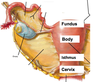
Describe the structure of the Vaginer?

A

Extends between the vestibule and the cervex of the uterus.

Fornex forms the recess between the cervex and the wal lof the vagina

Opening into the vestibule is partially colsed by membranous folds valled hymen.

25
Q

What is the innervation of the vaginer?

A
26
Q

What is the blood supply, structure, and innervation of the uturus?

A

Anteverted at the junction of the vagina and the cervex.

Anteflexed at the junction of the cervex and body.

Made of the fundus body ismus and cervex. Clinically the cervex is the most important.

Blood Supply= uterine artery and ovarian artery

27
Q

What is the struture, position, innervation, and blood supply of the ovary?

A

Lie on the posterior edge of the broad ligament and is bound by external and internal iliac vessles. Posterior and superior is the ureter. Superially is the iliac brim

Uterine ligament attaches it to the uterus

**Not covered by peritoneum

Revieve blood from the ovarian arteries which are contained in the suspensory ligament and anastomose with the uterine artery

Drained by ovarian veins

28
Q

What is the structure, blood supply, position, and innervation of the Uterine tube?

A
29
Q

What are the different parts fo the uturus?

What are their innervation?

A

Fundus- rounded part that is most superior and anterior

Body- main part of the ururus, triangular and contunuous with the lamina of the uterine tube

Isthmus- The constriction between the body of the uterus and the cervex

Cervex- Narrowest part of the uterus that projects into the Vag, most important clinically due to cancer

30
Q

Describe the broad ligament, differentiate between the three areas.

A

Double layered fold of peritoneum

Mesometrium- attaches to the uterus (like endometrium)

Mesovarium- attaches to one border of the ovary

Mesosalphinz- attaches to the ovaduct. (Phinz means tube)

**Base is the most important

31
Q

Describe the location and contents of the Ovarian ligament?

A

Ovarian ligament attaches the uterine end of the ovary to the uterus

32
Q

Describe the suspensory ligament of the ovary in regard to its location and contents

A

Infidubulopelvic ligament Attaches the tubal end of the ovary to the pelvis. Carries all the blood supply lymphatics and autonomic innervation to the ovary.

Counterpart of the spermatic cord.

33
Q

What are the ovarian ligament and the round ligament of the uterus remnants of?

A

Gabernaculum

34
Q

What three bones form the pelvis?

A
  1. Hip Bones
  2. Sacrum
  3. Coccyx
35
Q

What are the outlets of the true pelvis?

Name 6

A
  1. Obturator Canal
  2. Obturator Foramin
  3. Lesser Siatic Foramin
  4. Greater siatic foramin
  5. Urogenital Canal
  6. Rectum
36
Q

What muscle of the pelvic floor that makes a loop around the rectum and aid in rectal leakage?

A

Puborectalis

37
Q

When does sigmoid colon become rectum?

(Bony Landmark)

A

S3 vertebre

38
Q

What are the three points of constriction for the ureter

A
  1. Where the pelvis of the ureter joins the ureter
  2. Where the ureter crosses the pelvis brim
  3. Where the ureter joins the bladder
39
Q

Where is the ureter in relation to the common iliac artery?

A

The ureter crosses the pelvic brim in front of the biforcation of the common iliac artery and descends lateral to the pelvic wall.

40
Q

Where is the uterine artery in relation to the ureter?

A

The uterine artery runs above and anterior to the ureter.

“Water Runs Under the Bridge”

41
Q

Is the ureter retroperitoneal?

A

Yes

42
Q
  1. What does the internal venous plexus of the anal canal drain into?
  2. What does the external venous plexus drain into?
A
  1. Portal Venous System
  2. Caval Venous System

**Site of Portal Caval Anastomoses. **

43
Q

What type of epithelium is above the pectinate line, below the pectinate line?

A

Above the pectinate line the epithelium is columar and cubodal. Below the pectinate line is simple squamous.

44
Q

What is the Lymph Drainage of the Visceral portion of the Anus?

What is the Lymph Drainage in the Somatic portion of the Anus

A

The Visceral portion of the anus drians into the Internal Iliac Nodes

The somatic portion drains into Superficial Inguinal Nodes

Remember the Somatic portion of the anus is technically part of the perinem.

45
Q
  1. What is the venous drainage of the anus above the pectinate line?
  2. What is the venous drainage of the anus below the pectinate line?
A
  1. Portal Venous System
  2. Caval Venous System
46
Q

What is the lower dialated part of the anus that stores feces?

A

Ampulla

47
Q

What vessles supply blood to the anus?

Are there any anastomoses?

A

Superior, Middle, and Inferior Rectal Arteries and Middle Sacral Artery.

Middle rectal artery supplys the posterior portion.

Superior and Inferior form an anastomoses.

48
Q

What is the difference in innervation above and below the pectinate line of the anus?

Does this affect pain due to hemhorides?

(From Board Review Book)

A

Above the pectinate line the sensory innervation is via the pelvic plexus and is Visceral

Below the pectinate line is the somatic fibers of the pudendal nerve.

**External hemhorides will be sensed by GSA fibers of the peudendal nerve and will hurt much worse than internal hemhorides which will relay GVA

49
Q
  1. What innervates the Internal Anal Sphincter?
  2. What innervates the External Anal Sphincter?

Subqutaneous, Superficial, Deep

A
  1. Smooth Muscle. Autonomic Innervation

Sympathetic -Lumbar Splanchnic- CONTRACTION

Parasympathetic - Pelvic Splanchnic- EXTENSION

**Running from Panda Bear huge sympathetic blast, you wouldnt want to stop to poop. -N. Panda-

  1. Skeletal Muscle that recieves somatic innervation from the inferior rectal nerve
50
Q

What are the major differences between the male and female urethra.

A

Male Urethra is much longer and is made of 3 parts (See Figure).

Male urethra integrates with prostate and ejaculatory ducts.

Male urethra has two constriction points. At the external sphincter and the opening of the urethra.

51
Q

What is the homolog of uterine and vaginal structure in the male?

A

Prostatic Utricle

52
Q

Where does ejaculate enter the ale urethra?

A

To the prostatic urethra through the ejaculatory ducts.

53
Q

What are the three major landmarks of the male urethra?

A

Urethral Crest

Prostatic Sinus

Seminal Colliculus

54
Q

What do girls like

A
55
Q

What are the abnormal uterine positions?

A
56
Q

What is a recess useful for bladder surgery?

A

Retropubic Space

57
Q

Which wall of the vaginer is shorter?

What is a plane important to the OB surgeon?

A

Anterior, The fornex forms a recess posterior to the cervex.

Retrovaginal septum is an avascular plane where vagina and anus can be easily separated.

58
Q
A