Patterns of Liver Injury - Chronic Hepatitis Flashcards
What is the clinical definition of chronic hepatitis?
persistence of liver injury with raised serum aminotransferase levels for more than 6 months
What are the causes of chronic hepatitis?
- chronic HBV and HCV
- autoimmune hepatitis (less common)
- drugs
- NASH can present as elevated levels of ALT (to a lesser extent)
What is the differential diagnosis for chronically elevated ALT?
chronic hepatitis or NASH
What are the presenting symptoms of chronic hepatitis?
- can be asymptomatic
- not generally unwell; typically outpatients
- non-specific sysmptoms like fatigue and loss of appetite
What are the risk factors for NASH?
- obesity
- diabetes
- metabolic syndrome
What is the hallmark histological feature of chronic hepatitis?
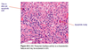
periportal inflammation/interface hepatitis:
- apoptosis of hepatocytes at the interface between portal tracts and lobular parenchyma
- associated with lymphoplasmacytic inflammation
- not specific for chronic hepatitis; must fit clinical dx
- degree = grade of chronic hepatitis
- determines rate of fibrosis

What is the major mode of cell death in chronic hepatitis?
apoptosis
Apoptosis is a hallmark feature of
acute and chronic hepatitis
How is chronic hepatitis diagnosed?
biopsy: degree of interface hepatitis (grade) and fibrosis (fibrous septa)

What forms the fibrous septa in chronic hepatitis?
- diffuse, persistent chronic inflammation
- causes deposition of abnormal collagen (scar tissue)
- pattern is septal fibrosis
- stellate pattern of bands or plates of scar tissue that radiate from portal tracts outward into the parenchyma
- can lead to cirrhosis

What is septal fibrosis?
stellate formations of scar tissue radiating from portal tracts

How does fibrosis develop in chronic hepatitis?
- portal tracts enlarge (contain more scar tissue/collagen)
- strands of collagen begin to extend outwards towards central veins
- occasionally link up = bridging fibrosis
- marker for treatment institution
- occasionally link up = bridging fibrosis
- portal to portal bridging of fibrous septa
- contracts, distorts liver anatomy and tf function
- lobules and sinusoids filling with collagen
- overtly symptomatic
- fibrosis = network of nodules separated by fibrous septae
What is fibrosis (histologically)?
network of nodules separated by bands of fibrous scar tissue
What is non-alcoholic fatty liver disease?
- previously crytogenic cirrhosis (unknown cause)
- important differential diagnosis of impaired liver function (children and adults)
- two components:
- steatosis (fat, nothing else), or
- steatohepatitis and fibrosis = NASH
What is NASH?
- non-alcoholic steatohepatitis
- steatohepatitis + fibrosis
- associated with obesity, metabolic syndrome, and diabetes
What is steatosis?
- aka fatty liver, fatty change, steatosis
- accumulation of abnormal amounts of lipid in hepatocytes
- two types:
- macrovesicular - large droplet, very common
- microvesicular - small droplet, very rare (acute fatty liver of pregnancy, Reye’s syndrome

What are the histological features of macrovesicular steatosis?
- large droplets of fat that displace nuclei to one side of cells
- well-delineated fat vacuoles

What is macrovesicular steatosis?
- caused by an abnormality of triglyceride synthesis
- increased synthesis or decreased excretion
- very common
How is macrovesicular steatosis diagnosed?
- severe: non-invasive imaging (ultrasound)
- LFT may be normal or mildly abnormal
- not possible to determine cause from histology alone, need clinical information
- related to obesity and alcohol
What is steatohepatitis?
- steatosis with inflammation and injury of hepatocytes
- significant risk of fibrosis –> cirrhosis
-
hallmark feature: hepatocellular ballooning degeneration
- also present in alcoholic steatohepatitis/alcoholic hepatitis

How are non-alcoholic steatohepatitis (NASH) and alcoholic steatohepatitis (ASH) differentiated?
-
clinically:
- is the patient drinking at a toxic level?
- do they have risk factors for NASH?
- essential histological features are identical (ASH tends to be more severe):
- hepatocellular ballooning degeneration (blue arrows) due to accumulation of water (hydropic swelling) - diffuse
- apoptosis

What is the pattern of fibrosis in steatohepatitis?
- pericellular fibrosis
- scar tissue surrounds individual or small groups of hepatocytes
- fills sinusoids, isolates hepatocytes
- same pattern in ASH and NASH

What is the pathogensis of NASH vs macrovesicular steatohepatitis?
- macrovesicular steatohepatitis occurs as a result of increased storage of triglyceride in the liver
- increased triglycerides due to insulin resistance, dietary factors, or poorly controlled diabetes
- FFAs diverted to liver for storage
- this is SAFE steatosis (no inflammation or fibrosis)
- NASH results when this pathway is overloaded and lipotoxic metabolites form
- cause cellular stress, inflammation, apoptosis, and necrosis
- develops into NASH - causes fibrosis and cirrhosis

