Pathology of Glomerular Disease Flashcards
What is NOT filtered in the glomerulus?
All proteins equal to or larger than albumin, including immunoglobulins They will stay in the plasma
What is a podocyte?
Cells in the Bowman’s capsule that wrap around capillaries of the glomerulus They interdigitate and look like finger processes
What is the function of a mesangial cell?
The primary function of mesangial cells is to remove trapped residues and aggregated protein from the basement membrane thus keeping the filter free of debris
What is the numbered arrows in this slide
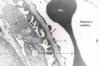
- Endothelium
- Basal lamina CT
- Podocytes
What is mesangial cell?
Tree like group of cells that support capillaries
What is glomerulonephritis?
Basic name for disease of the glomerulus
Even though it has “itis” doesn’t always mean the condition will be inflammatory
What is the difference between 1y and 2y glomerulonephritis?
1y - only affects the glomerulus
2 - other parts of body affected by the disease too
Give examples of 2y glomerulonephtitis.
SLE
Wegener’s
Aetiology of glomerulonephritis?
Some due to immunoglobulin depositon
BUT
Some are disease with no immunoglobulin deposition, such as diabetic glomerular disease
What are the 4 most common presentations of glomerulonephritis?
Haematuria
Heavy proteinuria
Slowly increasing proteinuria
Acute renal failure
What is heavy proteinuria known as?
Nephrotic syndrome
A 40 year old male presents to you with discoloured urine. What is the first test you would do?
Urine dipstick analysis
The dipstick shows positive for blood - what are the main causes of haematuria from most common to least?
- UTIs
- Urinary tract stone
- Urinary tract tumours
- Glomerulonephritis
What would you do next for the patient who presented with discouloured urine once blood has been confirmed in urine?
Send off a urine culture
Arrange a hospital appointmed for an ultrasound examination
The urine culture and ultrasaound exam came back as normal. Next steps?
Check his clotting
If clotting is okay do a renal biopsy
The renal biopsy shows increased levels of mesangial cells. What has occured?
- Immunoglobulin - mainly IgA - and complement componant C3 have accumulated in mesangial area of glomeruli
- IgA deposits irritate mesangial cells and cause proliferation
What is this disease called?
IgA glomerulonephritis
Aetiology of IgA glomerulonephritis?
Unknown
Excess IgA is sometimes present in serum showing an excess production but this isn not specific to IgA glomerulonephritis
Does the IgA get filtered into urine?
No - stays stuck in mesangial cells clogging up the mesangium NOT the fliter
If the filteration apparatus is not being targetted by IgA - how does blood get into the urine?
Unknown
Prognosis of IgA nephropathy?
Usally self limiting and will return to normal
BUT
A small % go into chronic renal failure as deposition of matrix does not stop and destroyes the glomeruli
Case 2 - A 50 year old male presents to you with 3 weeks of swollen legs and feelings of unwellness. What test is done and what will it show?
Send a blood biochemistry and haematology tests
Will show low serum albumin
The blood test showed low serum albumin. Next test? What does it show?
Urine dipstick to test for proteinuria
Shows a +ve result
Proteinuria is confirmed in case 2 patient - what next?
Refer to hospital nephrolagist who will measure specifially albumin levels in urine
What will this patients urine albumin level be?
Very high = a very heavy loss of albumin = a nephrotic syndrome
If this patient has a nephrotic sysndrome - where must the abnormality be? What tests will be done next?
In the glomerular filter
Check clotting and if ok do a renal biopsy
What will a renal biopy show in case 2?
- Thickened glomerular basement
- Spikes of new basement membrane matrix matierial under podocytes

What is case 2 called and what causes the thickened glomerus membrane and basal lamina spikes?
Known as membranous glomerulonephritis
Causes by IgG depostiting itself inbetween basal lamina and podocyte - so BL tries so spike and surround it to remove it
Why does IgG not just get filtered out?
Can’t - is too big
So if the glomerular membrane is now thickened - what causes albumin to present in urine?
IgG ativates a C3 complemet which punches holes in the filter, so even though it is thickened it is leaky and albumin gets through
Prognosis of membranous glomerulonephritis?
1/4 in chronic renal failure within 10 years
If you survive past 10 years without chronic renal failure - your chances don’t increase of avoiding it
Underlying cause of IgG production and accumulation which causes membranous glomerulonephritis?
Not known but may be an underlying maligancy
Also in some patients = antigen is phospholipase A2 receptor but not known why
Case 3 - 31 year old female, type 1 DM since 7 y/o and has had long periods of poor glycemic control. Has a developed retinopathy and albumin has slowly been increaing over the years in urine. Urine now reads as heavy protein leackage into urine. What is this a suspected case of?
Diabetic nephropathy
How would you confirm it was diabetic retinopathy?
Check clotting factors and go ahead with a renal biopsy
What is the pathology behind diabetic nephrology?
- Glycated molecules lead to matric deposition in the basal lamina underlying the endothelium AND in the mesangium
- This causes thickened BUt leaky basement membranes
- The mesangial matrix starts compressing the capillaries = reduced blood flow to glomerulus
Why are adhesions to the bowmans capsule seen in diabetic nephropathy?
Attempt to stop massive leaking of albumin into urine
Prognosis of diabetic nephropathy?
Has an inevitable decline - improving diabtetic control HALTS this decline but doen’t reverse it
So if the renal function gets so bad then even improving glycemic control won’t really help much as it will be too late
Case 4 - 50 y/o female been unwell for 3 weeks and has a cough. Serum creatinine measured at 500, last measurement was 60 a year prior. What considerations should we take when moving forward with investigations?
Is this acute or chronic? We don’t know as the pervious test was a year ago so renal function could have been slowly deteriorating or could be a sudden rise
It is established as an acute event since creatinine was measured a short while later and shot up. What tests would you do next?
Ultrasound to test for a renal tract lesion
Clotting and then biopsy
USS shows no renal tract lesion. What is this suspected to be and how will the renal biopsy support this.
Crescentic glomerulonephtitis
Renal biopsy will show:
- Early endothelial damage with fibrin deposition
- A cellular rpoliferation and influx of macroohages - shows up as a creasent around the glomerulus
Causes of cresentic glomerulonephritis? (Just list main one Paul Brown focused on)
Granulomatosis with polyangiitis
Also known as
Wegener’s granulomatosis
Explain what Wegener’s granulomatosis is.
A form of vasculitis - inflammation in vessels - usually affecting vessels in kidney, nose and lung
Furthe tests for wegeners?
Serum test shows presence of anti-neutrophil cytoplasmic antibodies
What are the anti-neutrophil cytoplasmic antibodies doing?
Not depositied in kideny but directed against 2 enzymes in primary granules of neutophils:
- Proteinase 2
- Myeloperoxidase
The antibodies result in tissue damage via the interactions with primed neutrophils and endothelial cells
Why do anti-neutrophil cytoplasmic antibodies form?
Unknown - could be form of autoimmunity
Prognosis of wegeners?
Fatal
Mean survival rate is 6 months if left untreated
Wegener’s - treatment
Cyclophosphamide - 75% complete remission
What were the 4 cases of glomerulonephritis discussed in this lecture?
- IgA glomerulonephritis
- Membranous glomerulonephritis
- Diabetic nephropathy
- Wegener’s