PACEs Flashcards
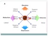
Aetiology of hypertension
PREDICTION
Primary: 95%
Renal: RAS, GN, APKD, PAN
Endo: Raised T4, Cushing’s, phaeo, acromegaly, Conn’s
Drugs: cocaine, NSAIDs, OCP
Raised ICP
CoA
Toxaemia of pregnancy (PET)
Increased viscosity
Overload with fluid
Neurogenic; DAI, spinal section
Aetiology of PUD
Acute: usually due to drugs (NSAIDs or steroids) or stress
Chronic: drugs, H. pylori, hypercalcaemia, ZE syndrome
CML
Key investigations
Complications
Findings - Signs of anemia, infection, HSM +- lymphadenopathy
Key investigations - Cytogenetic analysis - t(9:22) translocation - forming BCr-ABL fusion protein. Raised myelocyte count. Monitor for blast transformation (AML)
Rx - TKI - imatinib. BMT - curative in younger patients
Cx - AIHA, Infections (due to reduced immunoglobulins), BM Failure/infiltration
Smoking Cessation in history station?
3 As
Ask - enquire as to smoking status
Advise - best way to stop is with support and motivation
Act - provide details of where they can receive help (nhs stop smoking helpline, pharmacy, websites0
Hodkin’s Lymphoma
Classification system
Classification:
Ann Arbor:
1 - One LN group. 2 - >1 but same side of diaphragm. 3 - both sides of diaphraagm. 4 - extranodal inolvement +B for B symptoms
Mitral Stenosis
Aetiology
Murmur
Clinical Features
Dx
Severity Features
Complications (5)
Rx
Complications:
i) Pulmonary HTN, ii) Systemic Emboli - brain, GI, limbs, iii) ortner’s syndrome - RLN palsy, iv) Dysphagia - oesophageal compression, v) Bronchial obstruction
CLL
Complications
Complications - Paraproteinaemia
Causes of amyloidosis (5)
Features of amyloidosis (5)
Congenital - Familial Amyloidosis (AD - transthyretin mutation)
Primary Amyloidosis - AL Amyloid.
Spontaneous, Multiple Myeloma, Waldenstrom’s Macroglobulinaemia
Secondary Amyloidosis - AA Amyloid
Secondary to inflammatory diseases
SLE, RA, Ank Spond, IBD etc.
Dialysis Related - B2 Microglobulin
T2DM - amylin
Features: Renal (proteinuria, nephrotic syndrome, large kidney), Heart (restrictive, arrhythmias), Nerves - peripheral and autonomic neuropathy, carpal tunnel. GIT - macraglossia, malabsorption, hepatomegaly, obstruction. vascular - periorbital prupura
What is EAA
Extrinsic Allergic Alveolitis
- Acute/ Chronic
Acute = T3HS to allergen exposure
Chronic = Granuloma formation + obliterative bronchiolitis
Causes of jaundice
Conjugated
Unconjugated
Conjugated - Usually due to biliary obstruction (Stone, Tumour, Cirrhosis - alcohol, drug, viral, autoimmune/inflammatory)
Unconjugated - pre-hepatic (haemolysis), hepatic (reduced conjugation - CN, GS, hypothyroid, or Reduced uptake of BR- CCF, Drugs - rifa, contrast)
Causes of splenomegaly:
Haematological
Infective
Portal HTN
Connective Tissue
Others
Indications for splenectomy
Haematological - MF, MPS, CML,
Infective- Malaria, TB, EBV,
Portal HTN
Connective Tissue
Others - sarcoid (NCG), Gauchers (LSD), Amyloidosis, Primary immunodeficiency ( CVID)
Indications for splenectomy:
Trauma, Rupture
AIHA, ITP, HS, Hypersplenism (Pancytopenia, reticulocytosis - therefore not bM failure)
Seronegative Spondyloarthropathies/ arthropathies
Spondylo - features
Extra articular
PEAR- Psoriatis, Enteropathy, Ankylosis, Reactive
Spondylo:
Axial arthritis and sacroilitis, Asymmetrical large-joint oligoarthritis or monoarthritis
Enthesitis
Dactylitis
Extra articular: iritis, psoriaform rashes, oral ulcers, AR, IBD
Asthma:
Hx questions
Causes
Hx questions - Precipitants( dust, pollen, smoke, work, atopy? - hayfever, eczema), Diurnal variation, exercise tolerance, Effect on life
Causes - Atopic, Stress, Drug - NSAIDs, Environment - smoking, pollutants, occupation, Infection - post-viral, aspergillosis,
Haematological stains
Sudan Black
Tartate resistent acid phosphatase
Reduced leukocyte alkaline phosphatase
Increased leukocyte ALP
Sudan Black - myeloblasts
Tartate resistent acid phosphatase - Hairy cell leukaemia
Reduced leukocyte alkaline phosphatase - CML, PNH
Increased leukocyte ALP - PV, ET, MF4
Stroke
Causes
Classification -
ACA territory
MCA Territory
PCA territory
Vertibrobasilar
Location of motor symptoms?
Rx
MENDS
Ix SAH
Causes - Haemorrhagic (HTN, bleeding daithesis, thrombolysis, EtOh) Ischaemic - Atherosclerotic, embolic (AF, CAS) watershed, Vasculitis, Anti phospholipid snydrome, thrombophilia
RFs - age, diabetes, htn, smoking alcohol, HChol, PVD, AF, Raised PCT, OCP
Classification - Bamford clinical syndrome, Imaging allows vessel diagnosis, NIHSS/ Bartel - outcome scoring, dragon - prognosis after thrombolysis
ix - CT to exclude bleed but can also demonstrate thrombus in artery immediately and loss of grey white matter differentiation within an hour
DW MRI is the the best to visualise stroke
ACA - frontal and medial cerebrum. Leg effected greater than arm/face, abulia
MCA - lateral/external cerebrum. Face/Arm effected grater than leg. Homonmyous Hemianopia, higher cortical dysfunction (D- aphasia. ND - VS neglect/apraxia)5
PCA - occipital cortext. Mac Sparing HH
VB - Cerebellar, brainstem, occipital cortices. DANISH, HH, CN, Plegias, Sensory Sx
Motor symptoms:
Large artery territories - seizure, Homonymous hemianopia, higher cortical dysfunction
Internal capsule
Brainstem - accompanied by ipsilateral CN sign
Rx- Thrombolysis <4.5 hours, Thrombectomy, “permissive hypertension” to ensure cerebral perfusion. (CT 24hr to ensure no haemorrhagic transformation)
treat sequalae raised ICP if occurs secondary to vasogenic/cytotoxic oedema
Otherwise Aspirin 300 mg 14 days. Clopidogrel lifelong, statin if cholesterol >3.5 but not immediately due to risk of haemorrhagic transfomration
General Mx - Monitoring - Tight Glycaemic contro, BP control but not too agressive due to reduced CPP, regular neuro obs 5
Rx underlying causes - Carotid stenosis, AF, thrombophilia, infection, valve defect etc. Hypercholesterolaemia
Stroke requires a multi-faceted approach to management
MENDS
MDT- neuro, nursing, dietician, SALT, physio, OT, diisability
Eating - swallow screen, ?NGT, malnutrition risk
Neurorehab - physio, speech, cognitive
DVT prophylaxis
Sores - avoid pressure sores (waterlow)
Ix SAH:
CT - 90% positive
LP - Xanthocromia at 12 hours/ yellow looking
Liver Disease
Causes of cirrhosis
Cirrhosis scoring systemts
Cx of cirrhosis
Causes of budd chiari syndrome
Liver failure investigations
Signs of liver failure
Causes of portal htn
SAAG
Hepatorenal syndrome Rx
Cx of acute liver failure + rx
General management of LF
Abs in LF
Rx for ascites
Precipitants of hepatic encephalopathy
1st line for cirrhosis - transient elastography
ALT:AST >2 alcohol
Causes - Alcohol, NAFLD, Viral (viral serology), HH, Wilson’s, A1AT, Autoimmune (AIH, PSC, PBC), Ca (mets, primary), Drug (MTX, isoniazid, amiodarone, methyldopa), Vasc - CCF , budd chiari ,
Cirrhosis Scoring:
Childs pugh - ABCDE (Albumin, bilirubin, clotting, distension - ascites, encepholopathy)
MELD - now prefered. bilirubin, clotting,
Cx- portal hyertension (ascites, splenomegaly, varcies, encephalopathy), SBP, decompensation ( Jaundice, enceophalopathy, hypoalmbuminaemia, clotting abnormality, hypoglycaemia), HCC
Causes of budd chiari syndrome - hypercoaguability, local tumour, membranous obstruction (congenital)
Liver failure investigations - FBC, U&E, LFT, Clotting, Glucose, B12, Folate, albumin
Signs of liver failure
-Jaundice, oedema/AScites, enceophalopathy, bruising, varices, fecor hepaticus
FBC: infection, GI bleed, raised MCV (EtOH)
U+Es:
Reduced urea, raised creatinine: hepatorenal syndrome
Urea synthesised in liver: poor test of renal function
LFTs:
AST:ALT >2= EtOH
AST:ALT <1= viraL
Albumin: reduced in chronic liver failure
PT: prolonged in acute liver failure
- *Clotting:** Raised INR
- *Glucose**
ABG: metabolic acidosis
Cause:
Ferritin, a1AT, caeruloplasmin, autoAbs, paracetamol levels
infection - HAV, HBV, HCV, EBV CMV - PCR serology, virology, antibodies
Leptospirosis . Blood urine culture. imaging
Ascites - MC&S, cytology, SAAG, chemistry, AFB
Imaging - US And PV doppler
Transient Elastography
liver biopsy
Causes of portal htn
Pre hepatic - portal vein thrombosis, peritoneal carcinomatosis,
Hepatic- cirrhosis, HCC, schisto, sarcoid
Post hepatic - budd chiari syndrome, nephrotic syndrome , RHF, TR, constrictive pericarditis
TB,
SAAG
<1.1 = Exudative - nephrotic syndrome, malignancy, TB
>1.1 = Portal HTN - Cirrhosis, CCF, Budd chiari,
Hepatorenal syndrome (type 1 - fast and type 2 - slow)
Rx - IV albumin + terlipressin
Dialysis / hepatic transplant
cx of acute liver failure
- bleeding (vit k, FFP, platelets) , sepsis, ascites ( fluid restrict, frusemide, alosteron antag, tap,) , oedema, hypoglycaemia (IV glucose), encephalopathy (lactulose - reduces ammonia production, rifaximin), seizures (lorazepam), cerebral oedema (mannitol)
General Management
Cons - constant follow up, Dietician (must ensure high carb diet), avoidance of alochol and hepatotoxic drugs,
Mx - treat underlying cause, pabrinex (C, B1, B3, B6), diazepam if withdrawing,
NGT- high carbs, thiamine
PPI - against stress ulcers
Monitoring - Fluids - Monitoring of status and output. Avoid using NS due to RAS activation —> go for colloid/ HAS/ 5% dextrose
Daily Bloods
Daily wieghts
ABs
AIH - SMA, SLK, LKM
PBC - AMA
PSC - ANA, ANCA
Ascietes rx
- fluid restrict, frusemide, spiro, therapeutic tap, TIPSS
precipitants of hepatic encephalopathy
HEPATICS - Haemorrhage, Electrolyte- hypokaleamia, hypontaraemia, poisons (diuretics, sedatives), alcohol Tumour HCC, Infection,Constipation,Sugar (low)
Aortic Stenosis
DDx
Causes
Symptoms
Signs
Clinical indicators if severe
Ix
Mx
Indx for VR
Causes: senile calficiation, Congenital - bicuspid valve, supravalvular aortic stenosis (WS), Rheumatic fever
DDx- HOCM (increases with valsava manouvere), MR, Aortic Sclerosis (thickening but no narrowing), CAD
Symptoms:
Triad- dsypnoea, syncope, angina
LVF, Ahhyrthmias, Emboli, Death
Signs:
ESM (loudest at RSE) radiating to carotids
LVF, Soft A2, S4 Heart sound
Slow rising pulse
Severe:
LVF, Soft A2, S4 Heart Sound, NPP
Ix:
CXR - Cardiomeg, postestenotic dilatation, calcified valve ECG - LVH, Echo - thickened, immbole valve cusps. <1 VA, >40 VG, >4 VV. Catheterisation - assess coronaries, valve gradient.
Mx:
Conservative - optimise Rfs
Medical - Reduce preoload - CCB, Ace-i, Reduce afterload - diuretsic, improve myocardial perfusion - beta blockers
Surgical - Baloon valvuloplasty( interim measure) TAVI, Open valve replacement, xenograft/bioprosthesis
Indx for VR:
i) symptomatic ii) LVF EF <60% iii) CABG/ other valve op concurrently
ECG:
Normal: PR, QRS, QT
LVH?
Inferior View - Leads and Artery
Lateral View - Leads and ARtery
Anteroseptal - Leads and Artery
Anterolateral - Leads and ARtery
Posterior - Leads and ARtery
PE
Hypokalaemia
Hyperkalaemia
1st degree HB
Mobitz I
Mobitz II
Complete Heart block
Wellen’s syndrome
LBBB
RBBB
Pericarditis
PR <0.2 ms, QRS <0.12 ms, QT men <430 ms women <450 ms
LVH:
LBBB, complete AV block. - due to septal calcification
(lateral leads) v4-v6 - Tall R, ST depression, t wave inversion
Inferior View - II, III, AVF – RCA
Lateral View - I, AVL, V5, V6 – Left Circ
Anteroseptal - V2- V4 - LAD
Anterolateral - V2-V6 - Left Main Stem (LAD+ L Circ)
Posterior - V1-V3(Deep ST depression with R waves). RCA
PE - S1Q3T3 Classic, Sinus tachy cardia, RAD, RVH
Hypokalaemia - U waves, Absent T wave, prolonged PR, prolonged QT, st depression
Hyperkalaemia - tall t waves, absent p waves, broad qrs, prolonged PR –> sinusoidal
1st degree HB - PR >0.2 ms
Mobitz I - Wenkeback phenomenon, gradual PR prolongation til beat is missed then resets so next PR is shorter
Mobitz II - fixed non conduction of a p wave. Not preceded by PR lengthening or followed by PR shorting
Complete Heart block - P waves and QRS complexes are not related
Wellen’s syndrome - T wave inversion/ hyperkinetic t waves in V2-V3 highly suggestive of imminent LAD occlusion
LBBB - Wide QRS. SRS pattern in V1 and RSR pattern in V6. Wave comes down RV first then spreads to LV.
RBBB - Wide QRS. RSR pattern in V1. SRS pattern in V6.
Pericarditis - Shaddle shaped ST elevation, PR depression, Electrical alternans, Low voltage QRS
Heart Failure - Def
Ix
Medical Management
Causes - myocardial, pressure, arrhyhtmias
Low output
High Ouput HF causes
Causes of RVF
Signs of RVF
Causes of LVF
Criteria for HF diagnosis?
BNP
NYHA Classification of HF
Heart failure - inadequate CO to perfuse the body despite adequate filling pressures
Impaired Systolic function - dilation via starling’s effect –> failure to completely empty ventricle –>
RAS activation due to hypotension –> increase in afterload (peripheral vasoconstriction) / increase in preload–> salt/fluid retention (aldosterone)
Hypertrophied myocardium –> increased metabolic demand –> ischaemia
Ix
NT-Pro-BNP if no history of IHD –> positive then echo
History of IHD –> echo
CXR: A-Alveolar shadowing. B - Kerley Lines, C - Cardiomegaly D - upper lobe diversion E - pleural effusions F- fluid in fissures
Medical Management
Reduce preload - diuretics
Improve myocardial perfusion - Negative chronotropes (beta blockers)/ positive inortroped (digoxin)
Reduce afterload - CCB, ACE-i, hydralazine +nitrate (avoid in AS), aldosterone
Invasive
cardiac resync,
aortic baloon counter pulsation
heart transplant
Causes - myocardial, pressure, arrhyhtmias
myocardial:
IHD, Toxins - EtOH, Chemotherpay, Autoimmune- scleroderma, SLE, RA. infection - HIV, Infiltrative - malignancy, sarcoid, amyloid, HH. genetic- musc dystrophy
pressure:
HTN, Valve disease, restrictive pericarditis, volume
Arrhythmias - tachy / brady
Low output
Pump failure - Impaired systolic /diastolic function / arrhythmias
Excessive - preload (AR, MR, Fluid overload)
Excessive after load (Hypertension, Aortic Stenosis, HOCM)
High output
increased metabolic demand (RVF fails before LVF fails)
AATTPP –> Anaemia, AVM, Thyrotoxicosis , Thiamine Pregnancy, Paget’s
Causes of RVF
LVF, Cor Pulmonale, Tricuspid/pulmonary valve disease
Signs of RVF - Raised JVP, pulsatile hepatomegaly, pitting oedema, ascites
Causes of LVF - IHD, dilated cardiomyopathy, HTN, mitral and aortic valve disease
Signs of LVF - weight loss, muscle wasting, cyanosis, AF, cardiomegaly, S3, Wheeze, basal creps
Framingham Criteria
BNP <100. 96% NPV
NYHA - 1 - 4
1 breathlessness on unaccustomed exercise. 4 breathlessness at rest
Pulmonary HTN
Causes
Causes - Obstructive Airway disease (including OSA), Idiopathic, Sarcoid, Vasculitis, Heart Failure, PE
Diabetes Mellitus
T1DM vs T2DM
Complications:
Neuropathy
Autonomic
Vascular
Mx
T1DM - insulin, monitoring, transplant
T2DM - diet, exercise metformin, SFN, DPP4, GLP1, Acarbose, SGLT2, Insulin, transpl
T1DM - DR3/DR4. Anti - Gad/islet cell. Absent production of insulin.
T2DM - insulin resistance
neuropahy caused by- metabolic (glycosylation, ROS) and ischaemic damage to the vas nevorum
Can be - polyneuropathy (symmetrical), mono/ mono multip, amyotrophy (painful asymmetrical wasting of quads, loss of knee jerk) , autonomic neuropathy
Autonomic:
gastroparesis, urinary retention, postural hypotension, diarrhoea, Sexual dysfunction
Macro - MI, PAD, Stroke
Micro - retinopathy, nephropathy
Mx
MDT
Diet - Standard. Reduce total, refiend carbs, fats etc. Avoid binge drinking.
Hypoglycaemic meds - Biguianides (MFN), SFUs (Glibenclamide etc.), TZD (pioglit), DPP4 i ( Sitagliptin), GLP 1 mims- (Exanatide), SGLT-2 inhib (Dapiglafozin)
Statins - if >40 , T1DM >10 years, T1DM with cx,
Anti - HTN - <130/80. ACE-i best
4 Cs - Control (CBG, Hba1c), Complications (DKA, Honk, RF, Neuropathy, PAD, fundoscopy) , Coping, Competency
CNs Eyes:
II
III
III,IV, VI : Eye movements
V
VII
INO:
III,IV, VI : Eye movements
INO: MLF lesion (MS, stroke etc.) Impaired adduction of ipsilateral eye - i.e. conjugate nerve palsy. convergence is preserved
Neuro
LMN Signs/ Causes
LMN:
Weakness
Paralysis
Loss of reflexes
Causes:
Sinal cord infarction (fracture, dislocation, vasculitis, atheromatous)
