Ophthalmology Flashcards
Your patient awoke with this in the morning. It is painless. What is it, and what are 3 risk factors?

Sub-conjunctival haemorrhage
Sneeze, cough
hypertension
anticoagulants
What is this?

Hyphema
Haemorrhage of the anterior chamber - blood vessels at iris root
What is this? How would you assess any damage?

A foreign body - examine the pretarsal sulcus by everting the upper eyelid.
Remove with cotton applicator.
Apply fluorescein - an orange dye used with a blue light to visualise any abrasions
If there are abrasions - topical antibiotic?
What is this? List three things that might have caused it

Ptosis
Congenital - poor development of Levator palpabrae superioris
Acquired - Eyelid mass
neurological condition (Mysasthenia gravis, ophthalmoplegia, Horner’s (superior tarsal muscle) or third nerve paralysis)
What is this - be specific!

A horizontal strabismus - Right sided exotropia
What are the subtypes of strabismus?
Exotropia, esotropia, hypotropia, hypertropia
What is this? Be specific!

A horizontal stabismus - esotropia
Name two anti-muscarinic eyedrops
Cyclopentolate (think uveitis)
Tropicamide (best for reg eye exam)
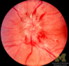
What is this? If unilateral, what might be be due to?

Papilloedema
Raised ICP
Idiopathic intracranial hypertension
What is this?
Optic disc oedema - the 4 Is
Inflammation
Infiltration (cancers)
ICP
Infarction
What is this?

Arterio-venous nicking
Hardened arteries due to chronic hypertension mean they distort the more pliant veins where they cross
What is this?

Copper wiring
Sign of chronic hypertension
due to fibrosis of intima and subintima
What is this?

Obliterative retinal vasculopathy
A thrombus has totally occluded this vessel - now looks like a pipe cleaner!
What is this?

A Hollenhorst spot - a platelet-fibrin-cholesterol embolus
it is likely to have originated at an artheroma of the carotid bifurcation
What is this?

Segemental retinal infarction resulting from Hollenhorst plaque - patient would report cloudy vision pertaining to this area of the retina
What is this?

Retinal vasculitis - ‘sheathing’ due to lympocytic infiltrates
MS
Sarcoidosis
What is this?

Myelinated nerve fibres
a congenital issue, does not affect the vision
What is this?

Cotton wool spots
(ischaemic bursting of axons in the nerve fibre layer)
Microinfarcts in the retinal nerve fibre layer
Hundereds of causes!
Diabetes, hypertension, hypercoagulable states, HIV, connective tissue diseases
What is this?
What is their ‘classic’ cause?

Roth spots
It’s a cotton wool spot (ischaemic bursting of axons in the nerve fibre layer) surrounded by haemorrhage (ischaemic bursting of pre-capillary arterioles)
Sub-acute bacterial endocarditis
What is this?

Hard exudates
lipid leakage from damaged capillaries
diabetes!
von Hippel-Lindau
retinal vein occlusion
What is this?

Retinal drusen
Remains of dead retinal pigment epithelial cells
age-related macular degeneration
biggest issue with be submacular neovascularisation
What is this?

Retinal neovascularisation
risk is they bleed easily or with minimal trauma
leads to retinal detachment eventually
near the optic disk? - retinal vein occlusion or, DIABETES!
near the extremities? Think sickle cell
What is this?

Boat haemorrhage
due to rupture of large superficial retinal veins between the retina and the vitreous
aka - subhyaloid haemorrhage OR pre-retinal haemorrhage. In practise, v difficult to tell the two apart
What is this?

Dot haemorrhage
Rupture of deep capillary
Diabetes!
What is this?

Flame haemorrhage
Rupture of superficial pre-capillary arterioles
Hypertension
What is this?

A sub-retinal haemorrhage - note the normal vessels above it
Label the arrows from left to right - top row then bottom of this normal fundus

Physiologic cup
Fovea
Neuroretinal rim
Retinal vein
Retinal arteriole
Macula
Diagnosis?

Phlyctenular conjunctivitis - senstization of the conjunctiva to an endogenous allergen
A sign of:
Primary TB
or more likely, S aureus
Retinopathies as a result of essential hypertension. What are they, in roughly the order that they might be seen
Copper wiring I
Silver wiring I
AV nicking II
Flame haem, cotton wool spots III
Papilloedema IV
Where, in terms of eye layers would you find drusen. What are they?
Between Bruch’s membrane and the retinal pigment epithelium (or within Bruch’s membrane.
Lipid deposits
What is the classic lesion seen with internucelar ophthalmoplegia?
Failure of adduction in each eye during horizontal gaze (signal, which goes from cortex to VI nuclei THEN III, via the medial longitudinal fasciculus, doesn’t get there as a result of a lesion in the MLF, and thus the medial rectus doesn’t adduct the eye.
Abducting eye also shows nystagmus
Convergence is NOT affected!!
