Neurovascular UL Flashcards
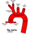
In typical anatomy, which branch off the arch of the aorta is the left subclavian artery?

Third

Describe the course of the axillary artery
Continuation of the “subclavian artery”. At midpoint of the clavicle it passes over the first digitation of serratus anterior at the outer border of the 1st rib.
It collects the prevertebral fascia as the “axillary sheath”.
Closely associated with the 3 cords of the brachial plexus (lateral cord laterally, medial cord medially and posterior cord posterior) behind pectoralis minor.
At lower border of teres minor becomes “brachial artery”
What are the branches of the axillary artery?
“St Thomas Loves Sweet Apple Pie”
3 parts (1,2,3)
Above pec minor (1) = superior thoracic A
Behind pec minor (2) = thoraco-acromial A + lateral thoracic A
Below pec minor (3) = Subscapular A + anterior circumflex humeral A + posterior circumflex humeral A
https://www.studyblue.com/notes/note/n/axillary-artery-branches/deck/11449501
Where does the superior thoracic artery arise and what does it supply?
First part of the axillary artery above the superior border of pectoralis minor.
Runs anteriroly to supply the pectoral muscles.
Where does the thoracoacromial artery arise and what does it supply?
Second part of the axillary artery (first branch)
Runs along the superior aspect pec minor, pierces clavipectoral fascia.
Four terminal branches:
clavicular + deltoid + acromial + pectoral
Where does the lateral thoracic artery arise and what does it supply?
Second part of the axillary artery (behind pec minor). Runs along inferior aspect of pec minor on serratus anterior fascia.
Supplies both pecs + breast tissue (females)
Where does the subscapular artery arise and what does it supply?
Third part of the axillary artery
(first branch distal to pec minor)
Largest branch
Runs down the posterior axillary wall, gives off circumflex scapular artery, before becoming the thoracodorsal A.
Thoracodorsal A runs with N to supply lats dorsi.
Where do the anterior and posterior circumflex humeral arteries arise and what do they supply?
Last two branches off the axillary artery.
Posterior is significantly larger.
Anterior circumflex humeral A runs deep to coracobrachialis and both heaps of biceps to supply long head tendon of biceps and joint capsule and then anastomosis with posterior circumflex humeral A to supply shoulder joint.
Posterior runs between subscapularis and teres major lateral to long head of triceps, accompanied by the axillary nerve.
Also supplies deltoid (with nerve) + lateral heads of triceps
AXILLA SLIDE (QUIZ FANTASTIC)
Define the borders of the axilla.
What are its contents?
https://www.meduhub.com/mod/view.php?id=53&type=15
Contents:
Axillary artery, axillary vein
Cords of brachial plexus (medial, lateral, posterior)
Lymph nodes, fat
Axillary tail of Spence (part of mammary gland)
Label clockwise


What muscles attach to the bicipital groove of the humerus?
“Lady between to majors”
Pec major (lateral), lats dorsi (floor of groove), teres major (medial)
How many groups of axillary lymph nodes and what are their names?
What do they drain?
Clinical importance
5 sets - APICAL
A = anterior (pectoral), P = posterior (scapular), I = none, C = Central, A = Apical, L = lateral (humeral)
Drain: UL, breast, chest above umbilicus

What structures pass via the bicpital groove of the humerus?
Long head of biceps brachii
Branch of anterior circumflex humeral artery
What spinal segments make up the brachial plexus?
What are some common variations?
Ventral rami of C5, C6, C7, C8 and T1 spinal nerve roots
“Pre-fixed” = inclusion of C4 and “Post-fixed” = inclusion of T2
What are the trunks of the brachial plexus?
Upper C5 + C6
Middle C7
Lower C8 + T1

What do the anterior and posterior divisions of the brachial plexus supply?
There are six divisions, three anterior and three posterior, which are branches off each trunk (upper, middle, lower).
Anterior divisions supply flexors. Posterior divisions supply extensors.
What are the cords of the brachial plexus?
Where do you find them?
What nerve roots/divisions do they contain?
Lateral cord = anterior divisions from upper and middle trunks (C5, C6, C7)
Medial cord = anterior divisions from lower trunk (C8, T1)
Posterior cord = posterior divisions from all trunks (C5-T1)
Found within the axilla, under pec minor, closely associated with the axillary artery. Lateral is lateral to artery, medial is medial and posterior is behind artery.

What are the branches of the lateral cord of the brachial plexus?
What do these branches supply?
LML
Lateral pectoral nerve, musculocutaneous nerve, lateral root of median nerve
Supply: flexor comparments
What are the branches of the medial cord of the brachial plexus?
What do these branches supply?
MMUMM
Medial pectoral nerve, medial root of median nerve, ulna nerve, medial cutaneous nerve of arm, medial cutaneous nerve of forearm
Supply flexor compartment

What are the branches of the posterior cord of the brachial plexus?
What do these branches supply?
ULTRA
Upper subscapular nerve, lower subscapular nerve, thoracodorsal nerve, radial nerve, axillary nerve
Supply: extensor compartments

Draw the brachial plexus

What is the name of the classical upper brachial plexus injury?
What is the mechanism of injury and clinical presentation?
What nerve roots and nerves does it involve?
Erbs palsy occurs with forcful lateral flexion of neck causing avulsion of C5 and C6 nerve roots (at Erb’s point where C5 and C6 nerve roots unite).
Mechanism: trauma or shoulder dystocia in birth.
Clinical presentation: “waiters tip” (Sh Add, Sh IR, elbow extension and pronation)
Involves suprascapular nerve, axillary nerve and musculoskeletal nerve.
What is the classical lower brachial plexus lesion?
What is the mechaism of injury?
What is the clinical presentation?
What nerve roots are involved?
Klumpke’s paralysis
Mechanism: breaking fall by hanging onto something or with pulling of arm out of birth canal
Clinical presentation: claw hand (supination, wrist and hand flexsion)
Nerve roots: C8, T1
What are the nerves that come off the roots of the brachial plexus? What do they supply? Clinical significance
Long thoracic nerve (C5-C7) = serratus anterior = damage causes winging of scapula
Dorsal scapular nerve (C5) = rhomboids = winging scapula

What are the nerves that come off the trunks of the brachial plexus? What do they supply? Clinical importance?
- *Suprascapular N** (C5) = infraspinatous and supraspinatous + sensation AC and GH joint = paralysis causes pain, difficulty with Abd and ER and wasting
- *Nerve to subclavius** C5+C6

Describe the superfical venous and sensory supply of the cubital fossa. Clinical importance?
Cephalic vein laterally with lateral cutaneous nerve of forearm
Basilic vein medially with medial cutaneous nerve of forearm
Connected by median cubital vein
(Makes up roof of cubital fossa with skin and superfical fascia, deep fascia with aponerosis of biceps tendon medially)
Clinical: phlebotomyn

Boundaries of cubital fossa
Roof = skin, superfical fascia, cephalic vein (lateral), basilic vein (medial), median cubital vein (central), medial and lateral cutaneous nerves of forearm, deep fascia with aponerosis of biceps (medially)
Lateral border = brachioradialis (medial border)
Medial border = pronator teres (lateral border)
Base is between two epicondyles and apex is meeting of brachioradialis and pronator teres
Floor = brachialis (upper portion), supinator (lower portion)
What are the contents of the cubital fossa?
Lateral –> Medial (RBBM)
Radial nerve and terminal branches
Biceps tendon (large)
Brachial artery and branches (ulnar and radial)
Medial nerve
(Ulna nerve is not the in the cutital fossa as it runs posterior to the medial epicondyle. Musculocutaneous nerve has become superfical as the lateral cutaneous nerve of forearm and is located superfically within the roof laterally)

Describe the path of the brachial artery. Where are two locations to find the brachial artery when dissecting?
Originates at the termination of the axiallary artery at the inferior border of teres major.
Continues down arm (medially to median nerve), halfway down arm median nerve cross infront (lateral –> medial). Gives off deep brachial artery, superior and inferior ulna collateral arteries. Then heads into cubital fossa, closest to the bicipital aponeosis under which it dives and terminates as the radial and ulnar arteries


