Neurology Flashcards

1
Q

Why do you get a fixed pupil from a head injury?

A

Compression of the parasympathetic fibres on the optic nerve due to raisefd ICP

2
Q

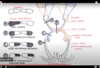
What does each spinal tract do?

A
  1. Dorsal column - Vibration and proprioception
  2. Spinaothalamic - pain, sensation and temp
  3. Corticospinal - Weakness
3
Q

What is Brwon-sequard syndrome?

A
  1. Hemisection of the spinal cord - anterior white commisure
    • Ipsilateral paralysis
    • Ipsilateral loss of proprioception and fine discrimination
    • Contralateral loss of pain and temperature
4
Q

ves How are headaches classified?

A
  1. Primary
  2. Secondary
  3. Cranial neuralgias
  4. Facial pain
  5. Other headaches
5
Q

What are the primary headaches?

A

Tension

Migraine

Cluster

6
Q

What are secondary headaches?

A

Headaches that arise from a cause, this includes:

Dental pain

Menengitis

Encephalitis

Traumatic head injury

Substance abuse

7
Q

WHAT IS A MIGRAINE?

A

Severe throbbing pain or a pulsing sensation, usually on one side of the head

8
Q

What causes migraines?

What are the triggers?

A

Exact cause unknown, but thought to be imbalances in brain chemicals

C - Chocolate
H - Hangovers
O - Orgasms
C - Cheese/Caffeine
O - Oral contraceptive pill
L - Lie-ins
A - Alcohol
T - Travel
E - Exercise

9
Q

What are the stages of a migraine?

A
  • Prodrome
  • Aura
  • Attack
  • Post-drome
10
Q

What are the symptoms of a migraine?

A

Migraine without aura
Most common
Headaches - one or both sides of the head
Sickness
Nausea
Photophobia
Phonophobia

Migraine with aura
Similar symptoms
Accompained by changes in vision, certain smells

11
Q

How is a migraine diagnosed?

A

Usually a diagnosis based on medical history, symptoms and a neurological examination

12
Q

What is the treatment for migraines?

A

Pain-relieving medications

  • Ibuprofen, paracetamol
  • Sumatriptan, rizatriptan

Preventive medications

  • Propanalol
  • Topiramate - Not in women who are child-bearing age
  • Verapamil - Blood pressure lowering meds
13
Q

WHAT IS A TENSION HEADACHE?

A

A tension headache is generally a diffuse, mild to moderate pain in your head that’s often described as feeling like a tight band around your head.

Most common type of headache

14
Q

What is the cause of tension headaches?

A

The cause of tension headaches is not known.

Experts used to think tension headaches stemmed from muscle contractions in the face, neck and scalp, perhaps as a result of heightened emotions, tension or stress.

15
Q

What are the symptoms of a tension headache?

A

Dull, aching head pain

Sensation of tightness or pressure across your forehead or on the sides and back of your head

Tenderness on your scalp, neck and shoulder muscles

16
Q

What are the different types of tension headache?

A

Episodic tension headaches

Episodic tension headaches can last from 30 minutes to a week. Frequent episodic tension headaches occur less than 15 days a month for at least three months. Frequent episodic tension headaches may become chronic.

Chronic tension headaches

This type of tension headache lasts hours and may be continuous. If your headaches occur 15 or more days a month for at least three months, they’re considered chronic.

17
Q

How do you diagnose a tension headache?

A

Medical history

Physical and neurological examinations

18
Q

What is the treatment for a tension headache?

A

Acute treatment

  • Aspirin, paracetamol or an NSAID are first-line

Prophylaxis

  • ‘Up to 10 sessions of acupuncture over 5-8 weeks’
  • Low-dose amitriptyline is widely used in the UK for prophylaxis against tension-type headache.
19
Q

WHAT IS A CLSUTER HEADACHE?

A

Cluster headaches, which occur in cyclical patterns or cluster periods, are one of the most painful types of headache.

A cluster headache commonly awakens you in the middle of the night with intense pain in or around one eye on one side of your head.

20
Q

What is the cause/triggers of a cluster headache?

A

Unknown

Possible triggers:
Alcohol

21
Q

What are the symptoms of a cluster headache?

A
  1. Pain typical occurs once or twice a day, each episode lasting 15 mins - 2 hours
  2. Clusters typically last 4-12 weeks
  3. Intense sharp, stabbing pain around one eye (recurrent attacks ‘always’ affect same side)
  4. Accompanied by redness, lacrimation, lid swelling
  5. miosis and ptosis in a minority
22
Q

How do you diagnose a cluster headache?

A

Clinical

23
Q

What is the acute treatment for a cluster headache?

A

100% Oxygen

Subcutaenous triptan

24
Q

What is the prophylaxtic treatment for a cluster headache?

A

Verapamil - Calcium channel blocker

Prednisolone

25
WHAT IS TEMPORAL ARTERITIS?
Giant cell arteritis is an inflammation of the lining of your arteries. Most often, it affects the arteries in your head, especially those in your temples. For this reason, giant cell arteritis is sometimes called temporal arteritis.
26
What is the pathophysiology of temporal arteritis? What condition is it closely linked to?
1. With giant cell arteritis, the lining of arteries becomes inflamed, causing them to swell. 2. This swelling narrows your blood vessels, reducing the amount of blood — and, therefore, oxygen and vital nutrients — that reaches your body's tissues 1. Polymyalgia rheumatica
27
What are the symptoms of temporal arteritis?
1. Persistent, severe head pain, usually in your temple area + jaw pain when you chew or open your mouth wide 2. Scalp tenderness 3. Fever 4. Fatigue 5. Unintended weight loss 6. Vision loss or double vision, particularly in people who also have jaw pain 7. Sudden, permanent loss of vision in one eye
28
What are the complications of temporal arteritis?
1. Blindness 2. Aortic aneurysm 3. Stoke
29
How do you diagnose temporal artertitis?
1. **Blood tests** * ESR * CRP 2. **Imaging** * Doppler ultrasound 3. **Biopsy** * Skip Lesions + Giant cells 4. **CK and EMG -** Normal
30
What is the treatment for temporal arteritis?
1. **High dose corticosteroid** * Prednisolone 2. May prescribe vitamin D supplements to help prevent bone loss
31
WHAT IS A STROKE?
A stroke occurs when the blood supply to part of your brain is interrupted or reduced, preventing brain tissue from getting oxygen and nutrients. Brain cells begin to die in minutes.
32
What are the causes of a stroke?
Ischaemic stroke Haemorrhagic stroke
33
What are the symptoms of a stroke?
1. Motor weakness 2. Speech problems (dysphasia) 3. Swallowing problems 4. Visual field defects (homonymous hemianopia) 5. Balance problems
34
How do you diagnose a stroke?
**Confirm diagnosis** 1. Non-contast CT is first line 2. Hyperdense sign in an artery **Estalish the site of pathology** 1. Carotid ultrasound 2. Echocardiogram 3. Cerebral angiogram **Identify factors which may influence management** 1. Chest X-ray - cardiac enlargement 2. Blood glucose - hyperglycaemia 3. Full Blood Count - polycythaemia, thrombocytopenia
35
How do you investigate a carotid bruit?
Duplex ultrasound
36
What is the treatment for a stroke?
**Ischaemic stroke** 1. Alteplase/tissue plasminogen factor within 4.5 hours 2. Once haemorrhagic stroke has been excluded give aspirin 300mg 3. Endovascular procedures - Thombectomy within 6 hours, an extended time of 6-24 hours may be considefed if CT shows salvagable tissue 4. **Thrombolysis and thrombectomy if under 4.5 hours** **Haemorrhagic stroke** 1. Blood thinners 2. Surgical clipping 3. Coiling
37
What is the long term management following a stroke?
1. Clopidogrel 75mg - FIRST LINE 2. Aspirin + dipyridamole for patinet who cannot tolerate clopidogrel
38
What is a pontine haemorrhage?
Pontine haemorrhage is a life-threatening condition. It often occurs as a complication secondary to chronic hypertension
39
How do patients with a pontine haemorrhage usually present?
1. Reduced Glasgow coma score 2. Quadriplegia 3. Miosis 4. Absent horizontal eye movements
40
What is the cause of lateral medullary syndrome?
Posterior inferior cerebellar artery
41
WHAT IS A TRANSIENT ISCHAEMIC ATTACK (TIA)?
A transient episode of neurologic dysfunction caused by focal brain, spinal cord, or retinal ischaemia, without acute infarction.
42
What is the cause of a TIA?
A transient ischemic attack has the same origins as that of an ischemic stroke, the most common type of stroke. In an ischemic stroke, a clot blocks the blood supply to part of your brain. In a transient ischemic attack, unlike a stroke, the blockage is brief, and there is no permanent damage.
43
What are the symptoms of a TIA?
1. Weakness, numbness or paralysis in your face, arm or leg, typically on one side of your body 2. Slurred or garbled speech or difficulty understanding others 3. Blindness in one or both eyes or double vision 4. Vertigo or loss of balance or coordination
44
How do you investigate a TIA?
1. **Brain Imaging** * **CT brain** should not be done 'unless there is clinical suspicion of an alternative diagnosis that CT could detect' * **MRI** is preferred to determine the territory of ischaemia, or to detect haemorrhage or alternative pathologies 2. **Carotid imaging** * Atherosclerosis in the carotid artery may be a source of emboli in some patients * **All patients** should therefore have an **urgent carotid doppler** unless they are not a candidate for carotid endarterectomy
45
What is the treatment for TIA?
**Immediate antithrombotic therapy:** * Give aspirin 300 mg - this is for 2 weeks **Continued antithrombotic therapy after the event:** * Clopidogrel is recommended first-line * Aspirin + dipyridamole given to patients who cannot tolerate clopidogrel
46
What do you give during a TIA?
Give aspirin 300 mg immediately, unless 1. the patient has a bleeding disorder or is taking an anticoagulant (needs immediate admission for imaging to exclude a haemorrhage) 2. the patient is already taking low-dose aspirin regularly: continue the current dose of aspirin until reviewed by a specialist 3. Aspirin is contraindicated: discuss management urgently with the specialist team
47
What is the management for a patient after a TIA if undetected at the time for before 7 and over 7 days?
1. **If the patient has had more than 1 TIA ('crescendo TIA') or has a suspected cardioembolic source or severe carotid stenosis:** * Discuss the need for admission or observation urgently with a stroke specialist 2. **If the patient has had a suspected TIA in the last 7 days:** * Arrange urgent assessment (within 24 hours) by a specialist stroke physician 3. **If the patient has had a suspected TIA which occurred more than a week previously:** * Refer for specialist assessment as soon as possible within 7 days 1. Advise the person not to drive until they have been seen by a specialist.
48
What is a patient is on blood thinners and has a TIA?
Needs urgent CT head
49
What condition can mimic the effects of a TIA?
Hypoglycaemia in diabetes
50
What are the rules after a stroke or TIA for driving?
1. Stroke or TIA: **1 month** off driving, may not need to inform DVLA if no residual neurological deficit 2. Multiple TIAs over short period of times: 3 months off driving and inform DVLA
51
What do midline lesions and hemisphere lesions cause in the cerebellum?
**Midline lesions** Can produce severe gait and truncal ataxia. As they extend they can also give fourth cranial nerve lesions and severe ipsilateral arm tremor, marked nystagmus **Cerebellar hemisphere** Lesions can produce classic ipsilateral limb ataxia (intention tremor, past pointing and mild hypotonia).
52
What are the causes of cerebellar disorders?
1. **Vascular** * **​​**Stroke 2. **Space-occupying lesion** 3. **Nutritional** * **​​​​**Thiamine * Vitamin E deficiency 4. **Infection** 5. **Truama**
53
What are the symptoms of cerebellar disorder?
**DANISH** Dysdiadochokinesia Ataxia (gait and posture) Nystagmus. Intention tremor Scanning dysarthria Heel-shin test positivity
54
What are the investigations for cerebellar disorder?
Blood tests - FBC, LFTs, cholesterol, protein electrophoresis, copper and caeruloplasmin, immunoglobulins and glycoproteins. Electroencephalogram (EEG). Electromyogram (EMG). Imaging - MRI is the modality of choice.
55
What is the treatment for cerebellar disease?
Treat underlying condition
56
WHAT IS EPILEPSY?
Epilepsy is a central nervous system (neurological) disorder in which brain activity becomes abnormal, causing seizures or periods of unusual behavior, sensations, and sometimes loss of awareness.
57
What are the different types of epilepsy?
**Focal seizures** **Generalised seizures**
58
What are the different focal seizures?
From one part of the brain **Focal seizures without loss of consciousness** Alter emotions or change the way things look, smell, feel, taste or sound. They may also result in involuntary jerking of a body part, such as an arm or leg, **Focal seizures with impaired awareness** Stare into space, repetative movements
59
What can focal seizures be further classified into?
**Motor** * *Non-motor** (e. g. déjà vu, jamais vu; ) **Features such as aura** | (e.g. Jacksonian march)
60
What is a Jacksonian march?
It characteristically starts by affecting a peripheral body part such as a toe, finger or section of the lip and then spreads quickly 'marches' over the respective foot, hand or face. In some with Jacksonian march seizures (as in this case), the electrical disorder spreads over larger areas of the brain, causing the seizure to develop into a tonic-clonic seizure.
61
What are the different generalised seizures?
**Absence seizures** Previously known as petit mal seizures. Staring into space or subtle body movements such as eye blinking or lip smacking **Tonic seizures** Tonic seizures cause stiffening of your muscles **Atonic seizures** Loss of muscle control, which may cause you to suddenly collapse or fall down. **Clonic seizures** Jerking muscle movements **Myoclonic seizures** Brief jerks or twitches of your arms and legs. **Tonic-clonic seizures** Grand mal seizures
62
What are the causes of epilepsy?
1. **Genetic influence** 2. **Head trauma** 3. **Brain conditions** * Stroke 4. **Infectious diseases** * Meningitis, AIDS and viral encepthalitis 5. **Prenatal injury** 6. **Developmental disorders** * Autism
63
How do you diagnose epilepsy?
1. ECG 1st line 2. MRI 3. FBC, U+Es and glucose 4. Brain imaging and EEG but **CLINICAL DIAGNOSIS**
64
What is the treatment for epilepsy?
1. **Generalised tonic-clonic seizures** * Sodium valproate * Second line: lamotrigine, carbamazepine 2. **Focal seizures** * Carbamazepine or lamotrigine * Second line: levetiracetam, oxcarbazepine or sodium valproate 3. **Absence seizures\* (Petit mal)** * Sodium valproate or ethosuximide 4. **Myoclonic seizures** * Sodium valproate * Second line: clonazepam, lamotrigine
65
What are the featurs of a temporal lobe seizure?
1. Hallucinations (auditory/gustatory/olfactory) 2. Epigastric rising/Emotional 3. **Automatisms (lip smacking/grabbing of clothes/plucking)** 4. Deja vu/Dysphasia post-ictal)
66
What are the features of a frontal lobe seizure?
1. Head/leg movements 2. Posturing 3. Post-ictal weakness 4. Jacksonian march
67
What are the features of a parietal lobe seizure?
Paraesthesia
68
What are the features of an occipital lobe seizure?
Floaters/flashes
69
What are the features of abscence seizures?
1. Absences last a few seconds and are associated with a quick recovery 2. Seizures may be provoked by **hyperventilation or stress** 3. The child is usually unaware of the seizure 4. They may occur many times a day 5. EEG: bilateral, symmetrical 3Hz spike and wave pattern
70
What is the management of abscence seziures?
Sodium valproate Ethosuximide
71
How can you differentiate between a pseudoseizure and an epileptic seizure?
Prolactin levels
72
What is todd's palsy?
Weakness of limbs after seizure
73
WHAT IS THE DIFFERENCE BETWEEN A VASOVAGAL AND TONIC-CLONIC SEIZURE?
Vasovagal shorter post-ictal
74
WHAT ARE THE FACTORS FAVOURING A NEAD?
1. Pelvic thrusting 2. Family member with epilepsy 3. Much more common in females 4. Crying after seizure 5. Don't occur when alone 6. Gradual onset
75
What are the causes of NEAD?
1. A heart condition that causes fainting 2. Diabetes or other metabolic disorders 3. Emotional pain 4. Mental pain 5. Being bullied 6. Physical or sexual abuse 7. A major accident
76
How do you diagnose NEAD?
1. **EEG** A patient with NES will not show unusual electrical activity in the brain on the EEG. 2. **MRI + CT** Epilepsy 3. **Blood tests** Diabetes
77
What is the treatment for NEAD?
CBT
78
What should a patient tell the DVLA with epilepsy?
**First unprovoked/isolated seizure** 1. **6 months off** if there are **no relevant structural abnormalities** on brain imaging and no definite epileptiform activity on EEG. 2. If these conditions are not met then this is increased to **12 months** **For patients with established epilepsy or those with multiple unprovoked seizures:** 1. → may qualify for a driving licence if they have been free from any seizure for **12 months** 2. → if there have been no seizures for **5 years (with medication if necessary) a ’til 70 licence is usually restored** 3. withdrawawl of epilepsy medication: should not drive whilst anti-epilepsy medication is being withdrawn and for **6 months** after the last dose
79
What is narcolepsy?
Suddenly causes a person to fall asleep at inappropriate times
80
What is cataplexy?
Sudden loss of muscle tone typically triggered by emotion such as laughing or crying
81
What are the different types of narcolepsy?
Narcolepsy that occurs with cataplexy is called type 1 narcolepsy. Narcolepsy that occurs without cataplexy is known as type 2 narcolepsy.
82
What is the cause of narcolepsy?
1. Associated with **HLA-DR2** 2. Associated with low levels of **orexin (hypocretin)**, a protein which is responsible for controlling appetite and sleep patterns
83
What are the symptoms of narcolepsy?
1. Typical onset in teenage years 2. Hypersomnolence 3. Cataplexy (sudden loss of muscle tone often triggered by emotion) 4. **Sleep paralysis** 5. **Vivid hallucinations on going to sleep or waking up**
84
How do you diagnose narcolepsy?
Multiple sleep latency EEG
85
What is the treatment for narcolepsy?
**Stimulants** 1. Modafinil 2. Armodafinil **Improves nighttime sleep** 1. Sodium oxybate
86
WHAT IS VASCULAR DEMENTIA?
It is not a single disease but a group of syndromes of cognitive impairment Caused by different mechanisms causing ischaemia or haemorrhage secondary to cerebrovascular disease.
87
What is the cause of vascular dementia?
**Stroke (infarction) blocking a brain artery** With both silent and apparent strokes, the risk of vascular dementia increases with the number of strokes that occur over time. One type of vascular dementia involving many strokes is called multi-infarct dementia. **Narrowed or chronically damaged brain blood vessels**. These conditions include the wear and tear associated with aging, high blood pressure, abnormal aging of blood vessels (atherosclerosis), diabetes, and brain hemorrhage.
88
What are the symptoms of vascular dementia?
1. Focal neurological abnormalities e.g. visual disturbance, sensory or motor symptoms 2. The difficulty with attention and concentration 3. Seizures 4. Memory disturbance 5. Gait disturbance 6. Speech disturbance 7. Emotional disturbance
89
Can vascular dementia and alzheimer's disease occur together?
Yes
90
How is vascular dementia diagnosed?
1. A comprehensive history and physical examination 2. Formal screen for cognitive impairment 3. Medical review to exclude medication cause of cognitive decline 4. MRI scan – may show infarcts and extensive white matter changes
91
What is the treatment for vascular dementia?
1. Often involves managing the risk factors 2. Lower blood pressure 3. Reduce your cholesterol levels 4. Prevent your blood from clotting 5. Help control your blood sugar
92
What is Horner's syndrome?
Horner's syndrome is a triad of features resulting from interruption of the sympathetic pathway from the hypothalamus to the orbit
93
What are the symptoms of horners syndrome?
1. Miosis (small pupil) 2. Ptosis 3. Enophthalmos\* (sunken eye) 4. Anhidrosis (loss of sweating one side)
94
How do you diagnose horner's syndrome?
1. A normal pupil is dilated by **cocaine and hydroxyamphetamine** 2. When the lesion is pre-ganglionic only **hydroxyamphetamine** causes dilatation 3. When the lesion is post-ganglionic only **adrenaline** causes dilatation
95
What is the treatment for horners syndrome?
Treat underlying condition
96
What is bell's palsy?
Bell's palsy may be defined as an acute, unilateral, idiopathic, ## Footnote **Facial nerve paralysis** **7th Nerve**
97
What are the symptoms of bell's palsy?
1. **Weakness** to total paralysis on one side of your face — occurring within hours to days 2. **Lower motor neuron** facial nerve palsy - **forehead affected**\* 3. Post-auricular **pain** (may precede paralysis) 4. Altered taste 5. Dry eyes 6. Hyperacusis
98
What is the cause of bell's palsy?
Latent herpes viruses (herpes simplex virus type 1 and herpes zoster virus), which are reactivated from cranial nerve ganglia
99
What is synkinesis?
Abnormal regrowth of nerve fibers. This may result in involuntary contraction of certain muscles when you're trying to move others (synkinesis) For example, when you smile, the eye on the affected side may close.
100
How do you diagnose bell's palsy?
Diagnosis of exclusion
101
What is the treatment of bell's palsy?
**Corticosteroids** Prednisolone **Antiviral drugs** Acyclovir **Articifical tears**
102
WHAT IS BULBAR PALSY?
Bulbar palsy refers to a range of different signs and symptoms linked to impairment of function of the cranial nerves IX - 9 Glossopharyngeal X - 10 Vagus XI - 11 Accessory XII - 12 Hypoglossal
103
What is the cause of bulbar palsy?
Lower motor neuron lesion in the medulla oblongata or from lesions of the lower cranial nerves outside the brainstem * **Vascular causes**: medullary infarction * **Degenerative diseases**: amyotrophic lateral sclerosis * **Inflammatory/infective**: Guillain–Barré syndrome, poliomyelitis * **Malignancy**: brain-stem glioma, malignant meningitis * **Toxic**: botulism * **Autoimmune**: myasthenia gravis
104
What are the symptoms for bulbar palsy?
1. Dysphagia (difficulty in swallowing) 2. Difficulty in chewing 3. Nasal regurgitation 4. Slurring of speech 5. Difficulty in handling secretions
105
How do you diagnose bulbar palsy?
EMA - electromagnetic articulography EPG - electropalatography
106
What is the management for bulbar palsy?
Treat the underlying cause Baclofen for spasticity Anticholinergics for drooling
107
WHAT IS SHINGLES?
Shingles is an infection that causes a painful rash
108
What is the cause of shingles?
Shingles is caused by the varicella-zoster virus — the same virus that causes chickenpox
109
What are the symptoms of shingles?
* Pain, burning, numbness or tingling * Sensitivity to touch * A red rash that begins a few days after the pain * Stripe of blisters that wraps around either the left or right side of your torso. * Sometimes occurs around one eye or on one side of the neck or face * Fluid-filled blisters that break open and crust over * Itching
110
How is shingles diagnosed?
History of pain on one side of your body Along with the telltale rash and blisters
111
What is the treatment of shingles?
Acyclovir (Zovirax) Famciclovir Valacyclovir (Valtrex)
112
WHAT IS WERNICKE'S ENCEPHALOPATHY?
Lack of vitamin B-1 (thiamine)
113
What is the risk factors for Wernicke's encephalopathy
1. Inability to afford medical care and proper food 2. Kidney dialysis, which reduces vitamin B-1 absorption 3. AIDS, which makes you more likely to develop conditions that lead to vitamin B-1 deficiency
114
What are the causes of Wernicke's encephalopathy?
* **ALCOHOL** * **Gastric bypass surgery**, which makes it difficult to meet nutritional needs due to limited food portions * **Gastric cancer**, which may limit the absorption of essential nutrients * **Colon cancer**, which can result in pain that causes you to put off eating * **Eating disorders**
115
What is the classic triad which occurs in wernicke's encephalopathy?
1. Ophthalmoplegia/nystagmus 2. Ataxia 3. Confusion
116
What are the symptoms of Wernicke's encephalopathy?
1. Nystagmus (the most common ocular sign) 2. Ophthalmoplegia 3. Ataxia 4. Confusion, altered GCS 5. Pripheral sensory neuropathy
117
How is Wernicke's encephalopathy diagnosed?
1. Decreased red cell transketolase 2. MRI
118
What is the treatment for Wernicke's encephalopathy?
1. Thiamine (IV vitamin B-1) 2. Treatment for alcoholism
119
What can happen if Wernicke's encephalopathy goes untreated?
**Develop Korsafkoff syndrome** 1. Addition of antero- and retrograde amnnesia 2. Confabulation
120
WHAT IS HUNTINGTONS DISEASE?
Huntington's chorea is a hereditary disease that is marked by chronic, progressive chorea and marked mental deterioration in middle life, usually in the fourth decade.
121
What is the cause of Hungtington's disease?
Autosomal dominant disorder CAG repeats
122
What are the symptoms of Hungtington's disease?
1. **Movement disorders** * Involuntary jerking or writhing movements (chorea) * Muscle problems, such as rigidity or muscle contracture (dystonia) 2. **Cognitive disorder** * Difficulty organizing, prioritizing or focusing on tasks * Lack of impulse control that can result in outbursts, acting without thinking and sexual promiscuity 3. **Psychiatric disorders** * Feelings of irritability, sadness or apathy * Social withdrawal
123
How is Huntington's disease diagnosed?
**Neurological examination** Motor symptoms, such as reflexes, muscle strength and balance Sensory symptoms, including sense of touch, vision and hearing Psychiatric symptoms, such as mood and mental status ## Footnote **Brain imaging** **Genetic testing**
124
What is the treatment for Huntington's disease?
**Control movement** 1. Tetrabenazine 2. Deutetrabenazine **Antipsychotic drugs** 1. Haloperidol 2. Fluphenazine **Antidepressants** 1. Citalopram **Psychotherapy**
125
WHAT IS PARKINSON'S DISEASE?
Parkinson's disease is a condition in which parts of the brain become progressively damaged over many years
126
What is the cause of Parkinson's disease?
Lack of dopamine Exact cause is unknown Due to genetic factors and environmental triggers
127
What are the symptoms of Parkinson's disease?
1. **Tremor** * unilateral tremor that improves with voluntary movement 2. Bradykinesia 3. Rigid muscles 4. Impaired posture and balance 5. Loss of automatic movements 6. Speech changes 7. Writing changes
128
How do you diagnose Parkinson's disease?
1. Mainly clinical 2. Can do a SPECT scan
129
What are the causes of parkinsonism?
1. Progressive supranuclear palsy 2. Multiple system atrophy 3. Drug-induced e.g. **antipsychotics, metoclopramide**\* 4. Wilson's disease
130
What is the treatment for Parkinson's disease?
1. If symptoms are affecting quality of life then levodopa first 2. If not affecting quality of life then dopamine agonist or MOA **Medications** 1. **Dopamine agonists** * Ropinirole, cabergoline, pramipexole - Associated with inhibition 2. **MAO B inhibitors** * **​​**Selegiline, rasagiline Prevent the breakdown of dopamine 3. **Catechol O-methyltransferase (COMT) inhibitors** * **​​**Entacapone Blocks an enzyme that breaks down dopamine 4. **Carbidopa-levodopa** **Deep brain stimulation**
131
Impulse control disorders are associated with what part of parkinsons?
1. Dopamine agonist therapy 2. A history of previous impulsive behaviours 3. A history of alcohol consumption and/or smoking
132
What drugs can worsen symptoms in parkinson's disease?
Haloperidol
133
What drug use useful for managing tremor in drug-induced parkinsonism?
Ropinirole
134
What food should you avoid when taking a monoamine oxidase inhibitor?
Cheese
135
WHAT IS ALZHEIMER'S DISEASE?
Most common cause of dementia Causes the brain cells to degenerate and die
136
What causes alzheimer's disease?
Exact cause is unknown Brain proterins fail to function properly, disrupt brain neurons and unleash a series of toxic events
137
What are the two proteins involved with alzheimers disease?
**Plaques** Beta-amyloid is a leftover fragment of a larger protein. When these fragments cluster together, they appear to have a toxic effect on neurons and to disrupt cell-to-cell communication. These clusters form larger deposits called amyloid plaques, which also include other cellular debris. **Tangles** Tau proteins play a part in a neuron's internal support and transport system to carry nutrients and other essential materials. In Alzheimer's disease, tau proteins change shape and organize themselves into structures called neurofibrillary tangles. The tangles disrupt the transport system and are toxic to cells
138
What are the symptoms of alzheimer's disease?
1. **Memory** * **​​**Early sign of the disease, difficulty remembering recent events 2. **Thinking and reasoning** 3. **Making judgements and decisions** 4. **Planning and performing familiar tasks** 5. **Changes in personality and behaviour** 6. **Preserved skills** * **​​**Reading or listening to books
139
What are the risk factors for developing alzheimer's disease?
**Age** **Family history and genetics** Apolipoprotein E gene (APOE) **Down syndrome** Three copies of chromosome 21, appear 10 to 20 years earlier **Woman** Live longer than men
140
How do you diagnose alzheimer's disease?
1. **Brain imaging -** MRI/CT * **Widespread cerebral atrophy, particulary the coretex and hippocampus** * Cortical plaques due to depositiuon of type A-beta-amyloid protein and intraneuronal neurofibriallry tau tangles 2. **​​​****Lab tests** * **​​**Thyroid disorders or vitamin deficiencies * Deficit of acetylcholine from damage to ascending forebrain projection 3. **PET**
141
What is the treatment for alzheimer's disease?
**Treat cognitive symptoms** * Cholinesterase inhibitors - preserve acetylcholine * *Donepezil, galantamine, rivastigmine** * N-methyl-d-aspartate (NMDA) inhibitor - glutamate * *Memantine** **Safe environment** **Alternative medicine** * Omega-3 fatty acids * Curcumin
142
WHAT IS FRONTOTEMPORAL LOBAR DEGENERATION?
Frontotemporal lobar degeneration (FTLD) is the third most common type of cortical dementia after Alzheimer's and Lewy body dementia
143
What are the different types of frontotemporal lobar degeneration?
1. Frontotemporal dementia (Pick's disease) 2. Progressive non fluent aphasia (chronic progressive aphasia, CPA) 3. Semantic dementia
144
What are the factors that favour frontotemporal lobar dementias?
1. Onset before 65 2. Insidious onset 3. Relatively preserved memory and visuospatial skills 4. Personality change and social conduct problems
145
What are the features of pick's disease?
1. Personality change and impaired social conduct. Other common features include 1. Hyperorality 2. Disinhibition 3. Uncreased appetite 4. Perseveration behaviours
146
What are the investigations for pick's disease?
1. **Focal gyral atrophy with a knife-blade appearance is characteristic of Pick's disease.** 2. **Macroscopic changes seen in Pick's disease include:-** * Atrophy of the frontal and temporal lobes 3. **Microscopic changes include:-** * Pick bodies - spherical aggregations of tau protein (silver-staining) * Gliosis * Neurofibrillary tangles * Senile plaques
147
What is the treatment of pick's disease?
1. NICE do not recommend that AChE inhibitors or memantine are used in people with frontotemporal dementia
148
WHAT IS NORMAL PRESSURE HYDROCEPHALUS?
Excess cerebrospinal fluid accumulates in the ventricles Called "normal pressure" because despite the excess fluid, CSF pressure as measured during a spinal tap is often normal.
149
What is the cause of normal pressure hydrocephalus?
Tumor, head injury, hemorrhage, infection or inflammation In most cases, the cause of the fluid buildup remains unknown
150
What are the symptoms of normal pressure hydrocephalus?
1. Difficulty walking 2. Mild dementia 3. Decline in thinking skills 4. Loss of bladder control
151
How do you diagnose normal pressure hydrocephalus?
**Brain imaging** Enlargement of the ventricles **Lumbar puncture** CSF normal or intermittently raised
152
What is the treatment for normal pressure hydrocephalus?
Ventriculoperitoneal shunting
153
What is hydrocephalus?
Hydrocephalus is the buildup of fluid in the cavities (ventricles) deep within the brain
154
What is the cause of hydrocephalus?
Hydrocephalus is caused by an imbalance between how much cerebrospinal fluid is produced and how much is absorbed into the bloodstream ## Footnote **Obstruction** **Poor absorption** **Overproduction**
155
What are the symptoms of hydrocephalus?
**Patients with hydrocephalus present with symptoms due to raised intracranial pressure, which include:** 1. Headache (typically worse in the morning, when lying down and during valsalva) 2. Nausea and vomiting 3. Papilloedema 4. Coma (in severe cases)
156
How do you diagnose hydrocephalus?
1. **CT head** - first line 2. **MRI** * May be used to investigate hydrocephalus in more detail 3. **Lumbar puncture**\* * Is both diagnostic and therapeutic since it allows you to sample CSF, measure the opening pressure, but also to drain CSF to reduce the pressure
157
What is the treatment for hydrocephalus?
1. An **external ventricular drain (EVD)** is used in acute, severe hydrocephalus and is typically inserted into the right lateral ventricle and drains into a bag at the bedside 2. A v**entriculoperitoneal shunt (VPS)** is a long-term CSF diversion technique that drains CSF from the ventricles to the peritoneum 3. In **obstructive hydrocephalus**, the treatment may involve surgically treating the obstructing pathology
158
What are the most common places for cancer to metastisise to the brain?
* Lung (most common) * Breast * Bowel * Skin (namely melanoma) * Kidney
159
What is the most common brain cancer in adults?
Glioblastoma multiforme
160
What can be used to treat oedema caused from a gliblastoma multiforme?
Dexamethosone
161
What is the most common peadiatric brain cancer?
Pilocytic astrocytoma
162
What is multiple sclerosis?
Inflammatory, demyelinating disease. Specific to the central nervous system. Has relapsing and remitting symtptoms.
163
What is the cause of multiple sclerosis?
1. **Vit D deficiency** 2. **Genetics** * Female * HLA DR2 3. **Environment** * Infections
164
What are the symptoms of multiple sclerosis?
1. Optic neuritis (impaired vision and eye pain) 2. Nystagmus, double vision and vertigo 3. Bladder and sexual dysfunction 4. Spasticity and other pyramidal signs 5. Sensory symptoms and signs * Reduced visual acuity after exercise = **Uhthoff's phenomenon** * **Lhermitte's sign** (electric shock-like sensation that occurs on flexion of the neck.)
165
How do you diagnose multiple sclerosis?
1. **CONTRAST MRI** * High signal T2 lesions * Periventricular plaques * Dawson fingers 2. **CSF** * Oligoclonal bands (and not in serum) * Increased intrathecal synthesis of IgG 3. **Visual evoked potentials** * Delayed, but well preserved waveform
166
What is the diagnostic criteria for MS?
Two or more CNS lesions disseminated in time and space Exclusion of conditions giving a similar clinical picture
167
What is the treatment for multiple sclerosis?
1. **Treatment to modify progression** * Primary-progressive - ocrelizumab 2. **For MS attacks** * Oral or IV Methylpredisolone for 5 days * Plasmapheresis
168
What is guillian-Barre syndrome?
Guillain-Barre syndrome describes an immune-mediated demyelination of the peripheral nervous system often triggered by an infection (classically Campylobacter jejuni).
169
What is the cause of guillian-barre?
1. The exact cause of Guillain-Barre syndrome isn't known. 2. The disorder usually appears days or weeks after a **respiratory** or **digestive tract infection** 3. **Classically Campylobacter jejuni**
170
What are the symptoms of guillian-bare?
Progressive, symmetrical weakness of all the limbs 1. The weakness is classically **ascending** i.e. the legs are affected first 2. Reflexes are reduced or absent 3. Sensory symptoms tend to be mild (e.g. distal paraesthesia) with very few sensory signs
171
How is guillain-barre diagnosed?
1. **Lumbar puncture** * Rise in protein with a normal white blood cell count (albuminocytologic dissociation) - found in 66% 2. **Nerve condution studies may be performed** * Decreased motor nerve conduction velocity (due to demyelination) * Prolonged distal motor latency * Increased F wave latency
172
What is the treatment for guillian-barre syndrome?
1. Plasma exchange 2. Immunoglobulin therapy
173
What is motor neuron disease?
Motor neurone disease (MND) is a neurodegenerative condition that affects the brain and spinal cord. MND is characterised by the degeneration of primarily motor neurones, leading to muscle weakness.
174
What are ther symptoms of motor neuron disease?
Clues which point towards motor neuron disease: 1. Fasciculations 2. The absence of sensory signs/symptoms\* 3. The mixture of lower motor neuron and upper motor neuron signs 4. Wasting of the small hand muscles/tibialis anterior is common Other features 1. **Doesn't affect external ocular muscles** 2. No cerebellar signs 3. Abdominal reflexes are usually preserved and sphincter dysfunction if present is a late feature
175
How do you diagnose MND?
**Nerve conduction studies** Normal motor conduction and can help exclude a neuropathy. **Electromyography** Reduced number of action potentials with increased amplitude. **MRI** Exclude the differential diagnosis of cervical cord compression and myelopathy
176
What is the management for motor neuron disease?
**Riluzole** Prevents stimulation of glutamate receptors Used mainly in amyotrophic lateral sclerosis Prolongs life by about 3 months **Respiratory care** Non-invasive ventilation (usually BIPAP) is used at night Studies have shown a survival benefit of around 7 months
177
What does the oxford stroke classification classify strokes based on?
1. Unilateral hemiparesis and/or hemisensory loss of the face, arm & leg 2. Homonymous hemianopia 3. Higher cognitive dysfunction e.g. dysphasia
178
What does total anterior circulaion infarcts involve within the oxford stroke classification?
**Involves middle and anterior cerebral arteries** 1. Unilateral hemiparesis and/or hemisensory loss of the face, arm & leg 2. Homonymous hemianopia 3. Higher cognitive dysfunction e.g. dysphasia
179
What does partial anterior circulation infarct involve within the oxford stroke classification?
Involves smaller arteries of anterior circulation e.g. upper or lower division of middle cerebral artery 1. Unilateral hemiparesis and/or hemisensory loss of the face, arm & leg 2. Homonymous hemianopia 3. Higher cognitive dysfunction e.g. dysphasia 2 of the above criteria are present
180
What is a lacunar infarct?
Involves perforating arteries around the internal capsule, thalamus and basal ganglia
181
What do lacunar infarcts present with?
**Presents with 1 of the following:** 1. Unilateral weakness (and/or sensory deficit) of face and arm, arm and leg or all three 2. Pure sensory stroke 3. Ataxic hemiparesis
182
What do posterior circulation infarcts involve?
Involves vertebrobasilar arteries
183
What do posterior circulation infarcts present with?
**Presents with 1 of the following:** 1. **SIGHT** - Isolated homonymous hemianopia 2. **MOVEMENT** - Cerebellar or brainstem syndromes 3. Loss of **consciousness**
184
What does lateral medullary syndrome (posterior inferior cerebellar artery) present with?
1. **Ipsilateral:** * Ataxia, nystagmus, dysphagia, facial numbness, cranial nerve palsy e.g. Horner's 2. **Contralateral:** * Limb sensory loss
185
What is weber's syndrome?
1. Ipsilateral III palsy (closed eyelid and eye outward and down) 2. Contralateral weakness
186
WHAT IS TUBEROUS SCLEROSIS?
Genetic condition that causes mainly non-cancerous (benign) tumours to develop in different parts of the body.
187
What is the cause of tuberous sclerosis?
Autosomal dominant
188
How do you diagnose tuberous sclerosis?
**Wood's light** Ultraviolet used to reveal hypopigmented patches on skin **CT or MRI** Detect tubers and calcification **Echocardiography** If any cardiac rhabdomyomas are suspected
189
What are the symptoms of tuberous sclerosis?
**Cutaneous features** 1. Depigmented '**ash-leaf' spots** which fluoresce under UV light 2. Roughened skin over lumbar spine (Shagreen patches) 3. Adenoma sebaceum (angiofibromas): butterfly distribution over nose 4. Fibromata beneath nails (subungual fibromata) 5. Café-au-lait spots\* may be seen **Neurological features** 1. Developmental delay 2. Epilepsy (infantile spasms or partial) 3. Intellectual impairment Retinal hamartomas: dense white areas on retina (phakomata)
190
What is the treatment for tuberous sclerosis?
**Medications** Anti-seizure **Surgery** Remove organs
191
What is sleep paralysis?
Sleep paralysis is when you cannot move or speak as you are waking up or falling asleep. It can be scary but it's harmless and most people will only get it once or twice in their life
192
What are the symptoms of sleep paralysis?
**Paralysis** This occurs after waking up or shortly before falling asleep **Hallucinations** Images or speaking that appear during the paralysis
193
What are the causes of sleep paralysis?
1. Insomnia 2. Disrupted sleeping patterns – for example, because of shift work or jet lag 3. Narcolepsy – a long-term condition that causes a person to suddenly fall asleep 4. General anxiety disorder 5. Panic disorder
194
What is the management of sleep paralysis?
Clonazepam
195
What does the third nerve do?
1. The third cranial nerve controls the movement of **four** of the **six** eye muscles. 2. These muscles **move** the eye **inward, up and down**, and they control **torsion** (rotating the eye downward and toward the ear on the same side) 3. The third cranial nerve also controls **constriction** of the **pupil,** the position of the upper eyelid, and the ability of the eye to focus
196
What are the causes of third nerve palsys?
1. Diabetes mellitus 2. Vasculitis e.g. temporal arteritis, SLE 3. Posterior communicating artery aneurysm 4. Cavernous sinus thrombosis 5. **Weber's syndrome**: ipsilateral third nerve palsy with contralateral hemiplegia -caused by midbrain strokes
197
What are the symptoms of a third nerve palsy?
Eye is deviated 'down and out' Ptosis Pupil may be dilated (sometimes called a 'surgical' third nerve palsy)
198
What are the symptoms of a fourth nerve palsy?
Palsy results in defective downward gaze → vertical diplopia
199
What are the symptoms of sixth nerve palsy?
Palsy results in defective abduction → horizontal diplopia
200
What is the treatment of a third nerve palsy?
Relief of pressure on the third nerve from a tumor or blood vessel (aneurysm) with surgery may improve the third nerve palsy.
201
What is myasthenia gravis?
Myasthenia gravis is an autoimmune disorder resulting in insufficient functioning acetylcholine receptors. Antibodies to acetylcholine receptors are seen in 85-90% of cases
202
What is the causes of myasthenia gravis?
1. Antibodies that attack acetylcholine receptors 2. Thymus gland maintains the production of these
203
Who is myasthenia gravis more common in?
Women
204
What are the symptoms of myasthenia gravis?
1. Progressive weakness with movement 2. **Extraocular muscle weakness** * **​​**Diplopia 3. **Proximal muscle weakness** * Face, neck, limb girdle 4. **Ptosis** 5. **Dysphagia**
205
What is myasthenia gravis associated with?
1. Thymomas in 15% 2. Thymic hyperplasia in 50-70% 3. Autoimmune disorders: pernicious anaemia, autoimmune thyroid disorders, rheumatoid, SLE
206
How is myasthenia gravis diagnosed?
1. **Single fibre electromyography** * High sensitivity (92-100%) 2. **CT thorax to exclude thymoma** 3. **CK normal** 4. **Autoantibodies - DIAGNOSIS** * 85-90% of patients antibodies to acetylcholine receptors. In the remaining patients, 40% positive for anti-muscle-specific tyrosine kinase antibodies 5. **Tensilon test** * IV edrophonium reduces muscle weakness temporarily - not commonly used anymore due to the risk of cardiac arrhythmia
207
What are the exaccerbating factors for myasthenia gravis?
1. Penicillamine 2. Quinidine, procainamide 3. Beta-blockers 4. Lithium 5. Phenytoin 6. Antibiotics: gentamicin, macrolides, quinolones, tetracyclines
208
What is the treatment for myastenia gravis?
1. **Long-acting acetylcholinesterase inhibitors** * Pyridostigmine is first-line 2. **Immunosuppression** * Prednisolone initially * Azathioprine, cyclosporine, mycophenolate mofetil may also be used 3. **Thymectomy**
209
What is a myasthenic crisis?
Myasthenic crisis is a life-threatening complication of myasthenia gravis. It’s when the muscles that you use to breathe get so weak that you can’t get air in and out of your lungs.
210
What are some causes of a myasthenic crisis?
1. Non-compliance with medication 2. Excessive activity 3. Infection 4. Drugs Penicillamine Beta-blockers Lithium Phenytoin **Antibiotics:** gentamicin, macrolides, quinolones, tetracyclines
211
What are the symptoms of a myasthenic crisis?
Laboured shallow breathing
212
What is the management of myasthenic crisis?
1. Plasmapheresis 2. IVIg
213
WHAT IS LAMBERT-EATON SYNDROME?
Lambert-Eaton myasthenic syndrome is seen in association with small cell lung cancerand to a lesser extent breast and ovarian cancer. It may also occur independently as an autoimmune disorder. Lambert-Eaton myasthenic syndrome is caused by an antibody directed against presynaptic voltage-gated calcium channel in the peripheral nervous system.
214
What are the features of Lambert-Eaton syndrome?
1. repeated muscle contractions lead to increased muscle strength (in contrast to myasthenia gravis) 2. in reality, this is seen in only 50% of patients and following prolonged muscle use muscle strength will eventually decrease 3. limb-girdle weakness (affects lower limbs first) 4. hyporeflexia 5. autonomic symptoms: dry mouth, impotence, difficulty micturating 6. ophthalmoplegia and ptosis not commonly a feature (unlike in myasthenia gravis)
215
What are the investigations for Lambert-Eaton syndrome?
incremental response to repetitive electrical stimulation
216
What is the management for Lambert-Eaton syndrome?
1. Treatment of underlying cancer 2. Immunosuppression, for example with prednisolone and/or azathioprine 3. 3,4-diaminopyridine is currently being trialled * Works by blocking potassium channel efflux in the nerve terminal so that the action potential duration is increased. Calcium channels can then be open for a longer time and allow greater acetylcholine release to the stimulate muscle at the end plate 4. Intravenous immunoglobulin therapy and plasma exchange may be beneficial
217
Where is the lesion for a bitemporal hemionopia?
Optic chiasm
218
What does congrous and incongrous mean?
A congruous defect is complete or symmetrical visual field loss Incongruous defect is incomplete or asymmetric
219
What is the cause of a bitemproal hemionopia?
Upper quadrant defect \> lower quadrant defect = Inferior chiasmal compression, commonly a **pituitary tumour** Lower quadrant defect \> upper quadrant defect = Superior chiasmal compression, commonly a **craniopharyngioma**
220
Where is the lesion for a homonymous hemianopia?
1. Incongruous defects: lesion of optic tract 2. congruous defects: lesion of optic radiation or occipital cortex 3. macula sparing: lesion of occipital cortex
221
Where is the lesion for a homonymous quadrantanopias?
Superior: lesion of temporal lobe Inferior: lesion of parietal lobe mnemonic = PITS (Parietal-Inferior, Temporal-Superior)
222
What are the dermotomes of the body?
223
What are the myotomes of the body?
A myotome is a group of muscles innervated by the ventral root a single spinal nerve
224
What should an individual tell the DVLA after scncope?
1. Simple faint: no restriction 2. Single episode, explained and treated: 4 weeks off 3. Single episode, unexplained: 6 months off 4. Two or more episodes: 12 months off
225
What is status epilepticus?
1. A single seizure lasting \>5 minutes, or 2. \>= 2 seizures within a 5-minute period without the person returning to normal between them
226
What are some causes of status epilepticus?
1. RULE OUT HYPOXIA AND GLUCOSE FIRST 2. Stroke 3. Imbalance of substances in the blood, such as low blood sugar 4. Drinking too much alcohol or having alcohol withdrawal after previous heavy alcohol use
227
What are the symptoms of status epilepticus?
1. Muscle spasms 2. Falling 3. Confusion 4. Unusual noises 5. Loss of bowel or bladder control 6. Clenched teeth 7. Irregular breathing 8. Unusual behavior 9. Difficulty speaking 10. A "daydreaming" look
228
How do you diagnose status epilepticus?
EEG Spinal Tap
229
What is the management for status epilepticus?
1. **ABC** * **​​**Oxygen * Check Blood Glucose 2. **PR Benzodiazepams Prehospital - IV if in hospital** * PR Diazepam or * IV lorazepam (can be repeated once after 10-20 minutes) 3. **If prolonged** * **​​**Phenytoin * General anaesthetic
230
What what level of the spinal cord does autonomic dysreflexia happen below?
T6
231
What do the different types of brain haemorrhages look like on imaging?
232
What is trigeminal neuralgia?
Pain syndrome characterised by severe unilateral pain
233
What are the red flag symptoms for trigeminal neuralgia?
1. Sensory changes 2. Deafness or other ear problems 3. History of skin or oral lesions that could spread perineurally 4. Pain only in the ophthalmic division of the trigeminal nerve (eye socket, forehead, and nose), or bilaterally 5. Optic neuritis 6. A family history of multiple sclerosis 7. Age of onset before 40 years
234
What is the cause of trigeminal neuralgia?
The vast majority of cases are idiopathic but compression of the trigeminal roots by tumours or vascular problems may occur.
235
How do you diagnose trigeminal neuralgia?
1. Type 2. Pain 3. **Triggers** * Eating, talking or even encountering a cool breeze.
236
What is the management for trigeminal neuralgia?
1. **Carbamazepine** is first-line 2. failure to respond to treatment or atypical features (e.g. \< 50 years old) should prompt referral to neurology
237
What is neurofibromatosis?
Neurofibromatosis is a genetic disorder that causes tumors to form on nerve tissue
238
What are the different types of neurofibromatosis?
**NF1 and NF2** Autosomal dominant **NF1** is also known as von Recklinghausen's syndrome. Chromosome 17 **NF2** Chromosome 22
239
What are the differences between type 1 and type 2 neurofibromatosis?
**NF1** 1. Café-au-lait spots 2. Axillary/groin freckles 3. Peripheral neurofibromas 4. Iris hamatomas (Lisch nodules)in \> 90% 5. Scoliosis 6. Pheochromocytomas **NF2** 1. Bilateral vestibular schwannomas 2. Multiple intracranial schwannomas, mengiomas and ependymomas
240
How do you diagnose neurofibromatosis?
1. \>6 cafe au lait spots 2. Eye exam 3. Hearing and balance exam 4. Imaging 5. Genetic testing
241
What is the treatment for neurofibromatosis?
1. Monitor 2. Surgery to remove tumours 3. Stereotactic surgery 4. Cochlear implants
242
What is the difference between neurofibromatosis vs tuberous sclerosis?
243
WHAT ARE THE SYMPTOMS OF A VESTIBULAR SCWANNOMA?
1. Vertigo 2. Hearing loss 3. Tinnitus 4. Absent corneal reflex
244
What is the imaging of choice for a vestibular schwannoma?
1. **Gadolinium-enhanced MRI of the cerebellopontine angle** is the investigation of choice. 2. Audiometry is also important as only 5% of patients will have a normal audiogram.
245
What are the symptoms of cerebellar disease?
D - Dysdiadochokinesia, Dysmetria (past-pointing), patients may appear 'Drunk' A - Ataxia (limb, truncal) N - Nystamus (horizontal = ipsilateral hemisphere) I - Intention tremour S - Slurred staccato speech, Scanning dysarthria H - Hypotonia
246
What are the causes of cerebellar disease?
1. Friedreich's ataxia, ataxic telangiectasia 2. Neoplastic: cerebellar haemangioma 3. Stroke 4. Alcohol 5. Multiple sclerosis 6. Hypothyroidism 7. Drugs: phenytoin, lead poisoning 8. Paraneoplastic e.g. secondary to lung cancer
247
What is a peripheral neuropathy?
A result of damage to the nerves outside of the brain and spinal cord (peripheral nerves)
248
Which peripheral neuropathies are predominantly motor loss?
* Guillain-Barre syndrome * Diphtheria * Lead poisoning * Hereditary sensorimotor neuropathies (HSMN) - Charcot-Marie-Tooth * Porphyria * Chronic inflammatory demyelinating polyneuropathy (CIDP)
249
What peripheral neuropathies are predominantly sensory loss?
1. Diabetes 2. Vitamin B12 deficiency 3. Alcoholism 4. Amyloidosis 5. Leprosy
250
What are nerve root does the bicep reflex?
C5-C6
251
What are the nerve roots that are responsible for the tricep reflex?
C7-C8
252
WHAT IS MENINGITIS?
Inflammation of the meningies lining the brain
253
What are the symptoms of meningitis?
1. Sudden high fever 2. Stiff neck 3. Severe headache that seems different from normal 4. Headache with nausea or vomiting 5. Confusion or difficulty concentrating 6. Seizures 7. Sleepiness or difficulty waking 8. Sensitivity to light 9. No appetite or thirst 10. Skin rash (sometimes, such as in meningococcal meningitis)
254
What bacteria cause meningitis?
**0 - 3 months** * Group B Streptococcus (most common cause in neonates) * E. coli * Listeria monocytogenes **3 months - 6 years** * Neisseria meningitidis * Streptococcus pneumoniae * Haemophilus influenzae **6 years - 60 years** * Neisseria meningitidis * Streptococcus pneumoniae
255
What viruses cause meningitis?
1. Herpes simplex virus 2. HIV 3. Mumps virus
256
What are the fungal causes of meningitis? What stain is used to detect this?
Cryptococcal meningitis India ink
257
What are the complications of meningitis?
1. **Sensorineural hearing loss (most common)** 2. Seizures 3. Focal neurological deficit 4. Infective * Sepsis * Intracerebral abscess 5. pressure * Brain herniation * Hydrocephalus
258
How do you diagnose meningitis?
1. Full blood count 2. CRP 3. Coagulation screen 4. Blood culture 5. Whole-blood PCR 6. Blood glucose 7. Blood gas 8. Lumbar puncture if no signs of raised intracranial pressure
259
What is the treatment of meningitis?
1. **IM Benzylpenicillin** at GP 2. **Antibiotics** \< 3 months: IV amoxicillin + IV cefotaxime \> 3 months: IV cefotaxime 3. **Steroids** Dexamethasone 4. **Give contacts some prophylaxis** Ciprofloxacin Rifampicin
260
What are the different components of bacteria, viral, TB and fungal meningitis? Appearance Glucose Protein White Cells
261
WHAT IS THE GLASGOW COMA SCORE?
Describe the level of consciousness in a person following a traumatic brain injury
262
What drug is given to close contacts with meningitis?
Rifampicin OR Ciprofloxacin within 7 days of contact
263
What are the different parts of the GCS?
**M**otor response - 6 **V**erbal reponse - 5 **E**ye opening response - 4
264
What points are given for each motor response in GCS?
6. Obeys commands 5. Localises to pain 4. Withdraws from pain 3. Flexion 2. Extension 1. None
265
What points are given to verbal response in GCS?
5 - Orientated 4 - Confused 3 - Words 2. - Sounds 1 - None
266
What points are given to eye opening in GCS?
* 4 - Spontaneous * 3 - To speech * 2 - To pain * 1 - None
267
Below what GCS score do you intubate?
Below 8
268
What is the Romberg's test?
Test used in an exam of neurological function for balance, and also as a test for driving under the influence of an intoxican
269
What is the Jendrassik maneuver?
Patient clenches the teeth, flexes both sets of fingers into a hook-like form, and interlocks those sets of fingers together. The tendon below the patient's knee is then hit with a reflex hammer to elicit the patellar reflex.
270
What is the Hoffmann's sign?
1. Involuntary flexion movement of the thumb and or index finger when the examiner flicks the fingernail of the middle finger down 2. A positive Hoffmann's sign is suggestive of **corticospinal tract dysfunction** localized to the **cervical segments of the spinal cord** 3. In this regard, it is analogous to the Babinski sign.
271
What is the hoover's sign?
The purpose of the test is to distinguish between leg paresis that is psychogenic from that which is genuine.
272
What is an extradural haematoma?
An extradural (or ‘epidural’) haematoma is a collection of blood that is between the skull and the dura
273
What is normally the cause of an extradural haemotoma?
It is almost always caused by trauma and most typically by ‘low-impact’ trauma (e.g. a blow to the head or a fall).
274
What artery is affected in an extradural haemotoma?
Middle meningeal artery
275
What are the symptoms of the extradural haemotoma?
1. ‘Lucid interval’ 2. This is lost eventually due to the expanding haematoma and brain herniation. 3. As the haematoma expands the uncus of the temporal lobe herniates around the tentorium cerebelli and the patient develops a fixed and dilated pupil due to the compression of the parasympathetic fibers of the third cranial nerve.
276
What is seen on a CT in extradural haemotoma?
Biconvex (or lentiform), hyperdense collection around the surface of the brain. They are limited by the suture lines of the skull.
277
What is the treatment for an extradural haemotoma?
Craniotomy and evacuation of the haematoma
278
What is a subdural haematoma?
A subdural haematoma is a collection of blood deep to the dural layer of the meninges. The blood is not within the substance of the brain and is therefore called an ‘extra-axial’ or ‘extrinsic’ lesion. They can be unilateral or bilateral.
279
How will an acute subdural haematoma look on CT?
1. CT imaging is the first-line investigation and will show a crescentic collection, not limited by suture lines. 2. They will appear hyperdense (bright) in comparison to the brain. 3. Large acute subdural haematomas will push on the brain (‘mass effect’) and cause midline shift or herniation.
280
What is the management for an acute subdural haematoma?
Small or incidental acute subdurals can be observed conservatively. Surgical options include monitoring of intracranial pressure and decompressive craniectomy.
281
What is the cause of a chronic subdural haematoma?
Rupture of the **small bridging veins** within the subdural space rupture and cause slow bleeding. **Elderly and alcoholic** patients are particularly at risk of subdural haematomas since they have brain atrophy and therefore fragile or taut bridging veins. **Infant**s also have fragile bridging veins and can rupture in shaken baby syndrome.
282
What do chronic subdural haematomas look like on CT?
On CT imaging they similarly are crescentic in shape, not restricted by suture lines and compress the brain (‘mass effect’). In contrast to acute subdurals, chronic subdurals are hypodense (dark) compared to the substance of the brain.
283
What is the management of a chronic subdural haematoma?
1. Conservative OR 1. If the patient is confused, has an associated neurological deficit or has severe imaging findings then **surgical decompression** with **burr holes** is required.
284
What is Wernicke's (receptive) aphasia?
The ability to grasp the meaning of spoken words and sentences is impaired, while the ease of producing connected speech is not very affected. herefore Wernicke's aphasia is also referred to as 'fluent aphasia' or 'receptive aphasia'. Comprehension is impaired
285
Where is the lesion for wernicke's aphasia?
Superior temporal gyrus It is typically supplied by the inferior division of the l**eft middle cerebral artery**
286
What is Broca's (expressive) aphasia?
Partial loss of the ability to produce language (spoken, manual, or written) A person with expressive aphasia will exhibit effortful speech. Comprehension is normal
287
Where is the lesion in broca's aphasia?
Due to a lesion of the inferior frontal gyrus. It is typically supplied by the superior division of the left MCA
288
What are the symptoms of conduction aphasia?
Speech fluent, repetition poor
289
What are the side effects of sodium valporate?
P450 inhibitor 1. Gastrointestinal: nausea 2. Increased appetite and weight gain 3. Alopecia: regrowth may be curly 4. Ataxia 5. Tremor 6. Hepatotoxicity 7. Pancreatitis 8. Thrombocytopaenia 9. Teratogenic 10. Hyponatraemia
290
What is the treatment for neuropathic pain?
1. Amitriptyline 2. Duloxetine 3. Gabapentin 4. Pregabalin if the first-line drug treatment does not work try one of the other 3 drugs 1. Tramadol Rescue therapy
291
What are the side effects of levo-dopa?
* **Dyskinesia** * **'on-off' effect** * **Postural hypotension** * Cardiac arrhythmias * Nausea & vomiting * Psychosis * Reddish discolouration of urine upon standing
292
What is the recognition of Stroke in the Emergency Room scale used for?
Effective in the initial differentiation of acute stroke from stroke mimics.
293
What are the red flags for headaches?
1. Compromised immunity 2. Age under 20 years and a history of malignancy 3. A history of malignancy known to metastasis to the brain 4. Vomiting without other obvious cause 5. Worsening headache with fever 6. Sudden-onset headache reaching maximum intensity within 5 minutes - 'thunderclap' 7. New-onset cognitive dysfunction 8. Change in personality 9. Headache triggered by cough, valsalva (trying to breathe out with nose and mouth blocked), sneeze or exercise 10. Orthostatic headache (headache that changes with posture)
294
What are the contraindications for triptan use?
1. **Ischaemic heart disease** 2. **Cerebrovascular disease** * E.g Stroke, aneurysm
295
What is idiopathic intracranial hypertension?
Pseuodtumour cerebri describes raised intracranial pressure in the absence of a mass lesion or of hydrocephalus
296
What are the risk factors for idiopathic intracranial hypertension?
1. Fat 2. Famle 3. Forty 4. Pregnancy Drugs\*: oral contraceptive pill, steroids, tetracycline, vitamin A, lithium
297
What are the features of idiopathic intracranial hypertension?
1. Headache 2. Blurred vision 3. Papilloedema (usually present) 4. Enlarged blind spot 5. Sixth nerve palsy may be present
298
What is the management of idiopathic intracranial hypertension?
1. **Weight loss** 2. **Diuretics** * e.g. acetazolamide 3. **Topiramate** (anti-convulsant) is also used, and has the added benefit of causing weight loss in most patients 4. **Repeated lumbar puncture** 5. **Surgery** * **​​**​Optic nerve sheath decompression and fenestration may be needed to prevent damage to the optic nerve. * A lumboperitoneal or ventriculoperitoneal shunt may also be performed to reduce intracranial pressure
299
WHAT ARE THE FEATURES OF ENCEPHALITIS?
1. Fever, headache, psychiatric symptoms, seizures, vomiting 2. Focal features e.g. aphasia 3. Peripheral lesions (e.g. cold sores) have no relation to presence of HSV encephalitis
300
What is the cause of encephalitis?
HSV-1 responsible for 95% of cases in adults Typically affects temporal and inferior frontal lobes
301
What are the investigations for encephalitis?
1. **CSF** * Lymphocytosis, elevated protein * PCR for HSV 2. **CT** * Medial temporal and inferior frontal changes (e.g. petechial haemorrhages) - normal in one-third of patients * LOW DENSITY 3. **Note - MRI is better** 4. **EEG pattern** * Lateralised periodic discharges at 2 Hz
302
What is the management of encephalitis?
IV aciclovir
303
What is given to reduce the effects of heparin?
Protamine sulphate
304
What is given to reverse the effects of DOACs?
Beriplex
305
What is given to reverse the effects of warfarin?
Vitamin K
306
WHAT IS A SQUINT?
A squint is a condition in which the visual axes of each eye are not directed simultaneously at the same object.
307
What are the investigations for a squint?
1. Facial appearance 2. Corneal reflections - symmetrical on pupil 3. Head tilt 4. Test ocular movements 5. Cover test
308
What is the management of a squint?
1. Referral to secondary care 2. Eye patches can be useful to prevent ambylopia
309
WHAT IS CEREBRAL PALSY?
Disorder of movement and posture due to a non-progressive lesion of the motor pathways in the developing brain
310
What are the causes of cerebral palsy?
1. Antenatal (80%): e.g. cerebral malformation and congenital infection (rubella, toxoplasmosis, CMV) 2. Intrapartum (10%): birth asphyxia/trauma 3. Postnatal (10%): intraventricular haemorrhage, meningitis, head-trauma
311
What are the different types of cerebral palsy?
1. Spastic (70%): hemiplegia, diplegia or quadriplegia 2. Dyskinetic 3. Ataxic 4. Mixed
312
What are the clinical features of cerebral palsy?
1. Abnormal tone early infancy 2. Delayed motor milestones 3. Abnormal gait 4. Feeding difficultie.
313
What diseases are associated with cerebral palsy?
1. Learning difficulties (60%) 2. Epilepsy (30%) 3. Squints (30%) 4. Hearing impairment (20%)
314
What is the management for cerebral palsy?
1. MDT appraoch 2. Spasticity include oral diazepam, oral and intrathecal baclofen, botulinum toxin type A, orthopaedic surgery and selective dorsal rhizotomy 3. Anticonvulsants, analgesia as required
315
WHAT IS CAUDA EQUINA SYNDROME? Where is the damage?
Compression of nerve roots Spinal damage distal to L1
316
What vertebrae are involved with cauda equina?
L4 and L5
317
What is the treatment for cauda equina?
1. Surgical decompression by full laminectomy
318
What is the difference between cauda equina and sciatica?
In cauda equina you get 1. Saddle anaesthesia 2. Bladder dysfunction 3. Sexual dysfunction 4. Decreased anal tone Sciatica is just 1. Back pain
319
What are the symptoms of cauda equina?
Foot drop Numbness Saddle anesthesia Bladder and Bowel incontinence
320
What are the tests for cauda equina syndrome?
1. URGENT MRI - Within 6 hours
321
What is a myelopathy and what causes it? What is the treatment?
1. Myelopathy= CORD gives UMN 1. Oseophytes 2. Disc prolapse (slower onset) 3. Tumour (slow onset) 1. Surgical decompression
322
What is a radiculopathy and what causes it? What is the treatment?
**Radiculopathy** 1. NERVE ROOT gives LMN **Caused by:** 1. Disc prolapse 2. Osetoarthrits **Treatment:** 1. Conservative
323
WHAT IS SCIATICA?
Pain in the distribution of the sciatic nerve, ie felt in the thigh and, MOST IMPORTANTLY, below the knee A pain that is not felt below the knee is not sciatica
324
What is the treatment for sciatica?
1. Conservative treatment for 6-8 weeks. Keep active Provide - paracetamol, NSAIDs, codiene, morphine 2. Steroid injections If pain doesn't improve after 6-8 weeks then you should refer to MSK 1. Surgery Laminectomy Discetomy
325
What are the investigations for sciatica?
1. MRI 2. Check for red flags e. g. malignancy, fractures, cauda equina syndrome 3. **Pain on straight leg raise**
326
What are the cuases of sciatica?
1. Disc herniation: Common sites of herniation are L4-L5 and L5-S1 2. Malignancy 3. Infection 4. Vascular compression
327
What are the risk factors for sciatica?
**Personal factors including:** Age (peak 45-64 years) Increasing risk with height Smoking Psychological stress **Occupational factors include:** Strenuous physical activity - for example, frequent lifting, especially while bending and twisting Driving Vibration of the whole body
328
What is the anatomy of the spine? What happens in cauda equina?
Nucelus pulposus surrounded by annulus fibrosis Herniates out and compresses spine
329
WHAT IS SPINAL STENOSIS?
Lumbar spinal stenosis is a condition in which the central canal is narrowed by tumour, disk prolapse or other similar degenerative changes.
330
What are the clinical features of spinal stenosis?
1. Back pain, buttocks, thighs or legs 2. Cramping of burning feeling 3. Problems with balance 4. Sensory loss 5. Muscle weakness
331
How do you differentiate neurogenic vs vascular claudication?
332
What are the investigations for spinal stenosis?
1. MRI
333
What is the treatment for spinal stenosis?
1. Laminectomy
334
WHAT IS A MONONEUROPATHY?
Disease of a single peripheral or cranial nerve is called a mononeuropathy. It may be associated with systemic illnesses such as diabetes mellitus, sarcoidosis, rheumatoid arthritis or polyarteritis nodosa. The most common cause worldwide is leprosy.
335
What are the causes of median nerve palsy?
1. Humerus fracture 2. Carpal tunnel syndrome
336
What are the clinical features of median nerve palsy?
1. Paralysis of thenar muscles, oppenens pollicis 2. Loss of sensation in the palmar aspect of lateral 3 1/2 fingers
337
What is seen on an EMG for carpal tunnel syndrome?
Motor + sensory: prolongation of the action potential
338
What is the treatment for carpal tunnel syndrome?
1. Corticosteroid injection 2. Wrist splints at night 3. Surgical decompression (flexor retinaculum division)
339
WHAT ARE THE CAUSES OF RADIAL NERVE PALSY?
1. Crutch palsy' - due to compression of the nerve above the spiral groove by crutches as the weight is borne in the axilla 2. Saturday night palsy' - due to compression of the nerve in the upper part of the arm as a result of resting the medial side of the arm against a sharp edge such as the back of a chair for a prolonged period. The person is usually intoxicated. It may also be seen after surgery when the anaesthetised patient is allowed to hang over the edge of the operating theatre table. 3. **Humeral fracture of the shaft** 4. Rarely, dislocation of the elbow
340
What are the clinical features of radial nerve palsy?
1. Weakness of forearm extension and flexion - triceps and brachioradialis 2. Wrist drop and finger drop - paralysis of the extensors of the wrist and digits 3. Weakness of the long thumb abductor and extensor muscles 4. Sensory loss on the dorsum of hand and forearm appropriate to the cutaneous distribution - see radial nerve anatomy
341
What is the treatment for radial nerve palsy?
1. Splint in extensor position 2. Should heal within 4 to 8 months
342
WHAT ARE THE CAUSES OF A ULNAR NERVE PALSY?
1. Elbow behind the medial epicondyle
343
What are the clinical features of ulnar nerve palsy?
1. Wasting and weakness of the small muscles of the hand and partial clawing of the ring and little finger 2. Hypothenar eminence 3. Weakness of thumb adduction and abduction 4. Loss of sensation of the little and ring finger
344
What is the treatment for ulnar nerve palsy?
1. Avoidance of full elbow flexion 2. Decompression of nerve
345
WHAT ARE THE CAUSES OF AXILLARY NERVE LESION?
1. Dislocation of shoulder 2. Break of humerus
346
What are the clinical features of axillary nerve lesion?
1. Loss of sensation over a small area of skin on the lateral aspect of upper arm 2. Loss of function of the deltoid muscle, inability to initate abduction of the arm from a position of zero abduction.
347
What is the treatment for axillary nerve palsy?
1. Splinting in a position of abduction at the shoulder
348
WHAT ARE THE CAUSES OF PERONEAL NERVE PALSY?
1. Acute trauma - direct blows and laceration 2. External pressure - Nerve compression, sitting cross-legged
349
What are the clinical features of common peroneal nerve palsy?
1. Foot drop 2. Weakness of dorsiflexion of the foot
350
What is the treatment for common peroneal nerve palsy?
1. Should self resolve if a result of compression
351
WHAT ARE THE CAUSES OF LONG THORACIC NERVE PALSY?
1. Often occurs during sport after a blow to the ribs 2. Mastectomy
352
What are the clinical features of long thoracic nerve lesion?
1. Winging of the scapula
353
What is the treatment for long thoracic nerve palsy?
1. Usually nothing, self resolving
354
WHAT IS SPINAL CORD COMPRESSION?
Spinal cord compression is characterised by a combination of a progressive history of neurological deficit and a sensory level on examination.
355
What are some caues of spinal cord compression?
1. Trauma 2. Congenital bone anomalies 3. Disc prolapse 4. Neoplasia 5. Abscess
356
What are the clinical features of spinal cord compression?
Depends on: 1. Site of lesion - intramedullary lesions produce within spinal segmental damage only; others produce cord both root and segmental damage 2. Extent of lesion - partial, e.g. Brown Sequard, or complete 3. Level of lesion - roots only damaged below L1 4. Speed of onset - rapidly progressive lesions usually produce permanent deficits 5. 'Spinal shock' - upper motor neurone disease - but with a flaccid paralysis, loss of reflexes, absent plantar reflexes
357
What are the investigations for spinal cord compression?
1. FBC, U&E, kidney function, LFTs 2. Chest x-ray 3. Spinal x-ray 4. **MRI - investigation of choice**
358
What is the treatment for spinal cord compression?
1. Analgesia 2. Bispohphonates 3. Radiotherapy 4. Vertebroplasty 5. **Corticosteroids - particularly in neoplastic spinal cord compression**
359
WHAT IS A SUBARACHNOID HAEMORRHAGE?
—Rupture of the arteries forming the circle of Willis —Often because of ‘Berry aneurysms’
360
What are the causes of subarachnoid haemorrhages?
1. Trauma 2. Berry aneurysm 3. Arteriovenous (AV) malformations 4. Idiopathic
361
What are the signs and symptoms of a subarchanoid haemorrhage? What is the headache called which they call a warning sign of a leaky vessel?
**Symptoms** 1. —Sudden onset severe headache (—‘Thunderclap headache’) 2. Meningism 3. Photophobia 4. Reduced consciousness 5. Vomiting 6. Collapse **Sign** 1. **Kernig's sign** 2. Terson syndrome (retinal, subhyaloid and vitreous bleeds) 3. 3rd oculomotor palsy (posterior communicating artery) 4. **Sentinel headache**
362
What are the investigations for a subarchanoid haemorrhage?
1. **CT** * Spider sign 2. **LP** * CSF bloody early * Becomes xanthochromic (yellow), breakdown of haem to bilibrubin * Normal or raised opening pressure 3. **Angio** * To determine location
363
What is the treatment for subarchanoid haemorrhage?
1. Neurosurgery!!!! 2. **Nimodipine (recued vasospasm)** 3. **If anneurysm deteced either coil or clipping**
364
What complication occurs following subarachnoid haemorrhage? Therefore what blood test should be done?
1. Hyponatraemia 2. Urea and electrolytes
365
WHAT ARE THE CAUSES OF RAISED ICP?
1. Idiopathic intracranial hypertension 2. Traumatic head injuries 3. Infection * meningitis 4. Tumours 5. Hydrocephalus
366
What are the features of raised ICP?
1. Headache 2. Vomiting 3. Reduced levels of consciousness 4. Papilloedema 5. **Cushing's triad** * Widening pulse pressure * Bradycardia * Irregular breathing
367
what investigations and monitoring is there for raised ICP?
1. **Neuroimaging (CT/MRI)** * ​​Effacement of the cerebral ventricles and loss of grey-white matter differentiation 2. **Invasive ICP monitoring** * Catheter placed into the lateral ventricles of the brain to monitor the pressure * May also be used to take collect CSF samples and also to drain small amounts of CSF to reduce the pressure * A cut-off of \> 20 mmHg is often used to determine if further treatment is needed to reduce the ICP
368
What is the management of raised ICP?
1. Investigate and treat the underlying cause 2. **head elevation to 30º** 3. **IV mannitol** may be used as an osmotic diuretic 4. **Controlled hyperventilation** * aim is to reduce pCO2 → vasoconstriction of the cerebral arteries → reduced ICP * leads to rapid, temporary lowering of ICP. However, caution needed as may reduce blood flow to already ischaemic parts of the brain 5. **Removal of CSF**, different techniques include: * drain from intraventricular monitor (see above) * repeated lumbar puncture (e.g. idiopathic intracranial hypertension) * **ventriculoperitoneal shunt** (for hydrocephalus)
369
WHAT ARE SOME CAUSES OF COMA?
1. Drug overdose 2. Metabolic causes 3. Acute central nervous system infection 4. Acute systemic infection 5. Cerebral vascular causes 6. Cerebral pressure causes 7. A variety of other causes, of which the principal is head injury
370
What is the definition is brainstem death?
Brainstem death is the point at which the brain has ceased to be capable of maintaining life. It is a concept which has developed from the advent of improved intensive care facilities and better resuscitation techniques.
371
What are the stages of certifying brainstem death?
1. Ensure certain pre-conditions - relating to the patient and to the staff involved 2. Perform the diagnostic tests - two sets, usually within 24 hours depending upon the primary pathology and the course of the disease 3. Record results - by hand and in the notes to avoid possible tampering
372
What is cortical brain death?
In cortical brain death, the cortex is destroyed. Often the brainstem is still intact, e.g. persistent vegative state, i.e. the patient can survive indefinitely if sufficient nutrition
373
How do you diagnose cortical brain death?
For this condition to be diagnosed there must be a demonstration of no perfusion of the cortex via cerebral angiography. It is not sufficient to just show no EEG activity.
374
WHAT IS A BRAIN ABSCESS?
Brain abscess is a focal collection of pus within the brain parenchyma. Abscesses can develop as a result of:
375
What are the causes of brain abscesses?
1. **Haematogenous spread** * Usually, from a primary site in the heart, lung or distal bones - for example: * Subacute bacterial endocarditis - usually causing multiple abscesses 2. **Direct implantation of organisms** * Usually, trauma, neurosurgery 3. **Local extension from adjacent foci** * Suppurative otitis media } mainly to temporal lobe 4. **Mastoiditis** } less to cerebellum 5. **Impaired immunity / diabetes**
376
What are the symptoms of a brain abscess?
1. Headache - dull 2. Fever 3. Focal neurology - oculomotor nerve palsy or abducens nerve palsy
377
What are the investigations for a brain abscess?
1. CT
378
What is the treatment for a brain abscess?
1. **Surgery** * A craniotomy is performed and the abscess cavity debrided * The abscess may reform because the head is closed following abscess drainage. 2. IV antibiotics: IV 3rd-generation cephalosporin + metronidazole 3. Intracranial pressure management: e.g. dexamethasone
379
WHAT ARE MYOPATHIES?
Myopathies are diseases of muscle. Like neuropathies, myopathies are characterised by: 1. Muscle weakness 2. Muscle atrophy​ 3. The pattern of muscle weakness seen in myopathies is usually proximal and symmetrical - this compares with a distal, or nerve or root distribution seen in neuropathies. 4. Tone normal or reduced 5. Reflexes normal or reduced Sensory loss is not seen in myopathies.
380
What is steroid myopathy? What is the treatment?
1. Proximal muscle weakness Also contibute by hypokalaemia 1. Minimise corticosteroid excess
381
WHAT IS MYOTONIC DYSTROPHY?
Inherited myopathy with features developing at around 20-30 years old. It affects skeletal, cardiac and smooth muscle
382
What is the cause of myotonic dytrophy?
Autosomal dominant
383
What are the clinical features of myotonic dystrophy?
1. Myotonic facies (long, 'haggard' appearance) 2. Frontal balding 3. Bilateral ptosis 4. Cataracts 5. Dysarthria
384
What are the complications of myotonic dystrophy?
1. Cardiac arrhythmias 2. Abnormalities of glucose/insulin metabolism 3. Reproductive dysfunction
385
What are the investigations for myotonic dystrophy?
1. Slightly raised CPK enzyme levels 2. Conduction abnormalities on ECG 3. **Characteristic myopathic EMG, with so-called dive bomber noise** 4. Cataracts may be seen by slit lamp examination 5. If urinary glucose is positive, it is followed by glucose tolerance test to confirm diabetes
386
What is the treatment for myotonic dystrophy?
1. **Membrane stabilisers** to reduce myotonia, including procainamide, **phenytoin**, quinine, quinidine, acetazolamide 2. Identification and treatment of cataracts and diabetes mellitus 3. Genetic counselling 4. Avoiding sedative drugs as sensitivity is increased
387
WHAT ARE THE SYMPTOMS OF STATIN MYOPATHY?
1. Myalgia 2. Myositis 3. Rhabdomyolysis 4. Asymptomatic raised creatine kinase
388
WHAT IS DUCHENNE MUSCULAR DYSTROPHY?
Duchenne muscular dystrophy is a common and possibly the best known muscular dystrophy. It follows an aggressive and progressive course.
389
What is the inheritance of duchenne muscular dystrophy?
X-linked
390
What are the clinical features of duchenne muscular dystrophy?
1. Progressive proximal muscle weakness from 5 years 2. **Calf pseudohypertrophy** 3. **Gower's sign**: child uses arms to stand up from a squatted position 4. Associated with **dilated cardiomyopathy** 5. 30% of patients have i**ntellectual impairment**
391
What are the investigations for duchenne muscular dystrophy?
1. CK 30 to 200 times higher than normal
392
What is the management of Duchenne muscular dystrophy?
**Largely supportive** 1. Mobility aids 2. Steroids 3. ACEI
393
WHAT ARE THE UPPER MOTOR SIGNS?
1. Weakness - the extensors are weaker than the flexors in the arms, but the reverse is true in the legs 2. Muscle wasting is absent or slight - muscle wasting is prominent in a lower motor neurone lesion 3. Hyper-reflexia and clonus - reflexes are absent or reduced in a lower motor neurone lesion 4. Spasticity 5. **No fasciculations - fasciculations occur in a lower motor neurone lesion** 6. Extensor plantar responses
394
WHAT ARE THE LOWER MOTOR SIGNS?
1. Weakness of the muscles innervated 2. Prominent muscle wasting - slight or absent in upper motor neurone disease 3. Absent or reduced reflexes - there are increased reflexes in upper motor neurone disease 4. Hypotonicity 5. Fasciculations - absent in upper motor neurone disease 6. Flexor or absent plantar responses
395
What does the trigeminal nerve do? What are the branches called?
Sensation to the face **Three branches** 1. Opthalmic 2. Maxillary 3. Mandibular
396
WHAT IS AN ESSENTIAL TREMOR?
Essential tremor (previously called benign essential tremor) is an autosomal dominant condition which usually affects both upper limbs
397
What are the features of an essential tremor?
1. postural tremor: worse if arms outstretched 2. improved by alcohol and rest 3. most common cause of titubation (head tremor)
398
What is the management of an essential tremor?
1. propranolol is first-line 2. primidone is sometimes used
399
WHICH DERMATOME INNERVATES THE THUMB AND INDEX FINGER?
C6
400
Which dermatome innervates the middle finger and palm?
C7
401
What is Erb-Duchenne paralysis?
1. Damage to C5,6 roots 2. Winged scapula 3. May be caused by a breech presentation
402
What is Klumpke's paralysis?
1. Damage to T1 2. Loss of intrinsic hand muscles 3. Due to traction
403
HOW CAN YOU QUICKLY CHECK THE FLUID DRIPPING FROM BACK OF THE NOSE TO SEE IF IT IS CSF?
1. Beta-2 transferrin 2. If not available glucose
404
ABOVE WHAT LEVEL DOES AUTONOMIC DYSREFLEXIA OCCUR? What is it triggered by?
T6 Faecal impaction or urinary retention
405
WHAT IS THE TREATMENT FOR LOW PRESSURE HYDROCEPHALUS?
1. Caffeine 2. Fluids
406
When does low pressure hydrocephalus oocur?
After a lumbar puncture
407
WHAT ARE THE FEATURES OF RESTLESS LEG SYNDROME?
1. uncontrollable urge to move legs (akathisia). Symptoms initially occur at night but as condition progresses may occur during the day. Symptoms are worse at rest 2. paraesthesias e.g. 'crawling' or 'throbbing' sensations 3. movements during sleep may be noted by the partner - periodic limb movements of sleeps (PLMS)
408
What are the associations with restless leg syndrome?
1. there is a positive family history in 50% of patients with idiopathic RLS 2. iron deficiency anaemia 3. uraemia 4. diabetes mellitus 5. pregnancy
409
What is the management of restless leg syndrome?
1. simple measures: walking, stretching, massaging affected limbs 2. treat any iron deficiency 3. dopamine agonists are first-line treatment (e.g. Pramipexole, ropinirole) 4. benzodiazepines 5. gabapentin
410
WHAT IS CREUTZFELDT-JAKOB DISEASE?
Creutzfeldt-Jakob disease (CJD) is rapidly progressive neurological condition caused by prion proteins. These proteins induce the formation of amyloid folds resulting in tightly packed beta-pleated sheets resistant to proteases.
411
What are the features of Creutzfeldt-Jakob disease?
1. Dementia (rapid onset) 2. Myoclonus
412
What are the investigations for Creutzfeldt-Jakob disease?
1. CSF is usually normal 2. EEG: biphasic, high amplitude sharp waves (only in sporadic CJD) 3. MRI: hyperintense signals in the basal ganglia and thalamus
413
WHAT IS CHARCOT-MARIE-TOOTH?
Charcot-Marie-Tooth disease is a hereditary sensory and motor peripheral neuropathy. UMN signs are not present in these patients. Patients can present with lower motor neurone signs in all limbs and reduced sensation (more pronounced distally).
414
WHAT DOES A BASILAR ARTERY STROKE CAUSE?
'Locked-in' syndrome
415
WHAT IS SUBACUTE COMBINED DEGENERATION OF SPINAL CORD?
1. due to vitamin B12 deficiency 2. dorsal columns + lateral corticospinal tracts are affected 3. joint position and vibration sense lost first then distal paraesthesia 4. upper motor neuron signs typically develop in the legs, classically extensor plantars, brisk knee reflexes, absent ankle jerks 5. if untreated stiffness and weakness persist
416
What are the indications for urgent CT scan?
1. GCS \< 13 on initial assessment 2. GCS \< 15 at 2 hours post-injury 3. suspected open or depressed skull fracture. 4. any sign of basal skull fracture (haemotympanum, 'panda' eyes, cerebrospinal fluid leakage from the ear or nose, Battle's sign). 5. post-traumatic seizure. 6. focal neurological deficit. 7. more than 1 episode of vomiting
417
What are the indications for a CT scan within 8 hours?
1. age 65 years or older 2. any history of bleeding or clotting disorders 3. dangerous mechanism of injury (a pedestrian or cyclist struck by a motor vehicle, an occupant ejected from a motor vehicle or a fall from a height of greater than 1 metre or 5 stairs) 4. more than 30 minutes' retrograde amnesia of events immediately before the head injury 5. If a patient is on warfarin who have sustained a head injury with no other indications for a CT head scan, perform a CT head scan within 8 hours of the injury.