Cardiac Pathology Flashcards

1
Q

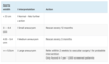
What are some examples of some common ECG abnormalities?

A
2
Q

What is the defintion of an aneurysm?

A

Dilation >150% of original diameter

3
Q

What is the difference between a true aneurysm and a false aneurysm?

A

True = abnormal dilation of vessel

False = collection of blood around a blood vessel that communicates with the lumen

4
Q

HOW CAN ISCHAEMIA OF HEART MUSCLE OCCUR?

A
  1. Reduced blood flow to the heart muscle (clot or atheroma)
  2. Increased distal resistance (LV hypertrophy)
  3. Reduced O2 carrying capacity (anaemia) or availability (hypoxia)
5
Q

What are some risk factors for IHD?

Modifaible and non-modifiable?

A

MODIFIABLE

  1. Smoking.
  2. Diabetes
  3. Hypertension.
  4. Hypercholesterolaemia.
  5. Sedentary lifestyle

Non-modifiable

  1. Gender.
  2. Family history.
  3. Personal history.
  4. Age.
6
Q

WHAT IS ANGINA?

A

Chest pain brought on either:

By exertion which resloves with rest

Or at rest

7
Q

What are the different types of angina?

A
  1. Stable angina
  2. Unstable angina
  3. Decubitus angina (precipitated by lying flat)
  4. Variant (Prinzmetal’s) angina: caused by coronary artery spasm.
8
Q

What are the causes of angina?

A
  1. Atheroma
  2. Anaemia
  3. Spasm
  4. Tachycardia
9
Q

What are the symptoms of angina?

A
  1. Chest pain/discomfort.
  2. Heavy, central, tight, radiation to arms, jaw, neck.
  3. Precipitated by exertion.
  4. Relieved by rest or GTN within 5 mins
10
Q

What are the tests for stable angina?

A
  1. CT Angiogram
    • Gold standard, shows luminal narrowing
  2. ECG
    • Pathological Q waves in particular, LBBB, and ST-segment and T wave abnormalities (for example, flattening or inversion).
  3. Bloods
    • May show anaemia
  4. CXR
    • May show increased heart size and pulmonary vessels
11
Q

What are the management options for stable angina pectoralis?

A
  1. Drugs
    • Aspirin to prevent clots
    • Statin to lower cholesterol
    • Glyceryl Trinitrate (SL) (GTN Spray)
    • BB (atenolol) (best in heart failure patients too)/CCB OR (verapamil/diltiazem) - FIRST LINE
      • If adding drug to beta blocker then nifedipine is drug of choice, if not tolerate then ivabradine
      • NEVER PUT BETA BLOCKER AND VERAPAMIL/DILTIAZEM TOGETHER
  2. If patient can’t tolerate beta blocker or calcium channel blocker then a long acting nitrate can be used
    • Ivabradine
    • Nicorandil - can cause ulcers
  3. Percutaneous Intervention (PCI)
  4. Coronary Artery Bypass Graft (CABG)
12
Q

WHAT IS ACUTE CORONARY SYNDROME PATHOLOGY?

A

Plaque rupture, thrombosis, and inflammation.

13
Q

What are the different acute coronary syndomes?

A
  1. Unstable angina
  2. (NSTEMI) Non-Q wave infarction, ST depression and T wave inversion
  3. (STEMI) Q wave infarction, ST elevation
14
Q

What are the different ECG changes for ACS?

A

STEMI
ST elevation and tall T waves, may be a new LBBB in larger MIs (STEMI)

NSTEMI
A retrospective diagnosis, will see ST depression

Ischaemia
ST depression and T wave flattening

15
Q

What are the poor prognostic factors for ACS?

A
  1. Age
  2. Development (or history) of heart failure
  3. Peripheral vascular disease
  4. Reduced systolic blood pressure
  5. EVIDENCE OF CARDIOGENIC SHOCK
16
Q

WHAT IS UNSTABLE ANGINA?

A

An acute coronary syndrome (ACS) that is defined by the absence of biochemical evidence of myocardial damage

17
Q

What is the clinical classification of unstable angina?

A
  1. Cardiac chest pain at rest.
  2. Cardiac chest pain with crescendo pattern.
  3. New onset angina.
18
Q

What are the test for unstable angina?

A
  1. FBC
    • Anaemia aggravates it
  2. Cardiac enzymes
    • Excludes infarction
  3. ECG
    • When in pain shows ST depression
  4. Coronary angiography
19
Q

What is the treatment for unstable angina?

A
  1. M - Morphine
  2. O - Oxygen (if sats <94%)
  3. N - Nitrates
  4. A - Aspirin

If STEMI then a second anitplatlet should be added (e.g. clopidogrel, ticagrelor)

Then patinets go for PCI

20
Q

WHAT IS A MYOCARDINAL INFARCTION?

A

Plaque rupture leads to a clot forming which then occludes one of the coronary arteries causing myocardial cell death and inflammation.

21
Q

What are the symptoms of a myocardial infarction?

How long does it need to last to be an MI?

A
  1. Acute central chest pain radiating to jaw or shoulder
  2. Nausea
  3. SOB
  4. Palpitations

Lasting >20 mins

22
Q

What are the signs of a myocardial infarction?

A
  1. Clammy and pale
  2. 4th heart sound
  3. Pansystolic murmur
  4. May later develop peripheral oedema
23
Q

What are the tests for a MI?

A
  1. ECG
    • ​​Classically, hyperacute (tall) T waves, ST elevation or new LBBB occur within hours of transmural infarction.
    • T wave inversion and development of pathological Q waves follow over hours to days.
  2. CXR:
    • Cardiomegaly, pulmonary oedema, or a widened mediastinum
  3. Cardiac enzymes
    • Troponin
    • Creatine kinase MB - If MI’s occur close together is better for for 4-5 days
    • Myoglobin
24
Q

What is the initial management for a MI?

A
  1. M - Morphine
  2. O - Oxygen (if sats <94%)
  3. N - Nitrates
  4. A - Aspirin

If from cocaine overdose then benzodiazepine should be added

If STEMI then a second anitplatlet should be added (e.g. clopidogrel, ticagrelor)

Then patinets go for PCI within 2 HOURS

IF NOT WITHIN 2 HOURS THEN FIBRINOLYSIS WITHIN 12 HOURS

Recheck ECG within 60-90 minutes to see if ST elevation gone - urgent PCI if not

25
What is the further management after an MI?
Drug Treatment: 1. Dual antiplatelet therapy (aspirin plus a second antiplatelet agent) 2. ACE inhibitor 3. Beta-blocker 4. Statin 1. **Diet:** advise a Mediterranean style diet, switch butter and cheese for plant oil based products. Do not recommend omega-3 supplements or eating oily fish 2. **Exercise:** advise 20-30 mins a day until patients are 'slightly breathless' 3. Sexual activity may resume 4 weeks after an uncomplicated MI
26
What are the complications of MI?
1. Cardiac arrest; cardiogenic shock; LVF. 2. Unstable angina 3. Bradycardias or heart block. 4. Tachyarrhythmias 5. Pericarditis 6. DVT & PE 7. Systemic embolism 8. Cardiac tamponade 9. Mitral regurgitation 10. Ventricular septal defec
27
What is Dressler’s syndrome?
1. Recurrent pericarditis 2. Pleural effusions 3. Fever 4. Anaemia and ESR increase 1–3 wks post-MI
28
What are the signs of a posterior MI?
Tall R waves in V1-V2
29
What are the contraindications to thrombolysis?
1. Active internal bleeding 2. Recent haemorrhage, trauma or surgery (including dental extraction) 3. Coagulation and bleeding disorders 4. Intracranial neoplasm 5. Stroke \< 3 months 6. Aortic dissection 7. Recent head injury 8. Severe hypertension
30
What are the symptoms of left ventricular free wall rupture?
1. This is seen in around 3% of MIs and occurs around 1-2 weeks afterwards. 2. Patients present with acute heart failure secondary to cardiac tamponade (raised JVP, pulsus paradoxus, diminished heart sounds). 3. Urgent pericardiocentesis and thoracotomy are required.
31
What are the symptoms of acute mitral regurgitation?
1. More common with infero-posterior infarction and may be due to ischaemia or rupture of the papillary muscle. 2. Acute hypotension and pulmonary oedema may occur. 3. An early-to-mid systolic murmur is typically heard. 4. Patients are treated with vasodilator therapy but often require emergency surgical repair.
32
WHAT IS SHOCK?
Circulatory failure resulting in inadequate organ perfusion
33
What is shock defined as?
1. Low BP 2. Evidence of tissue hypoperfusion.
34
What does a patient in shock look like?
Skin is pale, cold, sweaty and vasoconstricted Pulse is weak and rapid Pulse pressure reduced, MAP may be maintained Urine output reduced Confusion, weakness, collapse, coma
35
What is the main cause of injury from shock?
Prolonged hypotension can lead to life threatening organ failure
36
What are the different types of shock?
Hypovolaemic shock Cardiogenic shock **_Distributive shock_** Septic shock Analphylactic shock Neurogenic shock
37
WHAT IS HYPOVALEMIC SHOCK?
Low circulating blood volume
38
What can cause hypovalemic shock?
**Loss of blood** 1. Acute GI bleeding 2. Trauma 3. Ruptured AA **Loss of fluid** 1. Dehydration 2. Burns
39
How do you treat hypovalaemic shock?
1. Identify and treat underlying cause. 2. Raise the legs. 3. Give fluids
40
WHAT IS CARDIOGENIC SHOCK? When can it occur?
Cardiogenic shock is a state of inadequate tissue perfusion primarily due to cardiac dysfunction. —May occur suddenly or after progressively worsening heart failure
41
What are some causes of cardiogenic shock?
1. MI 2. Arrhythmias 3. PE 4. Tension pneumothorax 5. Cardiac tamponade 6. Myocarditis 7. Endocarditis 8. Aortic dissection
42
What are the symptoms for cardiogenic shock?
1. Low BP 2. High HR 3. High RR 4. Confusion 5. Pallor 6. Clammy 7. Pale peripheries 8. Reduced urine output
43
What are the investigations for cardiogenic shock?
1. **ECG** * Tachycardic 2. **Blood pressure** * Low 3. **JVP pressure** * RAISED
44
What is the management of cardiogenic shock? What do you need to monitor?
1. Treat the cause 2. Oxygen 3. Diamorphine IV for pain and anxiety 4. Correct arrhythmias, U&E abnormalities or acid–base disturbance —Monitor CVP, BP, ABG, ECG, urine
45
WHAT IS SEPSIS?
Sepsis exists when a systemic inflammatory response is associated with an infection
46
What is septic shock?
Septic shock exists when sepsis is complicated by persistent hypotension unresponsive to fluid resuscitation
47
What are the risk factors for septic shock?
1. Age 2. Diabetes mellituis (DM) 3. Immunocompromised 4. Alcoholics 5. Burns 6. IVDU 7. Pregnancy 8. Catheter
48
What are the symptoms for septic shock?
1. Low BP 2. High HR 3. Low sats (O2) 4. High resp rate (RR) 5. Lactate \>2 6. Unresponsive
49
What are the investigations for septic shock?
Cultures (2 peripheral blood, plus urine/sputum/CSF) LACTATE ABG and BP
50
What is the treatment for septic shock?
OXYGEN FLUIDS (check BP and ABG) IV ANTIBIOTICS (Tacozin and gentamicin, and vancomycin)
51
WHAT IS ANAPHYLATIC SHOCK?
Intense allergic reaction. Massive release of histamine and other vasoactive mediators causing haemodynamic collapse.
52
What type of hypersensitivity is anaphylatic shock?
Type-I IgE-mediated hypersensitivity reaction.
53
What are some causes of anaphylatic shock?
1. Drugs, eg penicillin, and contrast media in radiology 2. Latex 3. Stings, eggs, fish, peanuts, strawberries, semen (rare)
54
What are the signs and symptoms of anaphylatic shock?
1. Itching 2. Sweating 3. Diarrhoea and vomiting 4. Erythem 5. Urticaria 6. Oedema 7. Wheeze 8. Laryngeal obstruction 9. Cyanosis 10. Tachycardia 11. Hypotension
55
What is the management of anaphylatic shock?
1. Oxygen 2. Remove the cause 3. Adrenaline IM - 500 micrograms - 1 in 1000 Repeated every 5 minutes if necessary
56
WHAT IS NEUROGENIC SHOCK?
Distributive Disruption of the autonomic pathways within the spinal cord
57
What can cause neurogenic shock?
1. Spinal cord injury 2. Epidural 3. Spinal anaesthesia
58
What are the treatment options for neurogenic shock?
1. Dopamine and vasopressin (ADH). 2. Atropine is administered for slowed heart rate.
59
What organs are at risk of shock?
1. Kidneys - Acute tubular necrosis 2. Lung – Acute Respiratory Distress Syndrome (ARDS) (or “shock lung”) 3. Heart – myocardial ischaemia and infarction 4. Brain – confusion, irritability, coma
60
WHAT IS ACUTE RESPIRATORY DISTRESS SYNDROME?
1. Increase in alveolar permeability 2. Fluid accumilation in the alveoli 3. Not associated with cardiac causes 4. Impaired oxygenation
61
What are some causes of ARDS?
1. Infection: sepsis, pneumonia 2. Massive blood transfusion 3. Trauma 4. Smoke inhalation 5. Acute pancreatitis 6. Cardio-pulmonary bypass
62
What are the clinical features of ARDS?
1. Dyspnoea 2. Elevated respiratory rate 3. Bilateral lung crackles 4. Low oxygen saturations
63
What are the investigations for ARDS?
1. Chest x-ray 2. ABG
64
What is the management for ARDS?
1. ITU - Oxygenation/ventilation to treat the hypoxaemia 2. General organ support e.g. vasopressors as needed 3. Treatment of the underlying cause e.g. antibiotics for sepsis 4. Certain strategies such as prone positioning and muscle relaxation have been shown to improve outcome in ARDS
65
WHAT DOES CARDIOMYOPATHY REFER TO?
Primary heart muscle disease – often genetic. Three types
66
WHAT IS HYPERTROPHIC CARDIOMYOPATHY? https://www.youtube.com/watch?v=8RnkKB8xvwA
Heart muscle becomes thick, heavy and hypercontactile.
67
What is Hypertrophic cardiomyopathy (HCM) caused by? What inheritance is it?
Sarcomeric protein gene mutations. Autosomal dominant inheritance.
68
What is the epidemology of HCM?
Leading cause of death in the young
69
What is the pathology of heart walls in HCM?
1. Asymmetric septal hypertrophy 2. Intervenricular septum more than free wall takes up more room so less filling 3. More stiff and less compliant 4. Stroke volume goes down 5. LV outflow tract (LVOT) obstruction which pulls mitral valve towards it * Venturi effect
70
What type of heart failure if HCM?
Diastolic heart failure
71
What kind of heart sound is heard in HCM? Where is it also seen?
Crescendo-decrescendo murmur Aortic valve stenosis
72
What are the symptoms of HCM?
1. Sudden death 2. Fast arrythmias 3. Palpitations 4. Dyspnoea 5. Dizzy spells or syncope
73
What are the signs of HCM?
1. —Jerky pulse 2. Double apex beat 3. Systolic thrill at lower left sternal edge 4. Harsh ejection systolic murmur
74
What are the tests for HCM?
**_Echo_** Asymmetrical septal hypertrophy; small LV cavity with hypercontractile posterior wall; midsystolic closure of aortic valve **_ECG_** LVH; progressive T wave inversion; deep Q waves, AF
75
What is the treatment for HCM?
1. **Beta-blockers** or **verapamil** for angina + SOB. 2. **Amiodarone** for arrhythmias (AF, VT) 3. **DIGOXIN IS CONTRAINDICATED** 4. **Anticoagulate** for paroxysmal AF or systemic emboli.
76
What drug is contraindicated in HCM?
Digoxin Increase contraction force, increase obstruction
77
How is sudden cardiac death caused in HCM?
Ventricular arrythmia
78
WHAT IS DILATED CARDIOMYOPATHY (DCM)?
Causes all four chambers of the heart to enlarge
79
What is Dilated Cardiomyopathy (DCM) often caused by?
1. **Alcohol** 2. Increased BP 3. Haemochromatosis 4. Viral infection 5. Autoimmune
80
What happens in Dilated cardiomyopathy (DCM)? What type of heart failure is it?
1. Large space, thin walls 2. Weak contraction 3. Less blood pumped out in each beat 1. Biventicular congestive heart failure 2. Systolic heart failure
81
What type of heart sound is heard in Dilated Cardiomyopathy (DCM)?
1. Holosystolic murmur 2. S3 sounds also present, blood slamming into wall in diastole
82
What are the symptoms of Dilated Cardiomyopathy (DCM)?
1. Fatigue 2. Dyspnoea 3. Pulmonary oedema 4. Right ventricular failure 5. Emboli 6. Atrial fibrillation 7. Ventricular tachycardia
83
What are the signs of Dilated Cardiomyopathy (DCM)?
1. —Increased pulse 2. Decreased blood pressure 3. Increased JVP 4. Pleural effusion 5. Oedema 6. Jaundice, hepatomegaly, ascites 7. —Displaced diffuse apex beat, S3 gallop 8. —Mitral or tricuspid regurgitation
84
What are the tests for Dilated Cardiomyopathy (DCM)?
1. **Blood**: * Plasma BNP is sensitive and specific in diagnosing heart failure. 2. **CXR**: * Cardiomegaly, pulmonary oedema. 3. **ECG**: * Tachycardia, non-specific T wave changes, poor R wave progression. 4. **Echo**: * Globally dilated hypokinetic heart and low ejection fraction. Look for MR, TR, LV mural thrombus.
85
What is the treatment for Dilated Cardiomyopathy (DCM)?
1. **Stop drinking** 2. **Treatment of cardiac failure** * ACE inhibitors * Beta Blockers * Diuretics + Digoxin 3. **Anti-arrythmic drugs** * Amiodarone 4. **Cardiac transplantation and cardiomyoplasty**
86
WHAT IS RESTRICTIVE CARDIOPATHY?
Heart muscle becomes stiff and less compliant
87
What are the causes of RCM?
1. **Amyloidosis** 2. **Sarcoidosis** * Collection of immune cells 3. Radiation 4. **Loffler endomcarditis** * Eosinophils in lung tissue and heart tissue
88
What happens in amyloidosis?
Proteins that have been misfolded and become insoluble Deposit in tissue in organs making them become less compliant
89
What is the pathology of RCM?
1. Heart muscle stays same size 2. When blood comes in heart doesn't stretch 3. Less blood 4. Less pumped out 5. = Heart failure
90
What type of heart failure is RCM?
Diastolic heart failure
91
What are the signs of RCM?
1. These are mainly of right heart failure with increase JVP 2. Kussmaul’s sign (JVP rising paradoxically with inspiration) 3. Quiet heart sounds 4. S3 5. Diastolic pericardial knock, hepatosplenomegaly, ascites, and oedema.
92
What are the investigations for RCM?
**_ECG_** Low amplitude QRS
93
What is the treatment for RCM?
1. Treat underlying cause 2. Heart transplant
94
What do all cardiomyopathies carry?
An arrhythmic risk.
95
WHAT IS INHERITED ARRHYTHMIA (CHANNELOPATHY) CAUSED BY?
Ion channel protein gene mutations.
96
Which ions are involved with channelopathy?
1. Potassium 2. Sodium 3. Calcium channel.
97
What do channelopathies include?
1. Long QT 2. short QT 3. Brugada 4. CPVT
98
What do channelopathies normally present with and what do they have that is normal?
Recurrent syncope and have a structurally normal heart.
99
What is sudden cardiac death in young people normally due to? What disease is it most likely to be?
1. An inherited condition. 1. Cardiomyopathy or ion channelpathy.
100
What does sudden arrhythmic death syndrome (SADS) usually refer to?
Normal heart/arrhythmia.
101
WHAT IS AN ANEURYSM? https://www.youtube.com/watch?v=pEOqffiwE7k
Abnormal buldge in vessel
102
What are the risk factors for aneurysms?
1. Male 2. Over 60 3. Hypertension 4. Smoking
103
What are the typical causes of an anneurysm
1. Atheroma 2. Trauma 3. Infection 4. Connective tissue disorders 5. Inflammations
104
What is the pathology of an aneurysm?
Weakness in vessel wall Ballooning outwards of vessel wall due to pressure Laplace's law causes positive feedback loop Gives bigger aneurysm
105
When is an aneurysm official labelled an aneurysm?
When the diameter exceeds 1.5 times the normal size
106
What are the common sites for anneurysms What are the complications?
Common sites Aorta (infrarenal most common), iliac, femoral and popliteal arteries. Complications Rupture; thrombosis; embolism; fistulae; pressure on other structures.
107
What are the different types of aneurysms?
1. **True aneurysms** * Abnormal dilatations that involve all layers of the arterial wall. * THEN EITHER * Fusiform * OR * saccular (Berry aneurysms) 2. **False aneurysms (pseudoaneurysms**) * Blood in the outer layer only (adventitia) which communicates with the lumen (eg after trauma).
108
What happens when an aneurysm explodes?
Blood spurts out of the hole Less blood goes downstream Ischaemia of downstream cells
109
What are the features of an abdominal aortic aneurysm?
1. Severe, central abdominal pain radiating to the back 2. Pulsatile, expansile mass in the abdomen 3. Patients may be shocked (hypotension, tachycardic) or may have collapsed
110
What are the options for a unruptured anneurysm?
1. Elective surgery 2. Stenting
111
WHAT ARE THE CAUSES OF A AAA?
1. Several different groups of patients suffer from aneurysmal disease. 2. The commonest group is those who suffer from standard arterial disease, i.e. Those who are **hypertensive**, have **diabetes** and have been or are **smokers**. 3. Other patients such as those suffering from connective tissue diseases such as Marfan's may also develop aneurysms. In patients with abdominal aortic aneurysms the extracellular matrix becomes disrupted with a change in the balance of collagen and elastic fibres.
112
When is a AAA referred for surgery?
\>5.5cm
113
What are the different follow up times for each anneurysm size?
114
What are the treatments for a AAA?
1. Open surgery - needed if iliac arteru is too tortous 2. Endovascular aneurysm repair (EVAR) - longer follow-up, reintervention rate, not suitable for every type
115
What is a common complication of AAA repair? What is this caused by?
1. Trash foot * Disloged emboli travels around the body and lodges in distal vessels, causing ischaemia
116
WHAT IS AORTIC DISSECTION? https://www.youtube.com/watch?v=AZElPJtyxck
Tear in tunica intima, causes blood to pool between intima and media
117
What can cause aortic dissection?
1. **Chronic hypetension** 2. Increase blood volume 3. Coarctation 4. **Weakened aortic wall** 5. Marfan's 6. Ehlers-Danlos syndrome 7. **Aneurysms**
118
Where does aortic dissection normally occur?
Within the first 10cm of aorta
119
What can a aortic dissection cause?
1. **Blood back up into pericardial space causing** * Pericardial temponade 2. **Blood goes out intima and comes back into blood vessel through and hole** 3. **Blood flows does the aorta inbetween the layers and puts compression on other arteries** * Renal artery * Subclavian artery
120
What are the different types of aortic dissection?
Type A (70%) dissections involve the ascending aorta, irrespective of site of the tear, Whilst if the ascending aorta is not involved it is called type B (30%)
121
What are the symptoms of aortic dissection?
1. Sharp chest pain radiating to back 2. Hypotension 3. Shock **Type A** 1. **Weak pulses** 2. **Aortic regurgitation** 3. Weak pulse in downstream artery 4. Difference in BP between left and right arms
122
What are the investigations for aortic dissection?
1. **CXR** * Widend mediastinum 2. **CT Angiogram chest, abdo, pelvis** * ​​Suitable for stable patients and planning surgery 3. **Transoesophageal echo** * True lumen and flase aorta
123
What is the management of aortic dissection?
1. **Type A** * **IV labetalol AND** * Surgical management, but blood pressure should be controlled to a target systolic of 100-120 mmHg whilst awaiting intervention 2. **Type B\*** * Conservative management * Bed rest * Reduce blood pressure **IV labetalol** to prevent progression
124
HOW MANY LAYERS IS THE PERICARDIUM? WHAT IS THE STRUCTURE?
Two. Visceral single cell layer adherent to epicardium Fibrous parietal layer 2mm thick Acellular collagen and elastin fibres 50ml of serous fluid.
125
Why is the pericardium important? What is important about the small reserve volume?
1. Restrains the filling volume of the heart. 2. If the volume is exceeded the pressure is translated to the cardiac chambers.
126
WHAT IS CARDIAC TAMPONADE?
Fluid builds up in the pericardium and results in compression of the heart.
127
What is the cause of cardiac tamponade?
1. Any pericarditis 2. Aortic dissection 3. MI 4. Trauma 5. Trans-septal puncture at cardiac catheterization 6. Cancer
128
What are the signs of cardiac tamponade?
1. Pulse increase 2. BP decerease 3. **Pulsus paradoxus** * An abnormally large drop in BP during inspiration 4. JVP increase 5. Kussmaul’s sign 6. Muffled S1 and S2.
129
What is Beck's triad?
Symptoms of cardiac tamponade: 1. Falling BP 2. Rising JVP 3. Muffled heart sounds.
130
What are the investigations for cardiac tamponade?
1. **Beck’s triad** * Falling BP; rising JVP; muffled heart sounds. 2. **CXR** * Big globular heart (if \>250mL fluid). 3. **ECG** * Low voltage QRS ± **electrical alternans** 4. **Echo is diagnostic** * Echo-free zone around the heart ± diastolic collapse of right atrium and right ventricle.
131
What is the management of cardiac tamponade?
The pericardial effusion needs urgent drainage
132
What does chronic pericardial effusion have that is unique?
Pericardium slowly adapts to the increasing fluid and therefore reduces the effect on diastolic filling of the chambers.
133
WHAT IS PERICARDIAL EFFUSION?
Accumulation of fluid in the pericardial sac.
134
What are the causes of pericardial effusion?
Any cause of pericarditis 1. **Idiopathic.** 2. **Viral** * Coxsackie B virus * Enteroviruses * Herpesviruses (EBV, CMV, HHV-6), 3. **Dresseler's syndrome** * Happens after heart attack * When an MI happens lots of necrosis * This also affects the pericardium 4. **Autoimmune** * Rheumatoid arthritis * Scleraderma * SLE
135
What are the symptoms of pericardial effusion?
1. Dyspnoea 2. Raised JVP 3. Bronchial breathing at left base 4. Look for signs of cardiac tamponade
136
What are the investigations for pericardial effusion?
1. **CXR** * Enlarged, globular heart. 2. **ECG** * Low-voltage QRS complexes and alternating QRS morphologies (electrical alternans). 3. **Echo** * Shows an echo-free zone surrounding the heart.
137
What are the management options for pericardial effusion?
1. Treat the cause. 2. **Pericardiocentesis** may be **diagnostic** (suspected bacterial pericarditis) or **therapeutic** (cardiac tamponade).
138
WHAT IS ACUTE PERICARDITIS? https://www.youtube.com/watch?v=jqClJsqnFFA
Inflammatory pericardial syndrome with or without effusion.
139
What are the causes of pericarditis?
1. Idiopathic. 2. **Viral** * **Coxsackie B virus** * Enteroviruses * Herpesviruses (EBV, CMV, HHV-6), 3. **Dresseler's syndrome** * Happens after heart attack * When an MI happens lots of necrosis * This also affects the pericardium 4. **Autoimmune** * Rheumatoid arthritis * Scleraderma * SLE
140
What is the patholgy of pericarditis?
Fluid and immune cells move from tiny blood cells into the fibrous pericardium
141
What is the clinical presentation of somebody with pericarditis?
1. **Fever** 2. **Chest pain** * Worse with heavy breathing * Better with sitting up and leaning forward
142
What can a clinical diagnosis of acute pericarditis be diagnosed from?
**2 of 4 of:** 1. Chest pain. 2. Friction rub. 3. ECG changes. 4. Pericardial effusion.
143
What tests can you do to check if somebody has pericarditis?
1. ECG changes * The changes in pericarditis are often global/widespread, as opposed to the 'territories' seen in ischaemic events * 'Saddle-shaped' ST elevation * PR depression: most specific ECG marker for pericarditis 2. All patients with suspected acute pericarditis should have **transthoracic echocardiography TOE**
144
What does an ECG look like with a patient who has pericarditis?
**_First few weeks_** Diffuse saddle shaped ST segment elevation Decrease PR **_After that_** T waves flattened **_After that_** ECG returns back to normal
145
What is the management of pericarditis?
1. **Treat underlying cause** 2. **NSAID** * Ibuprofen PO or Aspirin PO 3. **Colchicine**
146
WHAT IS HEART FAILURE?
A symptomatic condition where breathlessness, fluid retention and fatigue are associated with a cardiac abnormality that reduces cardiac output A state where the heart is unable to pump enough blood to satisfy the needs of metabolising tissues
147
How do you calculate cardiac output?
CO = HR x SV
148
What is the most common cause of heart failure? What does this usually result from? What are some other causes?
1. Myocardial dysfunction. * Result of IHD. 2. Hypertension. 3. Alcohol excess. 4. Cardiomyopathy. 5. Valvular. 6. Endocardial. 7. Pericardial causes.
149
What is systolic heart failure?
Inability of the ventricle to contract normally, resulting in decreaed cardiac output. ## Footnote **Ejection fraction (EF) is \<40%**
150
What are the causes of systolic heart failure?
1. IHD 2. MI 3. Cardiomyopathy.
151
What is diastolic heart failure?
Inability of the ventricle to relax and fill normally Causing increased filling pressures. EF is \>50%.
152
What are the causes of diastolic heart failure?
1. Constrictive pericarditis 2. Tamponade 3. Restrictive cardiomyopathy 4. Hypertension
153
How may left and right sided heart failure occur?
Left ventricular failure (LVF) and right ventricular failure (RVF) may occur independently, or together as congestive cardiac failure (CCF).
154
What are some causes of left sided heart failure?
Hypertension
155
What are the symptoms of left sided heart failure?
1. Dyspnoea, 2. Poor exercise tolerance 3. Fatigue 4. Orthopnoea 5. Paroxysmal nocturnal dyspnoea (PND) 6. Nocturnal cough (±pink frothy sputum)
156
What are some causes of right ventricular failure?
1. LVF, 2. Pulmonary stenosis 3. Lung disease.
157
What are the symptoms of right ventricular heart failure?
1. Peripheral oedema (up to thighs, sacrum, abdominal wall), 2. Ascites 3. Nausea 4. Anorexia 5. Facial engorgement 6. Pulsation in neck and face (tricuspid regurgitation) 7. Epistaxis.
158
What tests can you do for heart failure?
1. **Blood Tests - FIRST LINE** * BNP 2. LFTS, FBC, U&Es, BNP, TFTs 3. **Cardiac enzymes** * Creatinine kinase, troponin I, troponin T 4. **CXR** 5. **ECG** 6. **Echocardiography**
159
What would you see on a chest XR for heart failure?
ABCDE 1. **A**lveolar oedema 2. Kerley **B** lines 3. **C**ardiomegaly 4. **D**ilated prominant upper lobe vessels 5. Pleural **E**ffusion
160
What are the systems involved with protection and survival of the heart?
**_The sympathetic system_** Increases afterload by causing peripheral vasoconstriction **_The renin-angiotensin-aldosterone axis_** Salt and water retention Increases afterload and preload (^ volume and vasoconstriction) **_Cardiac changes_** Ventricular dilatation Myocytehypertrophy
161
What does an increase in cardiac norepinephrine produce? What happens in the long term?
**_Increased adrenergic activation_** Direct toxicity to myocytes Increased HR and contractility causing extra strain on heart Increased vasocontriction, increased afterload, more strain on heart
162
What does angiotensin 2 produce?
Increase salt and water retention Increased preload, extra strain on heart
163
What is the ACUTE treatment for chronic heart failure?
1. **Recommended treatments for all patients** * IV loop diuretics * e.g. furosemide or bumetanide 2. **Possible additional treatments** 3. **Oxygen** * this should be given in line with British Thoracic Society guidelines, i.e. keep oxygen saturations at 94-98% 4. **Vasodilators** * Nitrates should not be routinely given to all patients * They may, however, have a role if concomitant myocardial ischaemia, severe hypertension or regurgitant aortic or mitral valve disease * The major side-effect/contraindication is hypotension 5. **Patients with respiratory failure** * CPAP 6. **Patients with hypotension (e.g. \< 85 mmHg)/cardiogenic shock** 7. **inotropic agents** * e.g. dobutamine * should be considered for patients with severe left ventricular dysfunction who have potentially reversible cardiogenic shock 8. **Vasopressor agents** * e.g. norepinephrine * normally only used if insufficient response to inotropes and evidence of end-organ hypoperfusion 9. **Mechanical circulatory assistance** * e.g. intra-aortic balloon counterpulsation or ventricular assist devices
164
What is the treatment for chronic heart failure?
1. **First-line treatment** * ACE-inhibitor and a beta-blocker 2. **Second-line treatment** * Aldosterone antagonist - e.g. spironolactone 3. **Third-line treatment** * ​​Ivabradine - if heart rate above 75 * Sucubitil-valsartan if EF \<35% * Digoxin * Hydralazine and nitrate * Spironolactone Loop diuretics **DO NOT** help
165
WHAT IS TETRALOGY OF FALLOT?
1. Pulmonary stenosis 2. RV hypertrophy 3. Overriding aorta 4. VSD
166
What are the physiology of Tetralogy of Fallot?
1. The stenosis of the RV outflow leads to the RV being at higher pressure than the left 2. Therefore blue blood passes from the RV to the LV 3. The patients are BLUE 4. Toddlers may squat
167
What is the presentation of infants in tetralogy of Fallot and why?
1. Infants may be acyanotic at birth 2. Pulmonary stenosis murmur as the only initial finding. 3. Cyanotic due to decreasing flow of blood to the lungs as well as right-to-left shunt across the VSD. 4. Toddlers may squat 5. Typical of TOF, as it increases peripheral vascular resistance and decreases the degree of right to left shunt. 6. Adult patients are often asymptomatic
168
What are the tests for tetrology of Fallot?
1. **ECG** * RV hypertrophy with a right bundle-branch block. 2. **CXR** * Boot-shaped heart 3. **Echocardiography** * Can show the anatomy as well as the degree of stenosis.
169
What is the management of tetrology of Fallot?
1. Surgical repair is often undertaken in two parts 2. Cyanotic episodes may be helped by beta-blockers to reduce infundibular spasm
170
WHAT IS A VENTRICULAR SEPTAL DEFECT?
Hole in the connecting venticles
171
What are the different pressures in the ventricles and what is the result in VSD?
1. High pressure LV 2. Low pressure RV 3. Blood flows from high pressure chamber to low pressure chamber 4. Therefore NOT blue 5. Increased blood flow through the lungs
172
What are the causes of VSD?
**_Congenital_** 1. Down's syndrome 2. Edward's syndrome 3. Patau syndrome 4. cri-du-chat syndrome **_Acquired (post-MI)._**
173
What are the ventricular septal defects clinical signs?
**_Large_** 1. Pan-systloic murmur varies in intensity 2. Small breathless skinny baby 3. Increased respiratory rate 4. Tachycardia 5. Big heart on chest X ray **_Small_** 1. Loud systolic murmur 2. Thrill (buzzing sensation) 3. Well grown 4. Normal heart rate 5. Normal heart size
174
What is the treatment for VSD?
1. **Large** * Require fixing in infancy (PA band, complete repair) 2. **Small** * Endocarditis risk * Need no intervention
175
What may VSD lead to?
May lead to Eisenmengers syndrome 1. High pressure pulmonary blood flow 2. Damages to delicate pulmonary vasculature 3. The resistance to blood flow through the lungs increases 4. The RV pressure increases 5. The shunt direction reverses 6. The patient becomes BLUE.
176
WHAT IS ATRIAL SEPTAL DEFECT?
1. Abnormal connection between the two atria (primum, secundum, sinus venosus) 2. Common 3. Often present in adulthood.
177
What are the two types of ASD?
1. Primum: present earlier, may involve AV valves 2. Secundum: may be asymptomatic until adulthood
178
What is the physiology of atrial septal defects i.e blood flow direction?
1. Slightly higher pressure in the LA than the RA 2. Shunt is left to right 3. Therefore NOT blue 4. Increased flow into right heart and lungs
179
What is the physiology between large and small atrial septal defects?
**Large** 1. Significant increased flow through the right heart and lungs in childhood 2. Right heart dilatation 3. SOBOE 4. Increased chest infections 5. If any stretch on the right heart should be closed **Small** 1. Small increase in flow 2. No right heart dilatation 3. No symptoms 4. Leave alone 5. NB. The shunt on small to moderate sized defects increases with age
180
What are some complications of ASD?
1. Reversal of left-to-right shunt, ie Eisenmenger’s complex 2. Paradoxical emboli (vein to artery via ASD; rare) 3. EMBOLI
181
What are the clinical signs of atrial septal defects?
1. Pulmonary flow murmur 2. Fixed split second heart sound (delayed closure of PV because more blood has to get out) 3. Big pulmonary arteries on CXR 4. Big heart on chest X ray
182
What is the treatment of ASD?
1. In children closure is recommended before age 10yrs. 2. In adults, transcatheter closure is now more common than surgical.
183
WHAT CAN ATRIA-VENTRICULAR SEPTAL DEFECTS INVOLVE?
Hole in the centre. Can involve the ventricular septum, the atrial septum, the mitral and tricuspid valves.
184
What is the symptoms of atria-ventricular septal defect? For a partial and complete defect?
**Complete defect** 1. Breathless as neonate 2. Poor weight gain 3. Poor feeding 4. Torrential pulmonary blood flow 5. Needs repair or PA band in infancy 6. Repair is surgically challenging **Partial defect** 1. Can present in late adulthood 2. Presents like a small VSD / ASD 3. May be left alone if there is no right heart dilatation
185
WHAT IS PATENT DUCTUS ARTERIOSUS?
Failure of ductus arterioles to close.
186
What are the clinical signs of ductus arteriosis?
1. Continuous ‘machinery’ murmur 2. If large, big heart, breathless 3. Heaving apex beat 4. Wide pulse volume 5. Large volume, bounding, collapsing pulse 6. Eisenmenger’s syndrome * Differential cyanosis (clubbed and blue toes, but pink not clubbed fingers)
187
What is the physiology of patent ductus arterioles with a large and a small?
**Large** 1. Torrential flow from the aorta to the pulmonary arteries in infancy 2. Breathless, poor feeding, failure to thrive 3. More common in prem babies –Need to be closed (surgically) **Small** 1. Little flow from the aorta to Pas 2. Usually asymptomatic 3. Murmur found incidentally 4. Endocarditis risk
188
How do you close the ductus arteriosus?
1. Indomethacin or Ibuprofen * Given to the neonate * Inhibits prostaglandin synthesis * Closes the connection in the majority of cases 2. If associated with another **congenital heart defect** amenable to surgery then **prostaglandin E1** is useful to keep the duct open until after **surgical repair**
189
WHAT IS COARCTATION OF THE AORTA?
Narrowing of the aorta at the site of insertion of the ductus arterioles.
190
What is the physiology for coarctation of the aorta?
**_Severe_** Complete or almost complete obstruction to aortic flow Collapse with heart failure Needs urgent repair **_Mild_** Presents with hypertension Incidental murmur Should be repaired to try to prevent problems in the long term
191
What are the clinical signs of coarctation of the aorta?
1. Infancy: heart failure 2. Adult: hypertension 3. Radio-femoral delay 4. Mid systolic murmur, maximal over back 5. Apical click from the aortic valve 6. **Notching of the inferior border of the ribs (due to collateral vessels) is not seen in young children**
192
What are the long term problems of coarctation of the aorta?
1. **Hypertension** 2. Early coronary artery disease 3. Early strokes 4. Sub arachnoid haemorrhage 5. Re-coarctation requiring repeat intervention 6. Aneurysm formation at the site of repair
193
What is the treatment of coarctation of the aorta?
1. **IV prostaglandins** are used in neonates to maintain a patent ductus arteriosus to allow adequate circulation until it is possible to attempt corrective surgery. 2. Surgery, or balloon dilatation ± stenting.
194
WHAT ARE THE PROBLEMS ASSOCIATED WITH BISCUSPID AORTIC VALVES?
1. Can be severely stenotic in infancy or childhood 2. Degenerate quicker than normal valves 3. Become regurgitant earlier than normal valves 4. Are associated with coarctation and dilatation of the ascending aorta
195
How can you treat bicuspid aortic valve?
Surgery
196
WHAT IS PULMONARY STENOSIS?
Narrowing of the outflow of the right ventricle
197
What is the treatment for pulmonary stenosis?
1. Balloon valvuloplasty 2. Open valvotomy 3. Open trans-annular patch 4. Shunt (to bypass the blockage)
198
WHAT IS HYPERTENSION?
Over 140/90
199
What are the two types of hypertension?
Essential hypertension (primary, cause unknown). ~95% of cases. Secondary hypertension ~5% of cases.
200
What are some causes of secondary hypertension?
1. **Renal** * CKD 2. **Endocrine disease** * Conn’s syndrome 3. **Others** * Coarctation, pregnancy
201
What are the tests for hypertension?
1. **24hr ambulatory BP monitor** 2. **Multiple home BP monitoring** 3. **Fundoscopy–in severe HTN** * Bilateral retinal haemorrhages * Papilloedema 4. **Overall CVD risk** * Fasting glucose * Cholesterol 5. **End Organ Damage** * 12 lead ECG –past MI, LV hypertrophy * Urine analysis –protein, blood
202
What are some lifestyle to lower blood pressure?
1. Stop smoking 2. Low-fat diet 3. Reduce alcohol and salt intake 4. Increase exercise 5. Reduce weight if obese
203
What is the pharmocological management of hypertension?
Step 1 \<55 years old and not afro-carribean - ACEi or ARB E.g. Ramirpil or losartan \>55 years old or afro-carribean - CCB - Amlodipine Step 2 Add the other from step 1 OR thiazide-like duirectic Step 3 ACEi or ARB + CCB + thiazide-like diuretic (Indapamide) Step 4 Low-dose spironolactone if K+ \<4.5 mmol/l Alpha-blocker (doxazosin) or beta blocker if K+ \>4.5 mmol/l
204
How can renal artery stenosis cause hypertension?
1. Less blood flow to kidneys 2. Secretes Renin 3. More water and salt retention 4. Higher BP
205
What is a pheochromocytoma? How does it cause hypertension?
Tumour on adrenal glands Constantly produce catecholamines (e.g. adrenaline) Increase heart rate and peripheral vascular resistance Increase BP
206
How does cushing's cuase hypertension?
1. Enhances adrenalines effect on blood vessels to constrict them 2. Also can act as a mineralacorticoid (aldosterone) 3. Increase BP
207
How does Conn's syndrome cause hypertension?
1. High aldosterone 2. Increased sodium and water retention 3. Increased BP
208
Over what value should an ACE inhibitor be stopped for potassium?
\>6 mmol/L
209
What is the first antihypertensive for diabetics?
Ace inhibitor
210
What is malignant hypertension?
Severe hypertension and bilateral retinal hemorrhages and exudates
211
WHAT IS RHEUMATIC FEVER?
Rheumatic fever develops following an immunological reaction to recent (2-6 weeks ago) Streptococcus pyogenes infection
212
What are the features of rheumatic fever?
**Major criteria** 1. Erythema marginatum 2. Sydenham's chorea: this is often a late feature 3. Polyarthritis 4. Carditis and valvulitis (eg, pancarditis) 5. The latest iteration of the Jones criteria (published in 2015) state that rheumatic carditis cannot be based on pericarditis or myocarditis alone and that there must be evidence of endocarditis (the clinical correlate of which is valvulitis which manifests as a regurgitant murmur) 6. Subcutaneous nodules **Minor criteria** 1. raised ESR or CRP 2. pyrexia 3. arthralgia (not if arthritis a major criteria) 4. prolonged PR interval
213
What is the management of rheumatic fever?
1. Antibiotics: oral penicillin V 2. Anti-inflammatories: NSAIDs are first-line 3. Treatment of any complications that develop e.g. heart failure
214
What is rheumatic heart disease?
Fusion of cusps.
215
WHAT ARE THE CAUSES OF AORTIC STENOSIS?
1. Congenital bicuspid valve 2. Degenerative calcification 3. William's syndrome 4. Rheumatic Heart disease
216
How does a person with aortic stenosis present?
1. Syncope 2. Angina 3. Dyspnoea 4. Sudden death
217
What physical signs would you hear on aortic stenosis?
1. **Narrow pulse pressure** * **​​**Slow rising carotid pulse (pulsus tardus) * & decreased pulse amplitude (pulsus parvus) 2. **Heart sounds** * ​​Soft or absent **S2**, **S4** gallop due to LVH. 3. **Ejection systolic murmur** * Crescendo-decrescendo character.
218
What tests would you do for someone with aortic stenosis?
1. **Echocardiography** * *Two measurements obtained are:** * Left ventricular size and function: LVH, Dilation, and EF * Doppler derived gradient and valve area (AVA). 2. **ECG** 3. **CXR** * LVH, calcified aortic valve
219
What is the management for aortic stenosis?
1. **General:** * Fastidious dental hygiene / care * Consider IE prophylaxis in dental procedures 2. **Medical** - limited role since AS is a mechanical problem. * Vasodilators are relatively contraindicated in severe AS 3. **Surgical Replacement** * Definitive treatment 4. **TAVI** * Transcatheter Aortic Valve Implantation - IF SYMPTOMATIC
220
What is the difference between aortic stenosis and aortic sclerosis?
Aortic sclerosis doesn't radiate to carotids
221
WHAT IS THE CAUSE OF MITRAL VALVE REGURGITATION?
1. Myxomatous degeneration (MVP) 2. Ischemic MR 3. Rheumatic heart disease 4. Infective Endocarditis.
222
What is the pathophysiology of mitral valve regurgitation?
1. Pure Volume Overload 2. Compensatory Mechanisms: Left atrial enlargement, LVH and increased contractility 3. Progressive left atrial dilation and right ventricular dysfunction due to pulmonary hypertension. 4. Progressive left ventricular volume overload leads to dilatation
223
What are the symptoms of mitral valve regurgitation?
1. **Mainly asymptomatic** 2. Exertion dyspnoea –exercise intolerance 3. Palpitations 4. Fatigue
224
What are the signs of mitral valve regurgitation?
1. Pansystolic murmur at apex radiating to axilla 2. Soft S1 3. Displaced hyperdynamic apex
225
What are the investigations for mitral regurgitation?
1. **ECG** * May show, LA enlargement, atrial fibrillation and LV hypertrophy with severe MR 2. **CXR** * LA enlargement, central pulmonary artery enlargement. 3. **ECHO** * Estimation of LA, LV size and function. Valve structure assessment TOE v helpful
226
What are the management for MR?
1. **Medical management in acute cases involves** * Nitrates, diuretics, positive inotropes and an intra-aortic balloon pump to increase cardiac output 2. **If patients are in heart failure** * ACE inhibitors may be considered along with beta-blockers and spironolactone 3. **In acute, severe regurgitation** * Surgery is indicated
227
What are the indications for surgery of severe MR?
1. ANY symptoms at rest or exercise with (repair if feasible) 2. **IF asymptomatic:** * If EF \<60%, LVESD \>45mm * If new onset atrial fibrillation/raised PAP.
228
WHAT IS THE CAUSE OF AORTIC REGURGITATION?
1. Bicuspid aortic valve 2. Rheumatic fever 3. Infective endocarditis
229
What is the pathophysiology of aortic regurgitation?
1. Combined pressure AND volume overload 2. Compensatory Mechanisms: LV dilation, LVH 3. Progressive dilation leads to heart failure
230
What are the symptoms of aortic valve regurgitation?
1. Exertional dyspnoea 2. Orthopnoea 3. Paroxysmal nocturnal dyspnoea
231
What are the signs of aortic regurgitation?
1. Collapsing (water hammer) pulse 2. Wide pulse pressure 3. Displaced hyperdynamic apex beat 4. Early diastolic murmur **_Notable eponyms_** 1. Corrigans sign (ear movements) 2. De Musset’s sign (head movements) 3. Duroziez’s sign (Femoral artery murmur) 4. Austin flint murmur (cardiac apex murmur) 5. Traube’s sign (pistol shot over femoral)
232
What would cardiac tests show you for aortic regurgitation?
1. **CXR** * Cardiomegaly 2. **ECHO** * LV dilation and hypertrophy
233
What is the management for aortic regurgitation?
1. **Vasdilators** * ACEi only if symptomatic or HTN 2. **Surgical** * Replace valve before LV dysfunction
234
WHAT IS MITRAL VALVE STENOSIS?
Obstruction of LV inflow that prevents proper filling during diastole
235
What are causes of mitral valve stenosis?
1. Rheumatic heart disease 2. Infective endocarditis 3. Mitral annular calcification.
236
What is mitral valve stenosis pathophysiology?
1. LA dilation incerease pulmonary congestion (reduced emptying) 2. Increased Transmitral Pressures * Leads to left atrial enlargement and atrial fibrillation. 3. Right heart failure symptoms: due to Pulmonary venous HTN
237
What are the symptoms of mitral valve stenosis?
1. Dyspnoea 2. Fatigue 3. Palpitations 4. Chest pain
238
What are the signs in mitral valve stenosis?
1. Malar flush on cheeks 2. Low volume pulse 3. Tapping, non displaced apex beat 4. Rumbling mid-diastolic murmur 5. Loud opening S1 6. P mitrale/bifid p wave
239
What tests can you do on mitral valve stenosis and what will they show?
**_MV ECHO_** The GOLD STANDARD for diagnosis Asses mitral valve mobility, gradient and mitral valve area **_ECG_** May show atrial fibrillation and LA enlargement **_CXR_** LA enlargement and pulmonary congestion Occasionally calcified
240
How would you manage a patient with mitral valve stenosis?
1. Patients with associated **atrial fibrillation** require anticoagulation * Currently warfarin is still recommended 2. **Asymptomatic patients** * Monitored with regular echocardiograms * Percutaneous/surgical management is generally not recommended 3. **Symptomatic patients** * Percutaneous mitral balloon valvotomy * Mitral valve surgery (commissurotomy, or valve replacement)
241
WHAT IS INFECTIVE ENDOCARDITIS?
Infection of heart valve/s or other endocardial lined structures within the heart.
242
What is the criteria used for defining infective endocarditis?
Duke criteria
243
What is the major and minor criteria for infective endocarditis?
1. **Major criteria** * **Positive blood cultures** * two positive blood cultures showing typical organisms consistent with infective endocarditis, such as Streptococcus viridans and the HACEK group, or * persistent bacteraemia from two blood cultures taken \> 12 hours apart or three or more positive blood cultures where the pathogen is less specific such as Staph aureus and Staph epidermidis, or * positive serology for Coxiella burnetii, Bartonella species or Chlamydia psittaci, or * positive molecular assays for specific gene targets * Evidence of endocardial involvement * positive echocardiogram (oscillating structures, abscess formation, new valvular regurgitation or dehiscence of prosthetic valves), or * new valvular regurgitation 2. **Minor criteria** * predisposing heart condition or intravenous drug use * microbiological evidence does not meet major criteria * fever \> 38ºC * vascular phenomena: major emboli, splenomegaly, clubbing, splinter haemorrhages, Janeway lesions, petechiae or purpura * immunological phenomena: glomerulonephritis, Osler's nodes, Roth spots
244
What is the most causative organism in infective endocarditis?
1. Staph aureus - MOST COMMON and in IVDU or prosthetic valves 2. Strep viridans 3. Staph epidermis - **prosthetic valves if \< 2 months post valve surgery**
245
Who is infective endocarditis most likely to affect?
1. The elderly (in an ageing population) 2. The young i.v. drug abusers 3. The young with congenital heart disease. 4. Anyone with prosthetic heart valves.
246
What is the clinical presentation of infective endocarditis?
1. Persistent fever 2. Emboli 3. New or changing murmurs
247
What are some peripheral signs of infective endocarditis?
1. Microscopic heamaturia 2. Petechiae 10 to 15% 3. Splinter hemorrhages 4. Osler’s nodes (small, tender, purple, erythematous subcutaneous nodules are usually found on the pulp of the digits) 5. Janeway lesions are erythematous, macular, nontender lesions on the fingers, palm, or sole 6. **Roth spots (boat shaped haemorrhages) on fundoscopy.**
248
What test can you do for infective endocarditis?
1. **Blood cultures - FIRST LINE** * ​​Three sets, taken from 3 different places, 3 times 2. **Echocardiography**, cardiac doppler, transoesophageal doppler 3. **Urine** * Microscopic haematuria and proteinuria are common 4. **Blood** * Coomb's test
249
How can you treat infective endocarditis?
**Native valve endocarditis (NVE):** 1. IV amoxicillin + consider low dose gentamicin 2. If penicillin allergic: IV vancomycin **Staph** 1. Flucloxacillin **Prosthetic valve endocarditis:** 1. IV vancomycin, gentamicin + rifampacin **Surgery if needed** 1. e.g. causing congestive cardiac failure
250
What prophylaxtic treatment is given for dental procedures in prosthetic valve replacement patients?
Good oral hygiene
251
WHAT IS PERIPHERAL VASCULAR DISEASE? https://www.youtube.com/watch?v=rTbIazck7rk&t=229s
Blood vessels outside of your heart and brain to narrow, block, or spasm.
252
What are the three patterns of disease seen in PVD?
1. Intermittent cludication 2. Critical limb ischaemia 3. Acute limb-threatening ischaemia
253
What is the classification of PVD called? What do the numbers mean?
**_—Fontaine_** —1 Asymptomatic —2 Intermittent claudication —3 Ischaemic rest pain —4 Ulceration/gangrene (critical ischaemia)
254
What is claudication?
Intermittent claudication is a cramp-like, muscular pain in the calf, brought on by exercise and relieved only by rest.
255
What are the symptoms of claudication?
1. Intermittent claudication: aching or burning in the leg muscles following walking 2. Patients can typically walk for a predictable distance before the symptoms start 3. Usually relieved within minutes of stopping 4. Not present at rest
256
What are the investigations for claudication?
**ABPI** Normal is 1-1.2; PAD is 0.5-0.9 gangene\<0.5 **Colour Duplex USS** Quick and non-invasive, can show vessels and blood flow within them **MR/CT angiography** Identify stenosesand quality of vessels. **Blood tests** Raised CK-MM, shows muscle damage
257
What is the management for claudication?
**Medications** 1st line - Clopidogrel **Risk factor modification** Quit smoking, treat HTN, lower cholesterol, improve DM control, lower fat diet. **Exercise programmes** Reduce claudication by improving blood flow. **PTA or surgery if severely stenosed**
258
WHAT IS CRITICAL LIMB ISCAHEMIA?
Critical limb ischaemia results from severe peripheral arterial disease
259
What are the causes of critical limb ischaemia?
260
What are the symptoms of critical limb ischaemia?
1. Pale 2. Pulseless 3. Painful 4. Paralysed 5. Paraesthetic 6. 'Perishing with cold'
261
What are the investigations for ctitical limb ischaemia?
Handheld arterial Doppler examination. If Doppler signals are present, an ankle-brachial pressure index (ABI) should also be obtained.
262
What is the management for critical limb ischaemia? How quick must this be done?
**Initial management** * ABC approach * Analgesia: IV opioids are often used * IV unfractionated heparin is usually given to prevent thrombus propagation * Vascular review Under 6 hours to salvage tissue **Definitive management:** * Intra-arterial thrombolysis * Surgical embolectomy * Angioplasty * Bypass surgery * Amputation: for patients with irreversible ischaemia
263
What is critical limb ischaemia due to?
May be due to a thrombosis (in ‘vasculopaths’), emboli, graft occlusion or trauma
264
What drugs cuase prolongation of QT interval?
1. Amiodarone 2. Tricyclic antidepressants, selective serotonin reuptake inhibitors (especially citalopram) 3. Methadone 4. Erythromycin 5. Haloperidol 6. Ondanestron
265
What are the normal S heart sounds?
1. **S1** * closure of mitral and tricuspid valves * soft if long PR or mitral regurgitation * loud in **mitral stenosis** 1. **S2** * closure of aortic and pulmonary valves * soft in **aortic stenosis** * loud in **pulmonary hypertension** * splitting during inspiration is normal
266
What are the pathological S heart sounds?
1. **S3 (third heart sound)** * Caused by **diastolic filling** of the ventricle * Considered normal if \< 30 years old (may persist in women up to 50 years old) * Heard in **left ventricular failure** (e.g. d**ilated cardiomyopath**y), constrictive pericarditis (called a pericardial knock) and **mitral regurgitation** 2. **S4 (fourth heart sound)** * May be heard in **aortic stenosis, HOCM**, **hypertension** * Caused by **atrial contraction** against a stiff ventricle * therefore coincides with the P wave on ECG * in HOCM a **double apical impulse** may be felt as a result of a palpable S4
267
WHAT IS ATRIAL FIBRILATION?
Atrial fibrillation (AF) is the most common sustained cardiac arrhythmia.
268
What are the different types of atrial fibrillation?
1. First detected episode (irrespective of whether it is symptomatic or self-terminating) 2. Recurrent episodes, when a patient has 2 or more episodes of AF. If episodes of AF terminate spontaneously then the term **paroxysmal AF** is used. Such episodes last less than 7 days (typically \< 24 hours). If the arrhythmia is not self-terminating then the term persistent AF is used. Such episodes usually last greater than 7 days 3. In **permanent AF** there is continuous atrial fibrillation which cannot be cardioverted or if attempts to do so are deemed inappropriate. Treatment goals are therefore rated control and anticoagulation if appropriate
269
What are the features of atrial fibrillation?
1. **Symptoms** * palpitations * dyspnoea * chest pain 2. **Signs** * an irregularly irregular pulse
270
What is the investigation for atrial fibrillation?
An ECG is essential to make the diagnosis as other conditions can give an irregular pulse, such as ventricular ectopics or sinus arrhythmia.
271
What is the management of AF?
1. **Rate/rhythm control** * Normall rate control unless have heart failure, new onset AF or reversible cause​ * ​A **beta-blocker** or a rate-limiting c**alcium channel blocker** (e.g. diltiazem/verapamil) is used first-line to control the rate in AF * Can also use: * **Digoxin** * **Only rhythm control if had for less than 48 hours for new onset** 2. **Reducing stroke risk** 3. **Catheter ablation** * **​​**Still need to continue DOAC
272
What is cardioverson for atrial fibrillation?
**Onset \< 48 hours** 1. If the atrial fibrillation (AF) is definitely of less than 48 hours onset patients should be heparinised. 2. Patients who have risk factors for ischaemic stroke should be put on lifelong oral anticoagulation. Otherwise, patients may be cardioverted using either: * Electrical - 'DC cardioversion' - uses R waves * Pharmacology - amiodarone if structural heart disease, flecainide or amiodarone in those without structural heart disease * Following electrical cardioversion if AF is confirmed as being less than 48 hours duration then further anticoagulation is unnecessary **Onset \> 48 hours** 1. If the patient has been in AF for more than 48 hours then anticoagulation should be given for at least 3 weeks prior to cardioversion 2. An alternative strategy is to perform a transoesophageal echo (TOE) to exclude a left atrial appendage (LAA) thrombus. 3. If excluded patients may be heparinised and cardioverted immediately.
273
What is used for atrial fibrilation post stoke?
1. Aspirin 300mg OD for 2 weeks THEN 2. Following a stroke or TIA - Warfarin or direct thrombin or factor Xa inhibitor should be gien 3. Anticoagulation should be given after 2 weeks
274
What drugs can be used in atrial fibrilation to cardiovert a patient into normal rhythm?
1. amiodarone 2. flecainide (if no structural heart disease)
275
HOW DO THROMBOLYSIS DRUGS WORK?
1. Thrombolytic drugs activate plasminogen to form plasmin 2. This in turn degrades fibrin and help breaks up thrombi 3. They in primarily used in patients who present with a ST elevation myocardial infarction
276
What are some examples of thrombolysis drugs?
1. Alteplase 2. Tenecteplase 3. Streptokinase 4. Fondaparinux
277
What are some contraindications to thrombolysis drugs?
1. active internal bleeding 2. recent haemorrhage, trauma or surgery (including dental extraction) 3. coagulation and bleeding disorders 4. intracranial neoplasm 5. stroke \< 3 months 6. aortic dissection 7. recent head injury 8. severe hypertension
278
What are the side effects of thrombolysis drugs?
1. haemorrhage 2. hypotension - more common with streptokinase 3. allergic reactions may occur with streptokinase
279
WHAT IS TAKAYASU'S ARTERITIS?
1. Takayasu's arteritis is a large vessel vasculitis. 2. It typically causes occlusion of the aorta and questions commonly refer to an absent limb pulse. 3. It is more common in younger females (e.g. 10-40 years) and Asian people.
280
What are the features of takayasu's arteritis?
1. systemic features of a vasculitis e.g. malaise, headache 2. unequal blood pressure in the upper limbs 3. carotid bruit and tenderness 4. absent or weak peripheral pulses 5. upper and lower limb claudication on exertion 6. aortic regurgitation (around 20%)
281
What is the association with takayasu's arteritis?
Renal artery stenosis
282
What is the management of takayasu's arteritis?
Steroids
283
WHAT IS THE ANTIHYPERTENSIVE OF CHOICE FOR DIABETIC PATIENTS?
ACE inhibitors/ARBs ARBs in black/afrocarribean patients
284
HOW TO STATINS WORK?
Statins inhibit the action of HMG-CoA reductase, the rate-limiting enzyme in hepatic cholesterol synthesis.
285
What range should statins stay within?
three times the reference range for transferase
286
What statin should be started with a high QIRSK?
Atorvastatin
287
WHAT IS THE DEFINTION OF POSTURAL HYPOTENSION?
Postural hypotension may be defined as a fall of systolic blood pressure \> 20 mmHg on standing
288
What are the causes of postural hypotension?
1. Hypovolaemia 2. Autonomic dysfunction: diabetes, Parkinson's 3. Drugs: diuretics, antihypertensives, L-dopa, phenothiazines, antidepressants, sedatives 4. Alcohol
289
WHAT IS THE TARGET INR FOR A PATIENT WHO SUFFERS FROM RECURRENT PE'S?
3-4
290
HOW CAN YOU CAPTURE A EPISODIC CARDIAC ARRHYTHMIA?
**The most common investigation is Holter monitoring** 1. Portable battery operated device 2. Continuously records ECG from 2-3 leads 3. Usually done for 24 hours but may be used for longer if symptoms are less than daily 4. Patients are asked to keep a diary to record any symptomatic palpitations. This can later be compared to the rhythm strip at the time of the symptoms 5. At the end of the monitoring a report is generated summarising a number of parameters including heart rate, arrhythmias and changes in ECG waveform
291
WHAT ARE THE SIDE EFFECTS OF THIAZIDE DIURECTS?
1. dehydration 2. postural hypotension 3. hyponatraemia, hypokalaemia, hypercalcaemia\* 4. gout 5. impaired glucose tolerance 6. impotence
292
WHAT IS THE ANTICOAGULAT OF CHOICE WITH A MECHANICAL HEART VALVE?
Warfarin
293
What are the results of pulmonary artery occlusion monitoring?
294
HOW DOES FONDAPARINUX WORK?
Activates antithrombin III
295
WHAT IS THE MANAGEMENT OF WARFARIN WITH A MAJOR BLEED?
1. Stop warfarin 2. Give intravenous vitamin K 5mg 3. Prothrombin complex concentrate - if not available then FFP\*
296
What is the management of warfarin with different INRs and bleeding?
297
When should warfarin be stopped before surgery?
5 days before, and once the INR is less than 1.5 the surgery can go ahead
298
WHAT ON AN ECG IS ALWAYS CONCERNING?
Left bundle branch block (LBBB)
299
Which conditions is associated with mitral valve prolapse?
Polycystic Kidney Disease
300
WHICH ARTERY SUPPLIES THE AV NODE?
Right coronary artery
301
What are the investigations for capturing episodic arrhythmias?
1. Holter monitor - FIRST LINE 2. External loop recorder 3. Implantabe loop recorder
302
What are the signs of hypercholesterolaemia on examination?
1. Xanthelasma 2. Corneal arcus 3. Tendon xantoma
303
What are normal variants for ECG?
1. Sinus bradycardia 2. Junctional rhythm 3. First degree heart block 4. Mobitz type 1 (Wenckebach phenomenon)