Neuroimaging Flashcards

1
Q

MRI TI, AKA?

A

Anatomical sequence because gray matter is gray and white matter is white.

This is as it appears in gross pathology.

2
Q

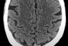
Landmark on CT and MRI for central sulcus?

A

Omega sign. Anterior is frontal lobe, posterior is parietal lobe.

3
Q

What level is this?

Identify structures.

A

Ventricles

Head of caudate, internal capsule, lentiform nucleus, thalamus, insular cortex, anterior temporal lobe.

4
Q

Identify sylvian fissure (AKA). What artery runs along here?

What does the sylvian fissure separate?

A

Lateral sulcus

Middle cerebral artery

Frontal/Parietal lobe from temporal lobe

5
Q

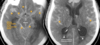
Identify Midbrain, basal cistern, medial temporal lobe “uncus”, temporal horn of lateral ventricle (what is specific for a normal appearance)?

Why is it important to local the medial temporal lobe?

What does it mean for the temportal horn of lateral ventricle to be dilated?

A

Uncus can herniate onto the midbrain

Temporal horn of lateral ventricle should normally be slit like.

Early sign of hydrocephalus if temporal horn of lateral ventricle is dilated.

6
Q

What level is this?

Identify brachium pontins, direction of connection between brachium pontis and cerebellum, 4th ventricle, and cerebello-pontine angle.

A

Mid pontine level

Brachium pontins Will hug the 4th ventricle

7
Q

Identify Medulla, 4th ventricle, and cerebellum

A
8
Q

Identify foramen magnum and spinal cord

A
9
Q

Steps for interpret Neuroimaging (5)

A

Identification - use landmarks

Symmetry

Density/Intensity

Pattern of Enhancement

Lesion location

10
Q

What appears dense on CT?

Sensitivity to detect blood?

A

Mineralized structures such as calcified bone or chronic calcified lesions.

>90% sensitivity to detect blood

11
Q

Identify hyperdense structures

A

Calvarium, lens, pineal gland, choroid plexus

12
Q

Diagnosis?

A

Neurocysticercosis

Calcified scolex in cysticercosis

Scolex - the anterior end of a tapeworm, bearing suckers and hooks for attachment.

13
Q

Diagnosis

A

Acute basal ganglia hemorrhage with intraventricular extension

14
Q

Provide simple description

Diagnosis

A

Chalk outlining the sulci

Diffuse subarachnoid hemorrhage which is extraxial + intraventricular extension

15
Q

For suspected extraxial epidural hematoma what can be done when reading the image?

A

Adjust to bone window to observe for calvarial sutures and fractures

16
Q

Identify hyperdensities

A

Hyperdense MCA sign in the sylvian fissure

Between the cerebral peduncles in the interpeduncular cisterns -> Dense vessel sign (top of basilar artery)

17
Q

Hypodense typically represent (4)?

A

Chronic lesions

Fluids

Cystic component

Edema

18
Q

Define encephalomalacia

Define atrophy

A

Softening or loss of brain tissue after cerebral infarction, cerebral ischemia, infection, craniocerebral trauma, or other injury

Loss of NEURONS and the connections between them. Atrophy can be generalized, which means that all of the brain has shrunk; or it can be focal, affecting only a limited area of the brain and resulting in a decrease of the functions that area of the brain controls.

19
Q

CT characteristics suggestive of chronic stroke vs traumatic brain injury?

A

Isodense to CSF

Follows vascular territory vs not

20
Q

Is this intraxial or extraxial?

A

Fronto-temporal arachnoid cyst

21
Q

Timeline for an acute stroke to present with hypodensity (edema) on CT?

A

6-8 hours

22
Q

Describe

A

Vasogenic edema surround a metastatic lesion

23
Q

MRI Brain without contrast

What are the basic sequences (4) + Optional

A
  1. T1WI
  2. T2WI + Fluid Attenuated Inversion Recovery (FLAIR)*
  3. DWI + Apparent Diffusion Coefficient**
  4. Gradient Echo Sequence (GRE)
  5. T1 with Gadolinium contrast

WI- Weighted Image

* Most filling and useful sequences

** Designed to diagnose energy failure and ischemia

24
Q

What are some specialized MR sequences?

A

MR Angiogram and Venogram

Fat suppresion

Perfusion

Spectroscopy

Tractography

25
Detail
Neurovascular unit is an essential component to make up the blood brain barrier
26
Detail following ischemia
The cellular components of the nuerovascular unit becomes ischemic. Cannon maintain the sodium/potassium gradient leading to edema -\> Cytotoxic edema
27
Detail
T2WI is great to highlight fluid. This is an acute stroke.
28
Detail
Leaky tight junction -\> vasogenic edema (poor vessel integrity) Occurs with neoplasm, abscess, trauma, and hypertensive emergency
29
Describe
Heterogenous mass
30
How does CT and MRI FLAIR relate in a stroke? DWI?
Both require about 8 hours to demonstrate evidence of ischemia Can detect ischemia within 30 minutes of onset.
31
Why do we need DWI and ADC to confirm infarction? T2 Shine through
DWI relates to T2WI (FLAIR) thus what is hyperintense on T2WI will be hyperintense on DWI. T2WI (FLAIR) "shine through" to DWI Thus if ADC is hyperintense, it is confirmed to be a T2 shine through
32
Etiologies for T2WI (FLAIR) hyperintensities (6)
micro-hemorrhages gliosis damage to small blood vessel walls breaches of the barrier between the cerebrospinal fluid and the brain loss and deformation of the myelin sheath Ischemia
33
Discuss the nature of localization of pyogenic abscesses
Hematogenous spread following vascular territory territories thus majority of septic emboli locate in major vascular territories such as MCA -\> frontal and pareital lobes being the most common locations in distribution of MCA
34
What is a common location for metastatis?
Like bacteria hematogenous spread is common thus gray-white matter junctions are common. Arterioles are just small enough at the junction to halt any further progression of emboli or metastatic cells.
35
Most common brain metastasis (2) Others (4)
1. Lung 2. Breast Melanoma, Renal, Thyroid, Gastrointestinal
36
Describe how transependymal flow of CSF will appear on FLAIR. What's one reason this will occur?
Symmetrical smooth periventricular hyperintensities and hydrocephalus from eg. a posterior fossa mass -\> communicating hydrocephalus.
37
Describe Dawson Fingers. Why does this occur?
Ovoid periventricular lesions Central veins run perpendicular to the ventricles. Immune cells exit these veins and penetrate the brain parenchyma. Axial View.
38
Detail the difference.
Left: Ovoid periventricular hyperintense lesions - \> MS VERY COMMON - Right: Confluent periventricular and subcortical hyperintense lesions along with accompanying cerebral atrophy - \> small vessel ischemic changes
39
MR T1 hyperintense lesions caused typically by (4).
Paramagnetic substances (iron, copper, melanin, calcium) Fat Protein-rich lesions Subacute hematoma
40
MR T1 What is hyperintense in orbit?
Orbital fat Orbital melanoma
41
What MR sequence is this and what is the abnormality?
MR T1 and copper accumulation in the basal ganglia -\> Wilson Disease
42
What makes up the basal ganglia
Caudate nucleus, putamen, and globus pallidus in the cerebrum, the substantia nigra in the midbrain, and the subthalamic nucleus in the diencephalon
43
What MR Sequence and Dx
Colloid cyst (Protein rich mass) leading to communicating hydrocephalus. Note this presentation of complete obstruction of the interventricular foramen (foramen of monro) can occur so acutely it can lead to imminent death
44
What makes Blood on MR challenging to interpret.
Blood on MR evolves along with the intensity signals on T1 and T2.
45
Acute hematoma MR T1 vs T2 What is the form of Hgb?
(1-3 days) Deoxyhemoglobin
46
Subacute hematoma MR T1 vs T2 What is the form of Hgb?
(3-14 days) Methemoglobin (F3+, Ferr**IC** ion)
47
Chronic hematoma MR T1 vs T2 What is the form of Hgb?
\> 14 days (2 weeks) Hemosiderin - iron-storage complex that is composed of partially digested ferritin and lysosomes Essentially appears as a CSF filled hole. Rim of hypointense region on T2 is the ring of hemosiderin
48
mnemonic for what can be identified on T2
S2HI2NE Stroke, Subacute hematoma Hydrocephalus Ischemic changes, Inflammatory Neoplastic Edema
49
Mnemonic for what can be seen using MR T1 sequence
SH IMMeR Subacute hematoma Iron Metals Melanoma Rich in protein
50
What is notable about hypdense lesions on T2
The same kinds of lesions that appear hyperintense on T1 will be hypointense on T2 Protein-rich masses Flowing blood is not depicted but flow voids do appear Paramagnetic substances (iron, copper, melanin, calcium) In addition, acute hematoma
51
MR sequence? What are we highlighting here?
Flow voids ICA, MCA, PCOM, PCAs, Confluence of venous sinuses
52
Identify the pathology
Absent right ICA flow void - \> occlusion
53
Back pain Exacerbating movements Extension exacerbates _____ ?
Compressed cord It decreases the diameter of the spinal canal, thus worsens compression. Flexed at the hips will increase the diameter of the canal and mitigate back pain.
54
Back pain Exacerbating movements Flexion exacerbates _____ ?
Compressed root. Radiculopathy may occur when the nerve root is compressed in the intervertebral foramina due to inflammatory changs causef by a herniated disk. Flexion reduces the size of the intervetebral foramina.
55
Chief complaints with spinal cord/nerve root pathology (6)
Back pain Weakness Gait abnormality (from weakness or disturbed position sense or damage to descending cerebellar pathways) Neurogenic claudication (muscle cramp/spasm) Bladder, bowel dysfunction (damage to descending pathways or sacral fibers) Sensory level ("squeezing belt" with numbness and tingling below that level)
56
Detail dorsal column topographically
Vibration, proprioception, fine touch Arms are lateral, legs are medial
57
Detail Anterolateral system topographically
Spinal thalamic tract Pain, temperature, crude touch
58
Detail lateral corticospinal tract topographically
Legs are lateral, arms are medial
59
Symptoms of complete transverse lesion Etiologies
Sensory level is most specific finding, bilateral weakness, urinary dysfunction, dysautonomia, respiratory distress, segmental anterior horn syndrome. Note: Sensory fibers enter posterior cord, ascend 1-2 levels, then decussate in the anterior commissure. Thus sensory level typically present 1-2 segments **below** actual lesion. Transverse myelitis
60
Acute cord injury causes spinal shock - tone and reflexes?
Low tone and decreased reflexes. Over days to weeks spasticity develops and hyper-reflexia If descending micturition pathways are affected -\> urinary dysfunction
61
Damage to sympathetic pathways traveling in the intermediolateral columns can lead to ?
Serious dysautonomia
62
Dangers of cervical lesions
Phrenic nerve can be affected causing paralysis of diaphragm
63
Segmental anterior horn syndrome
Lower motor neuron pattern of weakness in the muscles innervated by that segment
64