Neurobiology of hearing Flashcards
LOs
- Describe the components and functions of the external, middle and inner ear
- Explain the roles of the tympanic membrane, the auditory ossicles (malleus, incus and stapes) and scala vestibule in sound transmission
- Describe the way that movements of molecules in the air are converted into impulses generated in the cochlea
- Explain how pitch, loudness and timbre are coded in the auditory pathways
- Describe the components of the auditory pathway from the cochlear hair cells to the cerebral cortex
- Compare causes of conductive and sensorineural hearing loss and tests used to distinguish between them
Ears let us to: (2)
detect sounds
maintain balance
via receptors for eharing and equilibrium in the ear
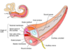
The anatomy of the human ears is divided into:
outer ear
middle ear
inner ear
which 3 structures are associated w hearing
which 3 structrues are associated w equillibrium
external ear, middle ear and cochlea
the external ear is composed of: (3)
- auricle (pinna) that captures sound waves
- external auditory meatus (ear canal)
- tympanic membrane
what is the stated hearing range for humans
over which range is hearing most clear
20 –> 20,000 Hz
2,000 –> 5,000
what is the auricle/ pinna
function?
paired structure on either side of the head
external ear
fucntion: gather and focus sound energy through the external auditory meatus to the tympanic membrane

lobule
the one part of the auricle that has no cartilage
aka ear lobe
concha
shell shaped structure of the cavity of the external ear

microtia- what is it and what are the grading systems
a congenital deformity where the pinna is underdeveloped
complete underdevelopment of the pinna is anotia
graded 1–> 4 with increasing severity
grade 1 severity- small ecternal ear and a small but present ear canal.
grade 2- partially developed ear (usually the top portion is underdeveloped) with a closed external ear canal (atresia) producing a conductive hearing loss.
grade 3- most common form with an absent external ear and small peanut-like vestige structure and canal atresia.
grade 4- complete absence of the external ear with canal atresia
thr concha and external auditory canal act as a resonator
effectively enchance the intensity of sound that reaches the tympanic membrane by about 10-15 dB
what is the ear canal/ external auditory meatus
Functions as an entryway for sound waves, which get propelled toward the tympanic membrane known as the eardrum.

3 major components of middle ear
general function of middle ear 2
- tympanic membrane (eardrum)
- auditory ossicles
- eustachian/ auditory tube
overall function:
- amplify the vibrations of the tympanic membrane to the oval window
- impedence matching
tympanic membrane
thin and pliable, therefore a sound, consisting of compressions and rarefractions of air particles, pulls and pushes at the membrane moving it inwards and outwards at the same frequency as the incoming sound wave.
It is this vibration that ultimately leads to the perception of sound
- greater the amplitude the greater the deflection of the membrane.
- higher the frequency of the sound, the faster the membrane vibrates.

auditory ossicles
what are the 3
3 of the smallest bones in the body.
they transfer vibrations of the tympanic membrane to the cochlea
- malleus (hammer)- forms a rigid connection with the incus
- incus (anvil)- forms a flexible connection with the stapes
- stapes (stirrups)- connects the oval window

how do the ossicles work to transmit sound to the cochlea
- The inward-outward movement of the tympanum displaces the malleus and incus
- the action of these two bones alternatively drives the stapes deeper into the oval window and retracts it
- resulting in a cyclical movement of fluid within the inner ear.
eaustachian tube
helps to ventilate the middle ear and maintain equal air pressure on noth sides of the tympanic membrane, inside the ,middle ear and outside the body, via nasopharynx (the nasal part of ther pharynx, lying behind the nose and above the level of the soft palate).
what is the vestibular system?
which 3 structures make it up?
the sensory apparatus of the inner ear that helps the body maintain its postural equilibrium.
Info furnished by the vestibular system is also essential for co-ordinating the position of the head and movement of the eyes.
Engages a number of reflex pathways that are responsible for making compensatory movements and adjustments in body position.
this info provideed is proprioceptive (inside)
1) semi-circular canals
2+3) the utricle and saccule
the semi-circular canals
- respond to rotational movements (angular acceleration)
- made of 3 pairs of ducts oriented at roughly 90* to each other for maximum ability to detect angular rotation
- canals don’t contribute to sense- the fleshy- fluid filled ducts inside
- mediate interactions between the vestibular system and the eye muscles via the cranial nerve- smooth eye movement left and right keeping visual fields stable when moving
Otolith organs
consist of the saccule and utricle
which are membranous sacs perpendicular to each other
AKA gravity receptors
both contain a maccula
both organs monitor the position of the head relative to vertical
what are macula?
patches of sensory hair cells in the otolith organs
what are hair cells?
how do they contribute to hearing and balance?
hair cells are topped by otoconia- small calcium carbonate crystals
due to the weight of the otoconia and a gelatinous layer, vibrations (hearing) and gravity will bend the hair cells.
bending of the hair cells leads to afferent activity to the brainstem
cochlea
The cochlea is the part of the inner ear involved in hearing. It is a spiral-shaped cavity in the bony labyrinth, in humans making 2.75 turns around its axis, the modiolus.

the cochlea is made up of 2 membranes and 3 compartments
what are they
- Raissner’s membrane
- Basilar membrane
- Scala vestibuli (vestibular ducts)
- Scala tympani (tympanic ducts)
- Scala media (cochlear duct)
raissner’s membrane
together with the basilar membrane creates a compartment in the cochlea filled with endolymph, which is important for the function of the spiral organ of Corti.
Thought to play an important role in otoacoustics as a wave on Reissner’s membrane can propagate along the whole extent of the cochlea

basilar membrane
forms divisions between the cochelar duct (scala media) and the tympanic membrane, allowing different frequencies to reach max amplitudes in different positions
Performs frequency selectivity by its filter bank so is effectively a continuous array of filters which decompose a complex waveform into its constituent frequency components.

what are the 3 compartments of the cochlea
- vestibular ducts- conducts sound vibrations to the cochlear duct
- tympanic ducts- together with the vestibular ducts aid in transducing the movement of air that causes the tympanic membrane and the ossicles to vibrate, to movement of liquid and the basilar membrane.
- cochlear duct- houses the organ corti that transforms fluid vibration into nerve impulse
oval window
receives vibration from the stapes and transmits this to the base of the basilar membrane
round window
vibrates with the opposite phase to vibrations entering the inner ear through the oval window.
It allows fluid in the cochlear to move, which in turn ensures that hair cells of the basilar membrane will be stimulated, and that audition will occur.
Organ of corti
Transduces auditory signals and minimizes the hair cells’ extraction of sound energy and cosists of 2 hair cells and a tectorial membrane
inner hair cells of organ of corti
detect the sound and transmit it to the brain via the auditory nerve
outer hair cells of the organ of corti
perform an amplifying role for sound
tectorial membrane
may be involved in longitudinal propagation of energy through the cochlea
hair cells and discharge
in unstimulated hair cells there is a low level of discharge of APs in the axons of the auditory nerve fibres
when the cell is stimulated, depolarisation results in an increase in the discharge rate of action potentials (excitation)
while hyperpolaristion results in a decrease in discharge rate of APs (inhibition)
endolymph
- fluid contained in the membranous labyrinth (semicircular ducts) of the inner ear and is involved in dynamic equillibrium
perilymph
Perilymph- extracellular fluid inside the perilympathtic space
what is the joint function of endo and perilymph?
whcih one resembles extracellular fluid and which one resembles intracellular fluid
regulate electrochemical impulses of hair cells
perilymph resembles extracellular fluid (Na+ predominates)
endolymph resembles intracellular fluid (K+ is the main cation)
endolymph has 2 other functions aside from regulating electrochemical impulses
- Hearing- fluid waves In the endolymph of the cochlear duct stimulate the receptor cells, which in turn translate their movement into nerve impulses that the brain perceives as sound
- Balance- angular acceleration of the endolymph in the semicircular canals stimulate the vestibular receptos of the endolymph
sound transduction
sound is a mechanical wave that needs a medium to propagate itself and is created by something that vibrates
vibration creates air particle movement due to change of air pressure.
- The sound wave travels thorugh air to the human ear via the pinna.
- sound waves (vibrations) are converted into action potentials by the inner row of hair cells in the cochlea (The apical end of the hair cell contains the sereocilia and they are arranged in order of ascending lengths from one side of the cell to the other. The membranes of the stereocilia contain mechanically gated cation channels. Extending from the gate of the ion channel to the adjacent, taller, stereocilium is a fibrous protein called a tip link.)
- When the stereocilia bend toward the longest stereocilium the tension in the tip link increases, pulling the gates on the ion channels open, and when they bend in the opposite direction the tension decreases and the gate closes.
- Stereocilia are bathed in endolymph of the cochlear duct (high in K+). When the gate on the cation channel opens, K+ rushes into the cellm], depolarising the membrane and causing voltage gated Ca2+ channels on the basal membrane of the hair cell to open, allowing Ca2+ to enter.
- Influx of Ca2+ stimulates the release of a NT by the hair cell, triggering an ap IN THE NEURONE THAT Synapses with the hair cell. The axons of these neurones form the cochlear nerve that transmits the AP’s to the auditory cortex of the brain.
In hair cells at rest, ~10% of the K+ ion channels are open resulting in a low frequency of AP’s travelling to the brain whrn it is perfectly quiet.
This allows for both an increase ein AP frequency when hair cells bend toward the longest stereocilium, and a decrease in frequency of action potentials when the hair cells bend the other way.

sound perception
Once the AP is generated and sent to the brain, it is the function of the auditory cortex (The primary auditory cortex (A1) is located on the superior temporal gyrus in the temporal lobe) to convert the AP into a perception and each regin of the cochlea is hardwired to its own specific region of the auditory cortex.
When that particular region of the brain receives input drom the ear we perceive the unique pitch associated with that frequency of sound wave
Each time an AP reached that specific segment of the auditory cortex we perceive the same sound.
Therefore, the pitch is determined by the region of the brain that receives input from the cochlea.
loudness
what determines it
is determined by the number of AP’s that reach the brain.
- Sound eaves of higher amplitudes cause the hair cells to vibrate more rigorously, which would cause more ion channels to open.
- This would result in a greater depolarisation of the hair cell, more ca2+ entry through the voltage gated ion channels and more NT release.
- End result is a greater frequency of ap’s going to the auditpry cortex, which is perceived as louder sound.
classification of hearing loss (2)
conductive
sensorineural
sensorineural hearing loss
Sensorineural hearing loss (deafness) is the most common type of hearing loss, it is usually due to the loss of cochlear hair cells but can be resultant from damage to the 8th cranial nerve or within the central auditory pathways.
It often impairs the ability to hear certain pitches while others are unaffected.

conductive hearing loss
refers to impaired sound transmission in the external or middle ear and impacts all sound frequencies.
Among the causes of conduction hearing loss are:
- plugging the external auditory canals with wax (cerumen) or foreign bodies
- otitis externa (inflammation of the outer ear “swimmers ear”)
- oitisis media (inflammation of the middle ear) causing fluid accumulation or scarring, or perforation of the eardrum.
Severe conductive deafness can result from otosclerosis in which bone is resorbed and replaced with sclerotic bone that grows over the oval window.

causes of hearing loss
- Aminoglycoside antibiotics such as streptomycin and gentamycin obstruct the mechanosensitive channels in the stereocilia of hair cells (especially outer hair cells) and can casue the cells to degenerate, producing sensorineural hearing loss and abnormal vestibular function.
- Damage to hair cells- by prolomged exposure to loud noise
- AID
- Traumatic injuries
- Acoustic neuromas
- Tumours of the eighth cranial nerve and cerebellopontine angle
- Vascular damage in the medulla
what is the unit of sound?
decibels dB
Normal hearing (up tp 20 dB HL)
Mild hearing loss (21 40 dB HL)
Moderate hearing loss (41 70)
Severe hearing loss (71 95 dB HL)
Profound hearing loss (95 >)
qualititative ways of describing hearing loss
Looking for qualities such as:
- Bilateral (noth ears) vs unilateral (one ear hearing loss)
- Symmetrical (Same level/ severity of eharing loss in both ears) vs asymmetrical hearing loss (different levels/ severity in each ear)
- High frequency/ pitched vs low frequency/ pitch
- Progressive versus sudden hearing loss
- Stable vs fluctuating hearing loss
type of hearing test
Pure tone audiometry (PTA)
This test involves hearing sensitivities across a range of frequencies (pitches), which are involved in speech perception. It involves listening to sounds via headphones by pressing a button every time a sound is heard.
Speech testing
Diagnostic test that assesses speech discrimination using single words
Involves listening to words and repeating what was heard to the audiologist who records the rsults
Bone conduction testing
Another type of pure-tone test that measures your inner ear’s response to sound.
A conductor will be placed behind the ear; it will send tiny vibrations through the bone directly to he inner ear.
Acoustic reflex testing
This test measures involuntary muscle contractions of the middle ear and is used to determine the location of the hearing problem (the ossicles, cochlea, auditory nerve etc) as well as the other type of hearing loss.
Auditory brainstem response (ABR)
Used to determine whether a specific type of hearing loss (sensorineural) exists
Also ised to frequently screen newborns for hearing problems
In an ABR test, electrodes are attached to the head, scalp or earlobes, and headphones to wear
Brainwave activity Is measured in response to sounds of varying intensities.
Bone conduction testing components (2)
Weber and Rinne test
Weber test
method
normal results
Conduction deafness one ear
Sensorineural deafness one ear
Base of vibrating tuning fork placed on vertex of skull (front central)
hears equally on both sides
conduction deaf- sound louder in diseased ear because background noise is absent on diseased side
sensorineural deaf- sound louder in normal ear
Rinne
method
normal results
Conduction deafness one ear
Sensorineural deafness one ear
Base of vibrating tuning fork on mastoid process until subject can no longer hear it then held in air next to it
Normal- hears vibration in air after bone conduction is over
Conduction deafness- vibrations in bone not heard after conduction is over
sensorineural deafness- vibration in air heard after bone conduction is over as long as nerve deafness is partial