Nerve tissue Flashcards

1
Q

What are the two types of cells in the Nervous system?

A

-neurons or nerve cells -supporting cells

2
Q

What is the difference between the nerve cells of the PNS and CNS?

A

PNS - Schwann cells surround nerve processes - Satellite cells - surround nerve cell bodies in ganglia -CNS - glial cells

3
Q

Describe some basic information of the neuron.

A

-structural functional unit of the NS -10 billion in humans -consist of central process- axon -peripheral process - dendrite

4
Q

Do neurons replicate?

A

no

5
Q

What is the name of the neuron cell body

A

Perikaryon

6
Q

Describe some of the structural components for the perikaryon.

A

-Nissl bodies -Mitochondria -large peripheral GA

7
Q

What are Nissl bodies?

A

-stacks of RER -extend into dendrites -depend on the cell body for maintenance

8
Q

Where can you find Nissl bodies?

A

In the dendrites but not the axons

9
Q

Describe the parts of the neuron.

A
10
Q

What carries information to the cell body

A

dendrites

11
Q

What has a greater diameter dendrite or axon?

A

dendrites

12
Q

Are dendrites myelinated?

A

no

13
Q

What are the purpose of arbonizations called dendritic trees on dendrites?

A

-allows for a greatly increase in the receptor surface of the neuron

14
Q

Where are axons origins on the nerve cell?

A

Axon Hillock

15
Q

What are the components of the Axon Hillock? What organelles are not present here?

A

In- microtubules, neurofilaments, mitochondria, vesicles, terminal arborizations Not present - Nissyl (RER) and Golgi

16
Q

What is the purpose of the axons?

A

carry information from the cell body.

17
Q

How many axons and dendrites per neuron?

A

1 axon and may have numerous dendrites

18
Q

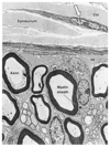
Axolemma

A

axon plasma membrane

19
Q

Axoplasm

A

axon cytoplasm lacks a golgi complex but contains SER, RER and elongated mitochondria - lacks a golgi apparatus

20
Q

Golgi Type I neurons

A
  • motor nuclei of CNS - axons more that 1 meter long ex. muscle
21
Q

Golgi Type II neurons

A
  • golgi cells in the cerebellum - have short axon - axon branched near target organ
22
Q

Where do sensory neuron travel?

A

receptor to CNS

23
Q

Where do motor neuron travel?

A

CNS or ganglia to effector cells

24
Q

What are interneurons?

A

connections between neurons and include internuncial, central and intercalated neurons, (99% of neurons). They regulate signal transmission between neurons.

25
Q

How are neurons classified?

A

by the number of processes

26
Q

Multi-polar neuron

A

one axon and 2 or more dendrites

27
Q

Bipolar neuron

A

one axon and one dendrite

28
Q

Unipolar (pseudo-unipolar in higher animals)

A

one process (axon) that divides close to cell body into two processes

29
Q

Distinguish between anterograde and retrograde transport.

A

Axon transport is bidirectional - ANTEROGRADE - cell body to periphery via kinesin - RETROGRADE - axon terminal & dendrites to cell body via dynein

30
Q

What are synapses?

A

-specialized junctions between neurons that facilitate transmission of impulses between neurons, axons and effector cells.

31
Q

How many synapses can occur on one neuron?

A

few to a thousand

32
Q

What is the name of the end of the axon.

A

Swollen bulb or BOUTON

33
Q

Two types of synapses

A

Electrical and Chemical

34
Q

What is an important structural component of the electrical synapse?

A

gap junctions which facilitate movement of ions between cells - action potential is directly from presynaptic cell to post synaptic cell.

35
Q

Cells that contain electrical synapse.

A

Nexus in smooth muscle Gap junctions in cardiac muscles

36
Q

What are the three components of chemical synapse?

A
  • -PRESYNAPTIC BOUTON -
  • SYNAPTIC CLEFT
  • -POSTSYNAPTIC MEMBRANE
37
Q

What facilitates information transfer at the presynaptic bouton?

A

synaptic vesicles which release transmitters into the synaptic cleft

38
Q

How big is the synaptic cleft?

A

20-30nm

39
Q

Patient comes in with gradually progressing stiffness, difficulty walking, and uncontrolled involuntary movement of legs and arms. What is a possible diagnosis and its mechanism of action?

A

Parkinson’s disease Physiology: loss of dopamine secreting cells in substantia nigra and basal ganglia Increase in the number of glia cells.

40
Q

What is a treatment for Parkinson’s disease?

A

synthetic dopamine

41
Q

What component of the nervous system is affected in someone with Myasthenia Gravis?

A

Neuromuscular junction where their is an autoimmune attack on acetylecholine receptors.

42
Q

What are some cells found in the PNS?

A

-Schwann cells -Satelite cells

43
Q

Cells of the CNS

A
  • Ependymal cells - Neuroglial cells : astrocytes, Oligodendrocytes, microglia
44
Q

Describe the nucleus of a neuronal cell body.

A

The nucelus is large, sperical and pale-staining and is centrally located in most neurons.

45
Q

What cell is responsible for myelination in the PNS?

A
  • Schwann cells
46
Q

What is another name for a sheath of Schwann?

A

Neurilemma

47
Q

What is in the Schwann cell cytoplasm?

A

nucleus few mitochondia and a small golgi region

48
Q

What area on a neuron are free of myelin?

A

axon hillock and terminal axon arborizations

49
Q

Nodes of Ranvier

A

gaps in the myelin sheath that allow for saltatory conduction of impulses. “jumping” from node to node.

50
Q

Describe the stages of myelination

A

Insert picture : a) A Schwann cell nestles itself around an axon. b) While continuously producing myelin, the Schwann cell winds a sheath-like protrusion around the axon yielding multiple layers. c) Finally, these layers are compacted into a tightly packed insulation coat.

51
Q

What is this image

A

Myelinated nerve fiber

52
Q

What are the components of the myelin cytoplasm?

A

-Inner collar of Schwann cytoplasm between axon and myelin - Schmidt-Lantermann clefts - small islands of cytoplasm within lamellae of myelin Perinodal cytoplasm - at node of ranvier Outer collar of perinuclear cytoplasm- around myelin Insert picture.

53
Q

Unmyelinated cells

A

enveloped by a single invagination of schwann cells. - axon is in a cleft of the cytoplasm of a schwan cell with a single mesoaxon

54
Q

Saltatory Conduction

A

myelinated axons conduct faster than unmyelinated ones. - called “jumping” from node to node - Speed is related to the thickness of myelin and axon.

55
Q

Guillian Barre Syndrome

A

results in loss of muscle co-ordination and cutaneous sensation when there is a large amount of lymphocytes, plasma cells and macrophage surround nerve fibers

56
Q

Multiple Sclerosis

A

Gradual degeneration of the myelin and destruction of oligodendrocytes

57
Q

What are Satellite cells? what do they look like? What do they surround?

A

-surround ganglionic neuronal cell bodies -Cuboidal shape - only able to see nuclei - originate from neural crest cells. - anagolous to schwann cells - insulation and nutrition

58
Q

What cells are analogous to Schwann Cells?

A

Satellite cells

59
Q

Neuroglia (Glial cells of CNS)

A

more numerous than neurons Three types: Oligodendrocytes, astrocytes, microglia

60
Q

What are the two types of astrocytes and how can you differentiate between the two of them?

A

Protoplasmic - prevalent in gray matter Fibrous- more common in white matter - both have elaborate processes extending between vessels and neurons - play a role in scar formation in the CNS

61
Q

What allows astrocytes to be viewed by immunohistochemistry?

A

they contain fibrillary acidic protein marker

62
Q

What cell may play a role in regulating the tight junction in the Blood Brain Barrier?

A

astrocytes

63
Q

What forms the Blood Brain Barrier?

A

capillary endothelium with occluding junctions with an envelop of astrocytes on the basal lamina capilaries

64
Q

Which area of the brain does not have astrocytes wrapping the basal lamina capillaries?

A

Choroid plexus and posterior pituitary

65
Q

Oligodendrocytes

A

cells that create myelin on CNS neurons. - align in rows between axons -each oligodrocyte gives off several tongue-like process that wraps around the axon.

66
Q

How many nerve fibers can one oligodendrocyte for myelin sheaths for?

A

3-50 nerves

67
Q

Microglia

A

mononuclear phagocytes -small numbers that proliferate during disease -secrete immunoregulatory cytokines

68
Q

Ependyma cells serve what roll in the CNS

A

line fluid filled cavities of the CNS - cuboidal to columnar shaped to allow for fluid transporting characteristics

69
Q

Choroid plexus

A

modified ependyma cells and capilary loops that produce cerebrospinal fluid.

70
Q
A

dendrites

cell body

neuroglia

axon

71
Q

Distinguish between neuron in the CNS and PNS

A
72
Q

What kind of nerves are labeled U and M?

A

Unmylenated and Mylenated

73
Q

What is the arrow pointing at?

A

Satellite Cells

74
Q

Describe some of the components of a Neuron cell body in a ganglion?

A

A neuron in the ganglion is surrounded by satellite cells and covered with Schwann cells on the axons.

75
Q
A
76
Q

What kind of cell is this?

A

Astrocytes

77
Q
A
78
Q

What is this arrow pointing at?

A

Ependyma - which is a cuboidal to colummnar cell that plays a role in production of CSF

79
Q

What are the choroid plexus?

A

Choroid plexus is invaginated folds of pia mater rich in dilated fenestrated capillaries covered by simple cuboidal cells

80
Q

Where is the choroid plexus found?

A

two lateral, 3rd and 4th ventricles and all the regions where epnymal cells liein contact with pia mater

81
Q

What is the function of the choroid plexus?

A

produces CSF

82
Q

What are each of the villus of CP covered with what layer of the CNS (pia, aracnoid or dura)

A

vascularized pia mater covered by cuboidal ependymal cells.

83
Q

What is the function of the CSF?

A

acts as a mechanical shock absorber and important for CNS metabolism

84
Q

What areas of the brain does the CSF fill?

A
  • ventricles
  • central canal of spinal cord
  • subarachnoid space
  • perivascular spaces
85
Q

What is this tissue and what are some important components of it?

A

Choroid Plexus with simple cuboidal cells.

86
Q

What is this tissue?

A

Choroid Plexus

87
Q

What is this tissue?

A

longitudinal section of myelinated peripheral nerve fibers

88
Q

What are the three components of connective tissue sheaths?

A
  • Endoneurium
  • Perineurium
  • Epineurium
89
Q

What is the Endoneurium?

A

Thin layer of reticular fibers produced by Schwann cells.

(Electron micrograph of a cross section through a nerve, showing the epineurium, the perineurium, and the endoneurium. The epineurium is a dense connective tissue rich in collagen fibers (Col) and fibroblasts (arrow). The perineurium is made up of several layers of flat cells tightly joined together to form a barrier to the penetration of the nerve by macromolecules. The endoneurium is composed mainly of reticular fibers (RF) synthesized by Schwann)

90
Q

Describe the perineurium?

A
  • surrounds bundles of nerve fibers
  • sheath of unique connectivie tissues
  • cells are joined by tight junctions
  • forms barrier to passage of most macromolecules
  • protects nerves from aggression
  • contractile cells
  • numerous actin filaments
91
Q

What part of sheath serves as a blood-neuron barrier. Separates the vascular tissue from the interstitial space between individual neurons

A

Perineurium

92
Q

Which component of the sheath protects nerves from aggression and are contractile?

A

perineurium

93
Q

What are the epineurium?

A
  • outmost tissue of the peripheral nerve
  • typical dense connective tissue that binds fascibles together
  • oftan contains adipose tissues and is vascular
94
Q

Does the perineurium have a vascular supply?

A

Vascular supply from the epineurium penetrates the perineurium

95
Q

What is this longitudinal section?

A

Peripheral nerve

96
Q

What are the two type of ganglia

A

Sensory and Autonomic

97
Q

Which ganglia is pseudounipolar with thick fascicles?

A

Sensory ganglia

98
Q

Which ganglia are multipolar, smaller and far apart?

A

Autonomic ganglia of the parasympathetic and sympathetic system

99
Q

Describe sensory ganglia?

A
  • craniospinal (dorsal root ganglion)
  • pseudounipolar neurons
  • larger
  • close together
  • thick fascicles
100
Q

Describe Autonomic ganglia

A
  • Sympathetic and parasympathetic
  • multipolar neurons
  • smaller
  • far apart
  • diffuse nerve fibers
101
Q

What kind of ganglia is this?

A

Sympathetic ganglia

102
Q

What kind of ganglion is this?

A

Sensory ganglion

C- capsule

So- soma (cell body)

Cc - capsule cells

F- nerve fiber

103
Q

What is the outflow of the sympathetic pathway?

A

Thoraco Lumbar outflow via norepinephrine

104
Q

What is the outflow of the parasympathetic pathway?

A

cranio-sacral outflow acetylcholine

105
Q

What are the two plexus of the Enteric Nervous system?

A
  • myenteric (Auerbach)
  • submucous (Meissner)
106
Q

What is the tissue?

A

Enteric nervous system- consits of ganglia and postganglionic networks

  • composed of unmyelinated nerve fibers intermingled with neuronal cell bodies.
107
Q

What is Hirschsprung Disease?

A
  • Congential mega colon.
  • arrest of one of the neural crest cells to one segment of the distal colon
  • no innervation of the colon
  • sign and symptoms: unable to pass stool at birth, with no vomiting, no abdominal distention.
    *
108
Q

What is the composition of grey matter?

A

neuron cell bodies and their dendrites along with axons and glia.

109
Q

Describe what white matter looks like?

A
  • mostly myelinated fibers
  • arranged in three pairs of funiculus - anterior, lateral and posterior
  • functionally related bundles called tracts
110
Q
A
111
Q

Label each component of the picture.

A

Cc - central canal

Gc- grey commissure

VH-ventral horn

DH-dorsal horn

WM- white matter

112
Q

What is in the cortex of the brain?

A

Grey matter- contains cell bodies, axons, dendrites, glial cells and synapses

113
Q

What are the nuclei?

A

Islands of grey matter found in deep portions of cerebrum and cerebellum

114
Q

What does white matter mostly consist of in the brain?

A

Myleniated axons of nerve cells, glial cells, vessels.

115
Q

Describe the 6 layers of the cerebrum?

A

Cerebral cortex which contains pyramidal cells in layers three and five.

116
Q
A
117
Q

What kind of tissue is dura mater?

A

dense irregular connective tissue

118
Q

What are the three layers of the brain?

A
  • pia - most intimate layer attached to the brain
  • arachanoid - delicate web like connective tissue
  • dura - composed of dense irregular connective tissue
119
Q

What are nuclie?

A

conglomeration of grey matter deep with in the cerebrum and cerebellum

120
Q

Where is the CSF found?

A

in arachnoid space

121
Q

What is the outer grey matter of the brain called?

A

cortex

122
Q

What kind of cells are interneurons?

A

multipolar

123
Q

What kind of neuron are sensory neurons?

A

unipolar

124
Q

What is the primary component of myelin?

A

lipids

125
Q

What is a basket cell?

A

type of neuron in the CNS

126
Q

What is an internuncial neuron?

A

interneuron

127
Q

What is another name for neuron cell body?

A

soma

128
Q

What is tightly attached to the periosteum of skull and spinal column?

A

Dura matter

129
Q

What is labeled as n?

A

multipolar neuron

130
Q

Where are synaptic vessicles found?

A

Terminal bouton

131
Q

What happens to the potassium in a resting neuron membrane?

A

The potassium is greater inside the cell than outside the cell.

132
Q

What happens to nissyl bodies after they are injured?

A

undergo chromatolysis

133
Q

Which structure forms trabeculae around which CSF flows?

A

arachnoid matter

134
Q

What features typify the theorny spine (gemmules) that project from dendrites?

A

Sites of symaptic contact with bouton terminal

135
Q

What cells are primarily found in white matter?

A

fibrous astrocytes

136
Q

Where is unmyelinated axons found in the CNS?

A

They can be found in grey and white matter.

137
Q

What is shown here?

A

spinal ganglion

138
Q

What is the most common cell in the CNS?

A

Oligodendrocyte

139
Q

What cell type surrounds neuronal perikaryn located in the ganglion?

A

satallite cells

140
Q

After transecting an axon, what changes occur distally?

A

retrograde degeneration aka complete axon degeneration

141
Q
A