Neoplastic Disorders, Infectious Disorders, Cutaneous signs of systemic Conditions Flashcards
What bacterial infections of the skin did we talk about?
Impetigo
Staphylococcal scalded skin syndrome
Cellulitis (Deep pyogenic infection)
Erysipelas
What viral infections of the skin did we talk about?
Verrucae (Warts), Human papilloma virus
Condyloma accuminatum
Herpes Simplex Virus (HSV-1/HSV-2)
Varicella Zoster Virus (Chicken pox/Shingles)
Molluscum contagiosum
What arthropod reactions of the skin did we talk about?
Scabies (Sarcoptes scabiei)
What fungal infections of the skin did we talk about?
Superficial fungus (Dermatophytosis) (Tinea)
Tinea versicolor
What epidermal, melanocytic, lymphoid neoplasms of the skin did we talk about?
Epidermal:
Sebhorrheic Keratosis
Actinic Keratosis
Squamous Cell Carcinoma
Keratoacanthoma
Basal Cell Carcinoma
Melanocytic Tumors:
Acquired and Congenital Melanocytic Nevi
Sporadic and Familial Dysplastic Nevi
Melanoma
Skin Lymphomas
Mycosis Fungoides
What is Impetigo?
With whom is it seen?
What causes it?
How does it present?
Where does it present?
Common superficial bacterial skin infection that is highly infectious.
Mostly seen in childhood or immunocomp adults
Staph aureus most common (Strep pyogenes less)
Small vesicles burst and replaced by thick yellowish crust (Honey colored)
Mouth, nose, extremities most commonly affected.

Impetigo
Histo: Spongiotic epidermis with neutrophilic infiltrate
Clinical: Honey colored thick yelloish dirty crust with margin of erethema
What is Staphylococcal Scalded Skin Syndrome?
In whom is it seen?
What causes it?
Clinical presentation?
Where is it clinically present?
What is it associated with?
Toxin-mediated type of exfoliative dermatitis causing intraepidermal splitting through the granular layer
Seen in infant and children
Caused by 2 exotoxins, ET-A and ET-B (Epidermolytic Toxin) from Toxigenic strains of Staph aureus
Sudden onset of skin tinderness and macular eruption followed by development of large flaccid bullae.
Face, neck, trunk, axillae, groin. MUCOUS NOT INVOLVED
Though rarely in adults, associated with renal disease/inability to clear the toxin and may result in fatal staphylococcal septicemia

Staphylococcal Scalded Skin Syndrome
Histological: Subcorneal splitting of the epidermis, a few acantholytic cells and sparse neutrophils present within blister
What is cellulitis (Deep pyogenic infection)?
Where is it common on the body?
What causes it?
What is the clinical presentation?
Diffuse inflammation of the connective tissues of the skin and/or the deeper soft tissues
More common on legs
Beta-hemolytic streptococci and/or coagulase positive staphylococci
Expanding area of erythema (tender)

Cellulitis (Deep pyogenic infection)
Histologically: In both cellulitis and erysipelas there is marked dermal edema and lymphatic dilatation. Also diffuse infiltrate of neutrophils accentuated around blood vessels
What is Erysipelas?
How does it clinically present?
Where does it commonly clinically present?
What organism commonly causes it?
Distinctive type of cellulitis, a bacterial skin infection involving upper dermis (Superficial cutaneous lymphatics)
Sharply outlined edematous erythematous tender and painful plaques
More common on lower exptremities and in elderly
S. pyogenes is most common

Erysipelas
Histologically: In both cellulitis and erysipelas there is marked dermal edema and lymphatic dilatation. Also diffuse infiltrate of neutrophils accentuated around blood vessels
What causes Verrucae (warts)?
What is the result of warts?
What are the different types of Verruca?
What are the different types of HPV?
What is the pathology?
Human Papilloma Virus (DNA virus)
Self limited, regressing spontaneously w/in 6mo-2yr
Verruca Vulgaris (hands commonly)/Verruca plana (face or dorsal surface of hands)/Verruca plantaris/Verruca palmaris
Low and High risk HPV (verruca caused by low-risk)
Verrucous epidermal hyperplasia/Koilocytosis of upper layer of epidermis/Keratohyaline granules and intracytopasmic aggreg

Verruca (warts)
Verrucous epidermal hyperplasia
Koilocytosis (cytoplasmic vacuolization) of the upper layer of epidermis
Infected cells show keratohyaline granules and intracytoplasmic aggregates
What is Condyloma Accuminatum?
What other HPV types are of note and why?
Clinical presentation?
Sexually transmitted disease caused by HPV 6 and 11
High risk HPV types 16, 18, 31, 33 may increase risk for cancer
Single or multiple papular lesions that are pearly, filiform, fungating, cauliflower, or plaquelike

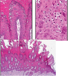
Condylomata acuminata
Histologically: characterized by marked acanthosis with a broad rounded exophytic growth. Surface of the lesion is hyperkeratotic parakeratotic
What is Varicella-zoster virus and what does it cause?
What causes it?
Pathology of Varicella
Pathology of Shingles
DNA Herpesvirus (lipid-enveloped DS) that causes Chickenpox and Shingles
HSV-1 (common in childhood, lips) and HSV-2 (genitalia, sexually transmit)
Varicella spreads through respiratory route. Rash progresses from macules to vesicles to pustules.
Shingles is recurrence of VZV in adulthood. Unilateral dermatomal distribution in thorax and lumbar

Herpes Simplex and VZV show same histologic changes
Acantholysis of epidermis
Multinucleated keratinocytes with intranuclear inclusions (Cowdry Type A inclusions)
Perineurial and intraneurial inflammation
What is a Tzank Smear
Rapid cytological diagnosis done by making a smear from the base of a freshly opened stain ( of HSV) and staining it with Giemsa stain.
Not as sensitive
What is Molluscum contagiosum?
Where is it seen?
Pathology?
Cutaneous infection caused by large brick shaped DNA poxvirus
Children acquire from close contact (eyelids, face, axilla) and immunocompromised patients (HIV). Penis vulva groin as STD
Pathology: Inverted nodule “crater-like”/Eosinophilic cytoplasmic bodies (Molluscum bodies or Henderson-Patterson bodies)

Molluscum Contagiosum
What is Scabies and how is it spread?
How does it present and where and when?

Contagious caused by the mite Sarcpotes Scabiei, transmitted via prolonged direct human contact and rarely by fomites
Presents as extremely pruritic papulovesicular eruption
Fingers/penis/umbilicus/waistband/axilla/hands
Erupts 4 wks after infestation

Scabies
Fertilized female deposits eggs in burrows in the epidermis. Extend at shallow angle through stratum corneium an dmay reach deeper epidermis.
What is tinea/dermatophytosis?
What are the 3 genera?
How does it present?
What test would you use to identify?
What are the different tineas?
Very common superficial cutaneous infections by fungus
Microsprum/Epidermophyton/Trichophyton
Scaly, erythematous plaques, annular
KOH prep rapid test to find branching septate hyphae
Tinea capitis (scalp)
Tinea corporis (trunk/back)
Tinea barbae (beard)
Tinea cruris (groin)
Tinea pedis (feet)

Tinea
Histologically: variable. Presence of sandwich sign (hyphae sandwiched between normal basket weave stratum corneum and a lower layer of stratum corneum with eitehr orthokeratotic or parakeratosis)
PERIODIC ACID SCHIFF (PAS) STAIN CAN REVEAL THE FUNGUS
What is Tinea versicolor?
Where is it common?
What causes it?
How is it presented?
A common superficial fungal infection.
Tropical climates and in summer months
Malassezia globosa
Multiple irregular areas of hypo or hyperpigmentation which are CIRCULAR AND MACULAR

Tinea versicolor
Histologically: Stratum corneum contains round budding yeasts and short septated hyphae imparting spaghetti and meatballs appearance.
Seen clearly in H&E and PAS preparations
What are Seborrheic keratoses?
Where are they commonly found on the body?
What causes it?
What is a condition involving multiple of these and what is it assoc with
Pigmented papules and plaques with “stuck on” appearance (warty)
Common on face, trunk, and upper extremities
Harbor activating mutations in fibroblast growth factor FGF receptor 3
Leser Trelat sign, associated with internal malignancies (stomach cancer)

Seborrheic Keratosis
Histologically: Hyperkeratotic, papillomatous, verrucous epidermis.
Presence of horn and pseudo horn cysts is characteristic
Variable melanin pigmentation accounting for brown coloration
What is Actinic Keratosis?
From what is it thought to arise?
How does it present?
Solar/senile keratosis
A result of chronic sun damage
Presents as Erythematous yellow/brown scaly lesions

Actinic keratinosis
Histoogically: Cytologic atypia of the basal layer of the epidermis. Corneal layer is thickened with retained nuclei (parakeratosis). Dermis contains thickened blue-gray elastic fibers (solar elastosis) the result of chronic sun damage.
What is Squamous Cell Carcinoma?
How does it present?
How do invasive lesions present?
What is the most common cause and other factors?
What % in situ develop invasive component and of these what have metastatic potential?
Common neoplasm in older people
Sharply defined red scaly plaque
Invasive tend to be nodular and may ulcerate
UVB most common. Others: ulcers, old burn scars, HPV, radiation, arsenic, immunosupp
5% invasive component. 30% of these w/metastatic potential

Squamous Cell Carcinoma in Situ
Histologically: Full thickness dysplasia, involving entire epidermis. Disorganization of the epidermal architecture, loss of maturation, and lack of polarity of cells.

Invasive squamous cell carcinoma
What are Keratoacanthomas?
How does it present?
Why is it treated?
Variant of SCC
Solitary flesh colored nodule w/ a CENTRAL KERATIN PLUG
Causes extensive local destruction

Keratoacanthoma
HIstologically: Exophytic lesions w/ invaginating mass of keratinizing well diff squamous epithelium at the sides and bottom o the lesion
Epithelium is characterized by well differentiated pale staining eosinophilic glassy cytoplasm showing striking tendency towards keratinization
What is basal cell carcinoma?
Does it metastasize?
What is it assoc w pathwise?
Presentation?
Most common human cancer secondary to chronic sun exposure. Locally destructive, slow growing tumor rarely metastasizes.
Metastasizes in immunocompromised
Dysreg of sonic hedgehog or PTCH pathway
Pearly papules w/ prominent dilated subepidermal blood vessels (telangiectasia)

Basal Cell Carcinoma
Histologically: characteristic clefting artifact b/w tumor nodules and stroma
What are melanocytic nevi?
What kinds are there?
What kinds are there histologically?
Seen in epidermis
Increase w sun exposure (acquired) and Present at birth (congenital)
Histologically: Junctional/Compound/Intradermal
What is dysplastic nevus syndrome?
How is the familial variant inherited?
What are the differences between Sporadic form and Familial form?
How does it present typewise?
Patients tend to develop large number of dysplastic nevi (80 or more)
Autosomal dominant disease with incomplete penetrance and is assoc w/ mutations of the CDKN2A gene
Sporadic has 2-10 dysplastic moles vs. hundreds for familial
and
lifetime risk of melanoma is 10% in sporadic, 100% in familial
Junctional or compound NOT INTRADERMAL
At what age and body location is Melanoma typically found?
Melanoma risk factors?
Adulthood. Men on back and women on legs
Factors: Sunlight exposure at early age is most important. Fair complexion/older age/dysplastic nevus/history of melanoma/Xeroderma pigmentosum
what are the ABCDEs of melanoma?
What are the growth phases for Melanoma
What is the most important predictor of metastatic disease?
What are the thicknesses that rarely metastasize vs. greater potential to?
Asymmetry/Borders uneven/Color heterogeneity/Diameter >6mm/Evolution or change
Early phase/Radial Growth phase/Vertical Growth phase
Depth of invasion (Breslow thickness) is the most important predictor of metastatic disease
Tumors < 1mm rare… >1.7mm potential
What are other indicators of metastatic potential (other than the most important factor of depth of invasion) for melanoma?
Where can metastases involve?
Ulceration, mitotic rate, angioinvasion
Metastases involve not only regional lymph nodes, but liver lungs brain, any other site.
What types of Melanoma are there?
Superficial spreading type- Most common, commonly in back and extremities
Nodular type- NO radial growth phase directly invading dermis. poor prognosis.
Lentigo maligna type- Commonly in head and neck area.
Acral Lentiginous type-Located on palm sole or beneath nail. Common in African Americans.
What is Mycosis Fungoides
How does it present?
What are the phases it goes through?
What syndrome is assoc?
A skin lymphoma, most common CTCL
Non-specific erythrodermic rash, progressing over time
Phases of patch, plaque, and nodules
Sezary syndrome is the blood involvement of T-cell lymphoma

Mycosis fungoides
What are the different skin conditions seen in diabetes mellitus?
Diabetic dermopathy: Lower legs/Atrophic, pink and hyperpigmented macules and plaques
Bullous diabeticorum: Acral, lower legs and lower arms. Tense blisters.
Necrobiosis lipoidica: Yellow atrophic plaques/shins/telangiectasia/multiple and bilateral/ulcerate esp in males
Acanthosis nigracans: Common in Type II DM. Marker for insulin resistance. Velvety hyperpigmented thickening of the skin. Also seen in other locations and skin tags. (AN 1- Familial/AN 2- Malignancy/AN 3- obesity insulin resistance and endocrinopathy assoc)
Other: Tinea/Candidiasis/Cellulitis/MRSA infections/Neuropathic ulcers
What are characteristics of Hyperthyroidism?
Fine, velvety, smooth skin
Warm and moist due to increased sweating
Hyperpigmentation – localized or generalized
Pruritus
Hair: Fine, thin hair
Mild, diffuse alopecia
Nails: Onycholysis (nail lifts off bed)
What are characteristics of Hypothyroidism?
Dry, rough, coarse skin
Cold and pale skin
Yellow discoloration from carotenemia
Thick scale on feet (keratoderma)
Generalized boggy and edematous skin (myxedema): rare
Hair: Dull, coarse, brittle, slow growing
Alopecia of the lateral third of the eyebrows
Nails: Thin, brittle, slow growing
What do people with Hyperthyroidism AND Grave’s disease have?
Pretibial myxedema:
Cutaneous infiltration of skin of shins with MUCIN
Peau d’orange, skin colored to brown red, firm
Can occur during Grave’s or following treatment of same
What is Addison’s disease?
What are the skin manifestations?
Primary adrenocortical insufficiency (Autoimmune)
Hyperpigmentation (MSH like effect of ACTH)
Striae of nails
Mucosal pigmentation
Loss of ambisexual hair in post pubertal women
Fibrosis and calcification of the cartilage (ear) rare
What is Cushing’s syndrome?
Symptoms?
Overproduction of Cortisol by adrenal gland
Moon facies
Buffalo hump
Truncal obesity
Spindly limbs
STriae distensae
Easy bruisability
Slow wound healing
Acne/hirsutism
What is Lupus Erythematosus?
What are the ARA criteria of SLE (systemic Lupus Erythematosus)? (Skin related and Multisystem)
Multisystem disorder that can have subsets of cutaneous lupus (3)
Malar (cheek) erythema/Discoid (chronic/thick) lesions/Oral ulcers/Photosensitivity
Arthritis/serositis/Nephropathy/CNS disorder/Hematologic disorder/Immunologic
What are the risk factors for Systemic Lupus Erythematosus
Female>male 6:1
Young>old
African american/dark skin>Caucasian
What is the marker for Acute Cutaneous Lupus Erythematosus?
What is the marker for Chronic cutaneous (discoid) Lupus Erythematosus?
What is the marker for Subacute cutaneous Lupus Erythematosus?
Butterfly rash (malar rash). Resolves in quickly in hrs to days w/out scarring. Brought on by sun but not always. Marker for Acute.
Discoid lupus top of head, cheek, and ear has scarring lesions
Polycyclic annular rash on back or arms found in Subacute cutaneous.
What is Neonatal lupus?
Mother’s antibodies cross placenta.
What is dermatomyositis?
What are the markers?
Why is having dermatomyositis concerning?
Inflammation of skin and muscle
Heliotrope rash. violet colored rash of upper eyelids
Photosensitive poikiloderma (Pigment altered skin thing and blood vessels)
Gottron’s papules (Characteristic flattop pink papules over extensor knuckles)
Nailfold capillary changes
Assoc with cancer in adults 10-50% (OVARIAN represented)
What is the skin manifestation of sarcoidosis?
When does cutaneous disease occur in sarcoidosis?
What is the cutaneous presentation of sarcoidosis?
Non-caseating granulomas (not a lot of lymphocytes around it) is characteristic in organs
Onset of disease is when cutaneous occurs
Periorbital granulomatous papules.
Diascopy reveals brow/red color.
Brown red papules and plaque/lesions can be small papules or larger plaques/A great pretender (looks like a lot of things)
What is diascopy?
Use glass slide and press down. Can reveal red brown of sarcoidosis. Can also reveal purpura
What is Lofgren’s syndrome?
A sarcoidosis syndrome charcterized by
Hilar adenopathy
Erythema nodosum
Fever
(Iritis, arthritis)
What is Porphyria Cutanea Tarda?
What does it present with?
Accumulation of porphyrins.
Caused by defective uroporphyrin decarboxylase which breaks down heme groups. Caused by liver disease or autosomal mutation.
Present with vesicles that heal with scarring on sun exposed areas particuarly on hands and face.
Hairgrowth of face
What cutaneous manifestation occurs with IBS?
Pyoderma gangrenosum
a sterile rapid ulceration necrosis of the skin caused by neutrophilic infiltration.
Also assoc with other diseases.
What is Dermatitis Herpetiformis
How does it present and Where does it occur?
Best Treatment?
Consequence of gluten sensitivity. Immune reaction in the small bowel which alters the mucosa, leading to antibody formation, leading to antibodies entering blood and attach to skin. Anti transglutaminase antibodies causing dermatitis herpetiformis
Little vesicles like herpes, super itchy. Presenting on extensor surfaces.
Avoid gluten. AFter 3 mo, ab levesl decrease mucosa of gut normalizes and symptoms of Dermatitis herpetiformis abate. Dapsone is effective treatment for relief of DH itchiness. But does not affect gut or autoantibodies.
What are the cutaneous reaction patterns we talked about?
Urticaria
Exanthematous Drug Eruptions/Morbilliform drug rashes
Drug reaction with Eosinophilia and systemic symptoms (DRESS)
Erythema Multiforme (EM)
Stevens-Johnson Syndrome and Toxic Epidermal Necrolysis
Vasculitis
Leukocytoclastic Vasculitis
Henoch-Schonlein Purpura
How does Urticaria present?
How is it happening?
Cause?
Treatment?
Pink edematous papules and plaques
Lesions are migratory and individual lesions last less than 24 hr
Angioedema and anaphylactic shock are the other forms of anaphylaxis
Caused by IgE mediated immediated hypersensitivity
Most common identified cause is Upper Respiratory Infection
Treat w/ oral antihistamines (nonsedating w/sedating H1 antihistimines)
Exanthematous drug eruptions/Morbiliform drug rashes mediated by?
How does it present?
When does it present?
Treatment?
Type IV (delayed) hypersensitivity
Monomorphic (individual lesions look similar to one another) macules and thin papules start on face and trunk then spread to extremities
2-14 days after drug initiation onset.
Spontaneously resolve over 1-2 wks (treatment is supportive for pruritis with topical corticosteroids and oral antihistamines)
DRESS (drug reaction w/ eosinophilia and systemic symptoms) resembles what but why is it different?
How is it presented?
Most common site of systemic involvement?
Treatment?
Resembles Exanthematous drug eruptions but commonly associated with fever
Facial edema, enlarged lymph nodes, arthralgias may be present
Liver is most common site of systemic involvement.
Drug discontinuation imperative. Systemic corticosteroids required to treat.
Erythema multiforme was similar to what?
Clinical presentation hallmark finding?
Tissue involvement?
Triggers?
Treatment?
Thought to be similar to SJS and TEN but now thought distinct
Target lesions characterized by 3 or more color zones and a dusky red or purple center as hallmar finding.
Ocular oral genital mucosa involvement
Infectious triggers for 90%, drugs 10%
Symptomatic treatment for pruritis or pain sufficient, systemic corticosteroids for severe disease
SJS (Stevens-Johnson syndrome) and Toxic epidermal necrolysis (TEN) presentation?
Difference between the spectrum of SJS/SJS TENOverlap/TEN
Tissue involvement?
Triggers?
Mortality?
Treatment?
Prodrome of fever, malaise, Upper respiratory symptoms before onset of cutaneous lesions.
Painful red patches evolve to bullae and areas of necrosis
Epidermal detachment <10%, 10-30%, >30% body surface area involvement
ANY MUCOSAL SURFACE may be involved (oral, conjuctiva, genital, GI)
Allopurinol, NSAID, sulfa, anticonvulsants, antibiotics implicated.
Mortality primarily from sepsis. Prompt discontinuation required. Support including IV immunoglobulin maybe
What is vasculitis?
Inflammation and destruction of blood vessels.Most cutaneous vasculitis is small vessel vasculitis with inflammation centered around arterioles and venules in the dermis.
LCV (Leukocytoclastic Vasculitis), what is it?
Common causes?
Hallmark cutaneous finding? Where is it?
Difference from urticaria?
Other findings?
Small vessel vasculitis of skin when neutrophils are predominant inflammatory cell seen on biopsy.
Common causes: infections (upper resp./Group A streptococal/hep b/hep c/HIV)/drug hypersensitivity (antibiotics)
Palpable purpura are the hallmark cutaneous finding, more commonly on legs
Lesions last longer than 24 hrs, unlike urticaria
Other findigns: nodules, ulceration, livedo reticularis (purple lacy netlike pattern)
What is Henoch-Schlonlein purpura (HSP)?
Diagnosis?
Triggers?
Hallmark?
Treatment?
Small vessel vasculitis predominantly affecting children
A leukocytoclastic vasculitis seen on biopsy but IgA immune complexes are a more specific finding when direct immunofluorescence is performed.
Triggered by infections, typically group A Streptococcal infection and other upper respiratory infections
Palpable purpra on the skin, especially buttocks and lower extremities are the main physical finding.
Self-limited and resolves over course of 2-4 wks