Neck Flashcards
Describe the surface anatomy assessment in physical exam
- observation and palpation
- normal v. abnormal
What is the most prominent spinous process at the base of the neck?
- C7
What is the keystone of the neck?
- hyoid
Describe how to palpate the hyoid?
- superior to the laryngeal prominence
What is the Adams Apple?
- laryngeal prominence
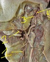
What are the landmarks on the hyoid?
- lesser cornu
- body
- greater cornu
Describe the functional role of cervical fascia
- vertical fascial compartments of neck
- “contain” infection, abscess, pathology
Where is the superfiscial cervical fascia?
- anterolateral cervical subcutaneous tissue
What is contained in the superficial cervical fascia?
- platysma
What are the deep cervical fascia layers?
- investing
- pretracheal
- prevertebral
- carotid sheath
- retropharyngeal space
Which layer of deep cervical fascia is indicated by the RED?
- investing
What is contained in the investing layer?
- SCM
- upper trap
What are the two portions of the pretracheal layer?
- muscular
- visceral
Which layer is indicated by the PURPLE?
- pretracheal - muscular portion
Which layer is indicated by the BLUE?
- pretracheal - visceral portion
What is contained in the muscular portion of the pretracheal layer?
- infrahyoid muscles
- suprahyoid muscles
What is contained in the visceral portion of the pretracheal layer?
- larynx
- trachea
- parhynx
What is indicated by the ORANGE?
- prevertebral layer
What is contained in the prevertebral layer?
- scalenes
- other prevertebral muscles
What is indicated by BROWN?
- carotid sheath
What is contained by the carotid sheath?
- common carotid a. (CCA)
- internal carotid a. (ICA)
- internal jugular v. (IJV)
- CN 10 (vagus n.)
What is the space between the prevertebral fascia and the visceral portion of the pretracheal fascia?
- retropharyngeal space
What is the clinical reasoning of the retropharyngeal space?
- allows for expansion of esophagus when swallowing
- trauma
- infection
What is indicated by GREEN?
- buccopharyngeal fascia
What are the two parts of the thyroid gland?
- R and L lobe
- isthmus
What is the function of the thyroid gland?
- growth and metabolism of human cells
- calcium homeostasis via calcitonin
Where is the thyroid in relationship to other neck muscles?
- deep to infrahyoid m.
Where are the parathyroid glands located?
- two on each lobe, one superior and one inferior
What is the function of the parathyroid gland?
- calcium/phosphate regulation
What are the muscles of the pharynx?
- constrictor pharyngeal (superior, middle, inferior)
- platopharyngeal
What are the regions of the pharynx?
- nasopharynx
- oropharynx
- laryngopharynx
Where is the nasopharynx?
- superior to the soft palate
Where is the oropharynx?
- soft palate to epiglottis
Where is the laryngopharynx?
- epiglottis to cricoid cartilage
What are the regions of the esophagus?
- first third
- middle third
- bottom third
What is the muscle type of the 1st third of the esophagus?
- voluntary, striated
What is the muscle type of the middle third of the esophagus?
- mixed between striated and smooth
What is the muscle type of the bottom third of the esophagus?
- involuntary, smooth
What are the borders of the 1st third of the esophagus?
- oropharynx to superior thoracic cavity
What are the borders of the middle third of the esophagus?
- superior thoracic cavity to diaphram
What are the borders of the bottom esophagus?
- diaphram to stomach
What are the structures in the endocrine viscera?
- tyroid
- parathyroid
What are the structure of the alimentary viscera?
- pharynx
- esophagus
What are the structures in the respirtatory viscera?
- larynx
- trachea
What innervates the larynx?
- CN 10 (vagus n.)
What are the branches of the vagus n. (CN10)?
- superior laryngeal
- internal laryngeal
- external laryngeal
- recurrent laryngeal
- inferior laryngeal
What does the right recurrent laryngeal n. loop under?
- right subclavian a.
What does the left recurrent laryngeal n. loop under?
- aortic arch
What does the inferior laryngeal n. innervate?
- all intrinsic laryngeal muscles EXCEPT CRICOTHYROID M.
What is seen on the c-spine lateral XR view?
- C1-C7
- C1 posterior arch
- C2-C7 SP
What is seen on the AP c-spine XR view?
- SP (not specific levels)
- tracheal shadow
What is the c-spine AP view used for?
- alignment
What is seen on an open mouth c-spine XR view?
- dens
- C2 body
What is seen on the c-spine oblique XR view?
- intervertebral foremen
Identify
- platysma
Identify
- clavicular end of SCM
- sternal end of SCM
Identify
- upper trapezius
Identify
- sternohyoid
Identify
- thyrohyoid
Identify
- superior belly of omohyoid
- inferior belly of omohyoid
Identify
- mylohyoid
Identify
- geniohyoid
Identify
- stylohyoid
Identify
- anterior belly of digastric
- posterior belly of digastric
Identify
- anterior scalenes
- middle scalenes
- posterior scalenes
- brachial plexus
- phrenic n.
Identify
- splenius capitis
Identify
- levator scapula
Identify
- rectus capitis lateralis
- longus colli/capitis
- rectus capitis anterior
Locate the thyroid
Locate the parathyroid glands
Identify
- constrictor
Identify
- platopharyngeal
What is the function of the larynx?
- voice production
Identify
- thyroid notch
Identify
- thyroid cartilage lamina
Identify
- thyrohyoid membrane
Identify
- laryngeal prominence
Identify
- epiglottis
Identify
- cricoid cartilage
What are the 3 primary branches off the aortic arch?
- brachiocephalic trunk
- left common carotid a.
- left subclavian a.
What are the two branches off the brachiocephalic trunk?
- right common carotid a.
- right subclavian a.
What is a key difference when distinguishing between the ECA and the ICA?
- ECA has branches
- ICA has NO branches
Identify
- external carotid a.
- common carotid a.
- internal carotid a.
What is contained in the carotid sheat?
- common carotid a.
- internal carotid a.
- internal jugular v.
- vagus n.
What is the carotid body?
- chemoreceptor
Where is the carotid body?
- CCA bifurcation
What is the carotid sinus?
- baroreceptor
Where is the carotid sinus?
- proximal ICA
Identify
- carotid body
- carotid sinus
What are the 4 branches (out of 8) of the ECA?
- superior thyroid
- facial
- maxillary
- superior temporal
Identify
- superior thyroid
- facial
- CCA
- ICA
- ECA
Identify
- superior thyroid
- facial
- maxillary
- superficial temporal
What is the route of the subclavian a?
- up and over 1st rib
- between anterior and middle scalenes
What are the 3 (out of 4) branches of the subclavian a?
- vertebral a.
- thyrocervical trunk
- costocervical trunk
Identify
- vertebral a.
- thyrocervical trunk
- anterior scalenes
- brachial plexus
- subclavian a.
What is the route of the vertebral a.?
- ascends in c-spine transverse foramen
- enters skul via foramen magnum
What two vessels enter the Circle of Willis?
- ICA
- vertebral a.
What branches off of the thyrocervical trunk?
- inferior thyroid a.
- ascending cervical a.
- transverse cervical a.
- suprascapular a.
Identify
- Thyrocervical trunk
A. inferior thyroid a.
B. ascending cervical a.
C. transverse cervical a.
D. Suprascapular a.
Identify
- thyrocervical trunk
A. inferior thyroid a.
C. transverse cervical a.
D. suprascapular a.
Identify
- costocervical trunk
A. deep cervical a.
platysma inervation
CN 7 - facial n.
SCM attachments
mastoid process to sternum and distal calvicle
SCM action
contralateral rotation and bilateral anterior head translation
SCM innervation
CN 11
upper trap action
contralateral rotation and bilateral anterior head translation
upper trap innervation
CN 11
sternohyoid attachments
sterum to hyoid
sternohyoid action
depresses hyoid
sternohyoid innervation
ansa cervicalis
sternothyroid attchment
sterum to thyroid
sternothyroid action
depresses hyoid
sternothyroid innervation
ansa cervicalis
thyrohyoid attachment
thyroid to hyoid
thyrohyoid action
depresses hyoid
thyrohyoid innervation
ansa cervicalis
omohyoid attachment
scapula to hyoid
omohyoid action
depresses hyoid
omohyoid innervation
ansa cervicalis
What are the two bellies of omohyoid?
- superior
- inferior
mylohyoid attachments
hyoid to mandible
mylohyoid action
elevates hyoid
mylohyoid innervation
CN 5
geniohyoid attachment
myoid to mandible
geniohyoid action
elevates hyoid
geniohyoid innervation
CN 12
stylohyoid attachment
hyoid to styloid process
stylohyoid action
elevates hyoid
stylohyoid innervation
CN 7
digastric attachment
mastoid to hyoid to mandible
digastric action
elevates hyoid
digastric innervation
anterior belly CN 5
posterior belly CN 7
What are the two bellies of digastric?
- anterior
- posterior
anterior scalenes attachment
C2-C7 TP to 1st rib
anterior scalenes action
lateral flexion
middle scalenes attachment
C2-C7 TP to 1st rib
middle scalenes action
lateral flexion
What exits between anterior and middle scalenes?
- brachial plexus
- phrenic n.
- subclavian a.
posterior scalenes attachment
C2-C7 TP to 2nd rib
posterior scalenes action
lateral flexion
longus colli/capitis action
deep neck flexor
Identify
- IJV
- EJV
- anterior jugular v.
Where do the cranial/cervical lymph nodes drain?
- into IJV junction with brachiocephalic v.
- R: right lymphatic duct
- L: thoracic duct
Identify
A. occipital
B. post auricular
C. pre auricular
D. jugulogastric
E. submandibular
F. submental
G. superficial cervical (ant and lat)
H. deep cervical (ant and lat)
I. supraclavicular/transverse cervical
Where does the brachial plexus exit the neck?
- between ant and middle scalenes
What are the three divisions of the cervical plexus?
- cutaneous branches
- phrenic n.
- ansa cervicalis
Identify
A. lesser occipital
B. greater auricular
C. transverse cervical
D. suprascapular
What do the cutaneous branches of the cervical plexus supply?
- sensation to skin of neck/lower head
Identify
A. lesser occipital
B. greater auricular
Identify
C. transverse cervical
D. suprascapular
What does the phrenic n. innervate?
- motor to diaphram
Identify
- phrenic n.
- vagus n.
What level is the phrenic n. from?
- C3-C5
Identify A
A1. superior root of ansa cervicalis
A2. inferior root of ansa cervicalis
What muscles to B and C innervate?
B1. superior and inferior belly
B2. sternohyoid
B3. sternothyroid
C1. thyrohyoid
Identify
A. CN 10 (vagus)
B. CN 11 (accessory)
C. CN 12 (hypoglossal)
What are the borders of the posterior triangle?
- SCM
- upper trap
- clavicle
What are the muscle contents of the posterior triangle?
- inferior omohyoid
- scalenes
- levator scap
- splenius cap
What are the NAV contents of the posterior triangle?
- brachial plexus
- EJV
- transverse cervical n.
- suprascap n.
- subclavian a.
- supraclav LN
- superficial cervical LN
- CN 11
Identify
- posterior triangle
What are the borders of the anterior triangle?
- SCM
- midline
- mandible
What are the sub-divisions of the anterior triangle?
- submandibular
- carotid
- muscular
- submental
Identify
anterior triangle
What are the borders of the submandibular triangle?
- mandible
- anterior digastric
- posterior digastric
What are the contents of the submandibular triangle?
- submandibular gland
- submandibular lymph nodes
- CN 12
Identify
submandibular triangle
What are the borders of the carotid triangle?
- SCM
- posterior digastric
- superior omohyoid
What are the contents of the carotid triangle?
- carotid sheath contents
- CN 11 & 12
- thyroid gland
- larynx
- pharynx
- cervical LN
Identify
carotid triangle
What are the borders of the muscular triangle?
- midline
- SCM
- superior omohyoid
What are the contents of the muscular triangle?
- infrahyoid muscle group
- thryroid
- parathyroid
Identify
muscular triangle
What are the borders of the submental triangle?
- hyoid
- R anterior digastric
- L anterior digastric
What are the contents of the submental triangle?
- submental LN
Identify
submental triangle