Musculoskeletal Anatomy Exam 2 (Final!) Flashcards
Carpal-Metacarpal Joints
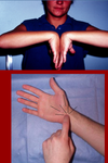
Saddle Joint
Carpal-Metacarpal Joints
nSaddle joint between trapezium & Metacarpal 1
•flexibility for thumb
n
nMetacarpals 4,5 more mobile than 2,3 due to differences in
conformation of carpal articular surfaces

Metacarpal-phalangeal &
Interphalangeal joints
Metacarpal-phalangeal &
Interphalangeal joints
Metacarpal-phalangeal (MCP)
Synovial joints
nCondyloid
nAllows flex-extension & adduction-abduction
n
Interphalangeal (IP)
Synovial joints
nPIPs and DIPs – proximal and distal interphalangeal joints
nBi-condyloid facets that
allow only flexion-extension and little adduction-abduction

Ligaments of fingers
Ligaments of fingers
Palmar:
nThickened anterior portions of the joint capsules of MCP & IP joints
Deep transverse
nInterconnect MCP joints, except thumb
•Allows for greater mobility of thumb

Collateral Ligaments
Collateral Ligaments
nCT bands on each side of joint
nSupport all MCP & IP joints
nAt MCP joints
•Flexion tautens collateral ligaments; this limits adduction-abduction
•Extension relaxes ligaments; this permits adduction-abduction
>>On sides

Palmar Surface of Hand
Palmar surface of hand

Palmar aponeurosis
nProtective covering over tendons
n
Superficial transverse ligament
nAcross palmar side of MCP jts.
Tendon coverings
Tendon coverings
nFibrous flexor sheaths: enclose flexor tendons in fingers
n
nSynovial sheaths around tendons
n
nFlexor retinaculum = transverse carpal ligament
Flex ret - roof of carpal tunnel

Synovial Sheaths
Synovial Sheaths

Synovial membranes are lined with 2 types of synoviocytes
1.Type A are macrophage-like
2.Type B secrete hyaluronic acid, increasing the viscocity
●
Synovial fluid consists of hyluronic acid, lubricin, proteinases and collagenases.
Synovial fluid displays thixotropic characteristics.
In addition to lubrication, synovial fluid also transports oxygen, nutrients, CO2 and waste products.
hypothenar and thenar muscles
hypothenar and thenar muscles
Thenar muscles
Abductor pollicis brevis
Flexor pollicis brevis
Opponens pollicis
All of these muscles originate from the flexor retinaculum, the scaphoid bone and the trapezium bone.
The Flexor pollocis brevis and the Abductor pollicis brevis insert on the proximal phalanx of thumb. The Opponens pollicis inserts onto the 1st metacarpal.
All these muscles are innervated by the median nerve
====
•
•The Opponens digiti minimi and the Flexor digiti minimi originate from the flexor retinaculum and the hook of the hamate. The Abductor digiti minimi originate from the Pisiform bone.
•
•The Abductor digiti minimi and the Flexor digiti minimi both insert onto the proximal phalanx of the 5th digit. The Opponens digiti minimi inserts onto the 5th metacarpal.
•
•All of these muscles are innervated by the Ulnar nerve
Adductor pollicis
adductor pollicis
•Adductor pollicis
•
•Oblique Head originates from the 2nd and 3rd metacarpals and the capitate bone.
•Transverse Head originates from the 3rd metacarpal
•
•Both heads insert onto the proximal phalanx of the thumb
•
•This muscle is innervated by the Ulnar nerve
Movements of the thumb vs. movements of the fingers
Movements of the thumb
nAbduction-adduction are perpendicular to palm
n
nFlexion-extension are parallel to palm
==
Movements of the
Fingers
Abduction - Adduction are referenced from the middle finger

Lumbricals & actions
Lumbricals
nLumbricals originate from tendons of flexor digitorum profundus; insert into dorsal expansions
•Lumbricals 1,2 innervated by median nerve
•Lumbricals 3,4 innervated by ulnar nerve
nFlexor pollicis longus does not have a lumbrical
==
Tata movements
nActions of the Lumbricals (with some help from the interossei:
•Flex MC-P joint
•Extend IP joints
n
n“Tata” movement

The Dorsal and Palmar interossei
The Dorsal and Palmar interossei
Both sets of muscles originate from the metacarpals
Both sets of muscles insert onto bases of proximal phalanges via the Dorsal Expansions
Both sets of muscles are innervated by the Ulnar nerve
====
nDorsal Interossei abduct fingers from midline
n
nPalmar interossei adduct fingers toward midline
•Pad & Dab
•
n1st dorsal interosseus and adductor pollicis both act to clench thumb against index finger for a strong pinching action
n
nLI 4 is located on 1st dorsal interosseus

Importance of finger ligaments
Importance of finger ligaments

Fiberous flexor sheaths act as a pulley system to assist action of flexor muscles/tendons on phalages
Link ligaments provide a connection between flexor tendon sheaths and dorsal expansions to coordinate movements
===
Trigger finger occurs when chronic irritation causes a nodule to form along the flexor tendon. This nodule prevents smooth action of the tendon and may even lock finger in flexed position
Picture: bootinear’s deform
Median nerve
median nerve
Cutaneous
n3 ½ digits and thenar eminence
n
Forearm Muscles
nPronator teres
nPalmaris longus
nFlexor carpi radialis
nFlexor digitorum superficialis and ½ profundus
nPronator quadratus
nFlexor pollicis longus
n
Hand muscles
nThenar muscles - except the adductor pollicis
nLumbricals 1,2
Power grip
nMuscles provide forceful grip
Carpal Tunnel
nSpace formed by flexor retinaculum / transverse carpal ligament spanning across carpal bones.
–Flexor retinaculum lies deep to palmar aponeurosis
n
nStructures in carpal tunnel
–Median nerve
–Flexors digitorum superficialis & profundus
–flexor pollicis longus
–
nStructures NOT in carpal tunnel:
–Ulnar nerve
–Radial and Ulnar arteries
–palmaris longus
–Flexor carpi ulnaris
–Flexor carpi radialis

Carpal tunnel syndrome
carpal tunnel syndrome
nMedian nerve is compressed
nCarpal tunnel size reduced due to
–local inflammation or tenosynovitis
–fluid overload
–dislocation of carpal bones
–arthritisnOften associated with diabetes mellitus which compromises nerve functionnSymptoms include:
–Loss of sensation over median nerve distribution
–Loss of function of thenars and lumbricals 1,2
§Thenar wasting: “ape hand“
==
Carpal Tunnel Syndrome
nCutaneous snesory loss on median nerve distribution
nThenar wasting
nWeakness of lumbricals 1,2

Testing the median nerve
Testing the median nerve
nTinel’s or Phalen’s test
nSensory Testing - tuning fork, two-point discrimination
nStrength Testing - Thenar musculature
nIt is possible to have a negative EMG study and still have Carpal Tunnel Syndrome (CTS)
Ulnar nerve
ulnar nerve

Cutaneous
n1½ digits, ulnar side of palm & dorsum of hand
Forearm Muscles
nFlexor carpi ulnaris
n½ Flexor digitorum profundus
Hand Muscles
nHypothenars
nAdductor pollicis
nLumbricals 3,4
nDorsal & Palmar Interossei
Precision grip
nIntrinsic hand muscles produce fine movements of the fingers
Tunnel of Guyon
Tunnel of Guyon or Ulnar Tunnel
nSpace between pisiform and hook of hamate, interconnected by pisohamate ligament (an extension of flexor carpi ulnaris tendon)
nFloor of tunnel is the flexor retinaculum
nRoof of tunnel is the palmar carpal ligament (extension of forearm deep fascia)
nContents of tunnel
–Ulnar nerve
§Ulnar nerve splits into deep and superficial branches
–Ulnar artery and veins

Ulnar nerve entrapment
Ulnar nerve entrapment
nClaw hand due to lack of MC-P flexion and IP extension in fingers
–Two fingers remain extended because of 2 lumbricals innervated by median nerve
nAtrophy of interossei
nCutaneous sensation loss of 1 ½ digits

Testing Ulnar Nerve
Testing Ulnar Nerve
nTinel’s over the Tunnel of Guyon
nSensation testing over Ulnar nerve distribution
nInstruct Pt to “cross one’s fingers”; this requires intact interossei function.
nEMG and nerve conduction studies

Testing Radial Nerve
Testing Radial Nerve
nWrist and elbow extension
nSensation around dorsum of thumb
nNerve conduction studies

Hypothenar muscles
Adductor pollicis
Adductor pollicis
Oblique Head originates from the 2nd and 3rd metacarpals and the capitate bone.
Transverse Head originates from the 3rd metacarpal
Both heads insert onto the proximal phalanx of the thumb
This muscle is innervated by the Ulnar nerve

Fluid in the Body
Fluid in the body
[*] Approx. 60% water by weight
[*] 2/3 intracellular, 1/3 extracellular
[*] 1/5 of extracellular is plasma
[*] 4/5 of extracellular is interstitial fluid
[*] When replacing fluids, the first 10kg = 4cc/kg/hr, the second 10kg = 2cc/kg/hr, then 1cc/kg/hr thereafter.
EX: 80Kg - 40 + 20, 60 @ 1cc/kg/hr = 120cc normal saline per hour