Musculoskeletal Flashcards
What are the functions of bone?
- STRUCTURE -> give structure and shape to the body
- MECHANICAL -> support and site for muscle attachment
- PROTECTIVE -> vital organs and bone marrow
- METABOLIC -> reserve of calcium (high surface area)
Describe the composition of bone.
o INORGANIC = 65%
- calcium hydroxyapatite (Ca10(PO4)6(OH)2)
- store house for 99% of Ca in the body, 85% of the phosphorous and 65% Na & Mg
o ORGANIC – 35%
- bone cells and protein matrix -> collagen, osteoid
Describe bone geography.

What are the 5 types of anatomical bones?
- flat
- long
- short/cuboid
- irregular (vertebrate)
- sesamoid
What are the 3 major classifications of bone?
- anatomical
- macroscopic
- microscopic
How is macroscopic bone classified?
o CORTICAL/COMPACT
- long bones
- 80% of skeleton
- appendicular
- 80-90% calcified
- mainly mechanical and protective
o TRABECULAR/CANCELLOUS/SPONGY
- vertebrae & pelvis
- 20% of skeleton
- axial
- 15-25% calcified
- mainly metabolic function
- Large surface
What is the classification of bone by microanatomy?
- woven -> immature
- lamallar -> mature
Describe the microanatomy of cortical bone.
- organised into concentric lamellae -> form in response to mechanical forces -> give bone strength in the cortical bone -> can be seen under a microscope due to its striped pattern
- blood vessels travel through and across the bone in Haversian canals -> come into the periosteum and travel up, down and across the bone
What do osteoblasts do?
- build bone by laying down osteoid
What do osteoclasts do?
- multinucleate cells of macrophage family -> resorb or chew bone
What do osteocytes do?
- osteoblast like cells -> sit in lacunae in bones -> look inert (imbedded in matrix)
What is RANK/RANKL?
- a ligand that is responsible for laying down of new bone via differentiation of the osteoclast
- up-regulated in response to stimuli -> infection, trauma etc
What is osteoprotegerin?
- inhibitor of RANk/RANKL -> inhibits osteoclastogenesis
When does osteoprotegerin fall?
- menopause -> oestrogen falls -> OPG falls -> more resorption
When might a bone biopsy be taken?
- evaluate bone pain or tenderness
- investigate an abnormality seen on x-ray
- bone tumour diagnosis (benign vs. malignant)
- determine the cause of an unexplained infection
- evaluation of therapy
Name the 2 types of bone biopsy.
- CLOSED -> needle (Jamshidi needle) -> core biopsy
- OPEN -> for sclerotic or inaccessible lesions -> requires general anaesthetic
What is the common site for bone biopsy?
- transilliac -> good site to get a sample of cortical and cancellous bone
What are the 3 bone labels?
- H&E
- Masson - Goldner Trichrome
- tetracycline
What are the 3 main categories of metabolic bone disease?
- Related to endocrine abnormality (Vitamin D, Parathyroid hormone)
- Non-endocrine (e.g. age-related osteoporosis)
- Disuse osteopenia (results from the reduced use of bones e.g. in injury)
How is osteoporosis definded?
- a patients with a bone mineral density T-score -2.5 or lower when using a DEXA scan
Describe the aetiology of osteoporosis.
- primary = age-related or post-menopausal
- secondary = drugs or systemic disease
What are the two classification of osteoporosis?
- high and low turnover
What is osteomalacia?
- defective mineralisation of normally synthesise bone matrix
- can be due to vitamin D or PO4 deficiency
o called Rickets in children
What are the signs and symptoms of osteomalacia?
- bone pain/tenderness
- fracture and microfracture
- proximal weakness
- bone deformity -> e.g. tibia bowing in rickets
What happens in someone with hyperparathyroidism?
o excess PTH -> bone resorption
- ncreased Ca and PO4 excretion in urine -> calcium hydroxyapatite is broken down into its constituents
- hypercalcaemia
- hypophosphatemia
- skeletal changes of osteitis fibrosa cystica if disease is allowed to progress
What 4 organs are affeced by PTH?
- parathyroid
- bones
- kidneys
- proximal small intestine
What are the causes of hyperparathyroidism?
- primary = parathyroid adenoma (85-90%) or chief cell hyperplasia
- secondary = chronic renal deficiency or vitamin D deficiency
What are the symptoms of hyperparathyroidism?
- stones -> Ca oxalate renal stones
- bones -> osteitis fibrosa cystica, bone resorption
- abdominal groans -> acute pancreatitis
- psychic moans -> psychosis & depression
What bone changes occur in hyperparathyroidism?
- small Brown cell tumours (lytic lesions) -> usually on the radial side of the digits and thumbs
What are the 3 stages of Paget’s Disease?
- osteolytic (osteoclast pre-dominant stage)
- osteolytic-osteosclerotic (osteoblasts will try to build bone)
- quiescent osteosclerotic (there is disorganised, mineralised bone
When does Paget’s disease usually arise?
- over the age of 40
What is the aetiology of Paget’s disease?
o aetiology is unknown but could be:
- familial -> cases show autosomal pattern of inheritance with incomplete penetrance
- mutation 5q35-qter -> sequestosome 1 gene (on the long arm of chromosome 5)
- parvomyxovirus type particles have been seen on EM in Pagetic bone but has been disproven so unlikely
- overuse or previous bone injury
What sites are commonly affects in Paget’s disease?

What are the symptoms of Paget’s Disease?
- often asymptomatic -> found by chance
o pain
o microfractures
o nerve compression (incl. Spinal N and cord)
o skull changes may put medulla at risk
o deafness – AFFECTS region of temporal bone
o haemodynamic changes (sometimes)
o cardiac failure haemodynamic changes, cardiac failure
o hypercalcaemia
o development of sarcoma in area of involvement, 1% (osteosarcoma)
What is osteopenia?
- a T-score of -1.5 to -2.5
What is the FRAX tool?
- a calculation using BMD, age, weight, sex and height to generate a risk score for a particular patient -> Nogg guidelines are then referred to
What are the radiological signs of osteoporosis?
- loss of cortical bone/thinning of cortex (white line around bones on an X-ray - prominent of vertebrate)
- loss of trabeculae
- insufficiency fractures
What are insufficiency fractures?
- stress fractures due to normal stress on abnormal bones -> not limited to osteoporosis
What are the common sites of insufficiency fractures?
- sacrum
- underside of the femoral neck
- vertebral bodies
- pubic rami
What are the signs of insufficiency fractures on imaging?
o X-ray/CT scan
- initially normal (if caught early) -> but can get periosteal reaction and callus
- more commonly seen is increased sclerosis around fracture lines
o MRI
- bone oedema (i.e. low signal on T1, high signal on T2 and STIR)
o Bone scan
- increased osteoblastic activity (i.e. increased uptake)
Describe what is being seen on these scans.
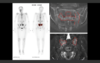
- CT scan = would expect the normal bone to have a similar sort of density -> there would be areas of increased density (SCLEROSIS).
- MRI = same as CT
- bone scan = increased bone turnover in the same area -> commonly seen in the pelvic area (Honda sign) -> also see areas of increased uptake due to a vertebral body fracture
o all this points to an insufficiency fracture and osteoporosis
What is a possible consequence of osteomalacia is it is untreated?
- secondary hyperparathyroidism
What are the radiological signs of osteomalacia?
- radiology of osteomalacia depends on AGE and CLOSURE of the growth plates
- if the patient is an adult with a MATURE skeleton and CLOSED growth plates, they will get: looser’s zones osteopenia, codfish vertebrae and bending deformities
- if the patient is a child (i.e. before growth plate closure), they develop rickets: radiological signs centres mainly to growth plates, changes of osteomalacia
What are Looser’s zones?
o Looser’s zones are pseudofractures at high tensile strength areas -> type of insufficiency fracture
o found at similar areas to other insufficiency fractures:
- medial proximal femur
- lateral scapula
- pubic rami
- posterior proximal ulna
- ribs
o usually, they look less clean than normal insufficiency fractures -> look like short lucent lines with irregular sclerotic margins -> because the bones cannot heal properly
What can be seen on these scans?

o Looser’s Zones
- short, lucent lines with very sclerotic, irregular margins
- in right diagram, the bone is very osteopenic -> the whole femur has bent compared to normal

What is codfish vertebrate?
- bioconcave deformities of vertebral bodies
- seen in osteoporosis and osteomalacia
What can be seen in this scan?

o codfish vertebrae
- bodies should be much squarer
- usually bones are also very osteopenic
What are the features of rickets?
o changes are focused around the growth plate
- indistinct/frayed metaphyseal margin
- widened growth plate without calcification
- cupping/splaying metaphyses due to weight bearing
- enlargement of anterior ribs
- osteopenia

What are the main radiological differences between primary and secondary hyperparathyroidism?
- primary = bone resorption
- secondary = bone resorption and increase density
What is bone resorption?
- where bone as been eroded away
- often centred in spaces that are subperiosteal, subchondral and intracortical
- if they are large, they become brown tumours.
Describe what can be seen in these scans.

- cortical margin looks like it is harder to define on an x-ray
- lucencies in the skull, and there is a lack of crisp cortical edges due to periosteal reactions -> lucencies are areas of intracortical resorption (can be dot like instead of large) – PEPPER POT SKULL
- large lytic bone lesions in patients with primary hyperparathyroidism (brown tumour)
- on the same scan, you’ll probably see bone resorption elsewhere
What are the radiological signs of renal osteodystrophy?
- Looser’s zones
- bending of bones
- osteopenia
- insufficiency fractures at end plates
- subperiosteal erosions and brown tumours
- sclerosis
- soft tissue calcification
- vertrea with increased density at endplates but reduced density in the middle -> called rugger jersey spine
What is renal osteodystrophy?
- a specialised type of primary hyperparathyroidism which can lead to secondary hyperparathyroidism
What is the diagnosis of this patients?

o renal osteodystrophy
- subperiosteal erosions and brown tumours are present
- sclerosis -> vertebral endplates giving a rugger jersey spine
What radiological signs of Paget’s exsist?
- cortical thickening
- bone expansion
- coarsening of trabeculae
- osteolytic
- osteosclerotic
- mixed lesions and osteoporosis circumscripta
What can be seen in this X-ray?
- patient is in the early phase of Paget’s
- fracture at the femoral head, and loosened bones around inferior pubic ramus
- at the same time, there is thickening of the bone cortex and trabeculae
What can be seen in this X-ray?

- this is a patient in a late phase of Paget’s
- left pubic ramus and left pelvic bone are very thick
- trabeculae is a lot coarser compared with the right side.
Explain what disease this patient is suffering from?

- Paget’s
- cortex is very thick -> disease is also affecting the mandible
- multiple lucencies in the skull (osteoporosis circumscripta)
What is a unique feature of Paget’s?
- Paget’s tends to only affect one bone, and doesn’t cross the joint -> might get poly-ostotic Paget’s (multiple bones affected)
- if a disease process affects adjacent bones -> it’s probably not Paget’s
What is rheumatoid arthritis?
- chronic joint inflammation -> specifically inflammation of the synovium (lining of synovial joints) -> SYNOVITIS
- if untreated it causes pain and destruction of the joint
What antibodies are associated with rheumatoid arthritis?
o autoantibodies
- rheumatoid factor
- anti-cyclic citrullinated peptide (CCP)
What is ankylosing spondylitis?
- a seronegative spondyloarthropathy
- is chronic inflammation to the enthesis (where tendons insert into bone) -> ENTHESITIS
- if untreated it leads to pain and spinal fusion and deformities -> exaggerated thoracic kyphosis
What antibodies are associated with ankylosing spondylitis?
- no autoantibodies -> is seronegative
What are seronegative spondyloarthropathies?
o spinal inflammation of the joints without the presense of rheumatoid factor
- ankylosing spondylitis
- reiters syndrome and reactive arthritis
- arthritis associated with psoriasis (psoriatic arthritis)
- arthritis associated with gastrointestinal inflammation (enteropathic synovitis)
What is systemic lupus erythematosus?
- chronic tissue inflammation in the presence of antibodies directed against self antigens -> PATHOGENESIS IS DRIVEN BY AUTOANTIBODIES AND IMMUNE COMPLEXES
- causes multi-site inflammation but particularly the to joints, skin and kidney
What antibodies are associated with systemic lupus erythematosus?
o autoantibodies
- anti-nuclear antibodies
- anti-double stranded DNA antibodies
What HLA molecule is associated with a higher genetic risk of rheumatoid arthritis?
- HLA-DR4
What HLA molecule is associated with a higher genetic risk of systemic lupus erythematosus?
- HLA-DR3
What HLA molecule is associated with a higher genetic risk of ankylosing spondylitis?
- HLA-B27
What class are HLA-A/B/C molecules?
- MHC Class I molecules
What class are HLA-DR molecules?
- MHC Class II molecules
Where are MHC Class I molecules found and what is there role?
expressed on = all nucleated cells
- antigen they present = endogenous -> e.g. viral particles, tumour antigens, self-peptides
- recognised by = CD8+ T cells
- response = cell killing/death
Where are MHC Class II molecules found and what is there role?
expressed on = antigen presenting cells -> e.g. B cells, dendretic cells
- antigen they present = exogenous -> e.g. bacterial particles, self-peptides
- recognised by = CD4+ T cells
- response = antigbody response
Name 4 rheumatoid disease which are do not have an associations with any autoantibodies.
- ankylosing spondylitis
- osteoarthritis
- reactive arthritis
- gout
Describe the 2 main autoantibodies associated with SLE.
o anti-nuclear antibodies -> seen in all SLE cases but isn’t specific for SLE
o anti-double stranded DNA antibodies -> specific for SLE but isn’t always seen -> its serum levels correlates with the disease activity
What is the current theory on SLE pathogenesis?

In sympatomatic lupus, what immune elements would be high and what would be low?
- low = complement levels
- high = serum levels of anti-ds-DNA antibodies
What are the functions of TNF-alpha?
- activate osteoclasts -> bone erosion
- activate synoviocytes -> joint inflammation and swelling (due to increased production of synovial fluid)
- activate chondrocytes -> cartilage degradation
What is the clinical use for anti-TNF-alpha?
- rheumatoid arthritis -> has had a a massively positive effect
What is the general treatment for SLE?
- treatment paradigm for lupus is to attack the tissue inflammation -> target B cells to have an affect on B cell hyper-reactivity is key feature of SLE
Name 2 drugs used to dampen B cell hyperactivity in SLE.
- rituximab -> a chimeric anti-CD20 antibody used to deplete B cells
- belimumab -> a monoclonal antibody against a B cell survival factor call BLYS
What painkillers are used commonly used in rheumatology?
- NSAIDs -> reduce pain, swelling/inflammation but don’t actually prevent joint damage
Define rheumatoid arthritis.
- chronic autoimmune disease characterised by pain, stiffness and symmetrical synovitis (inflammation of the synovial membrane) of synovial (diarthrodial) joints.
What are the key features of rheumatoid arthritis?
o polyarthritis -> swelling of the small joints of the hand and wrists is common
o symmetrical
o early morning stiffness in and around joints is common
o may lead to joint damage and destruction: ‘joint erosions’ on radiographs
o extra-articular disease can occur -> rheumatoid nodules or others which are rarer e.g. vasculitis, episcleritis -> because of rheumatoid factor (autoantibody) forming immune complexes
What is rheumatoid factor?
- IgM autoantibody against the Fc portion of IgG -> hence also called rheumatoid antibody
- can be detected in the blood -> isn’t a test as 1/3 of rheumatoid arthritis is negative
Is rheumatoid arthritis more common in men or females?
- females by 3:1
What is the big environmental factor for rheumatoid arthritis?
- smoking -> contributes to 25%
What joints are most commonly affected in rheumatoid arthritis?
- metacarpophalangeal joint (MCP)
- proximal interphalangeal joint (PIP)
- wrists
- knees
- ankles
- metatarsophalangeal joint (MTP)
What deformities will occur in severe rheumatoid arthritis?
- swan-neck deformity -> hyperextension at the PIP and hyperflexion at the DIP
- Boutonniere deformity -> hyperflexion at the PIP (boutonniere means ‘button-like’)

Define dactylitis.
- swelling of an entire digit
Is dactylitis a sign of rheumatoid arthritis?
- no -> rheumatoid arthritis is just swelling of the joints
What is tenosynovium and how is it linked to rheumatoid arthritis?
- tenosynovium wraps around tendons to allow them to move freely
- if you ask a patient with extensor tenosynovitis to raise fingers à you will see the swelling being pulled back -> confirms that the synovitis is around the tendons and not the joints
- tenosynovitis can damage the tendons and impair their function
What are sometimes found at the ulnar border in patients with rheumatoid arthritis?
- rheumatoid factor can produce immune complexes that can deposit in any tissue -> have a tendency to deposit in subcutaneous tissue and cause extra-articular manifestations -> commonly along the ulnar border of the forearm
- these extra-articular manifestations are relatively rare
- rheumatoid nodules are clinically relevant because if rheumatoid nodules are present, the patient is always rheumatoid factor positive
What extra-articular features are still common and which are uncommon?
o common -> fever (due to abnormal production of cytokines), weight loss and subcutaneous nodules
o uncommon -> vasculitis, ocular inflammation, neuropathies, amyloidosis, lung disease (nodules, fibrosis, pleuritic) and Felty’s syndrome (triad of splenomegaly, leukopenia and rheumatoid arthritis)
- a less common due to the better health of general population -> less smokers mainly
What is Felty’s syndrome?
- triad of splenomegaly, leukopenia and rheumatoid arthritis
What are the radiographic signs of rheumatoid arthritis?
- early -> juxta-articular osteopenia (bones look a little less dense around the joints)
- later -> joint erosions at margins of the joint
- later still -> joint deformity and destruction
Describe the synovial joint.
- the synovium is normally a single type-1 collagen cell lining (can be 1-3 cells deep)
- are macrophages and fibroblasts (which produce synovial fluid) within the synovial lining
- synovial fluid is viscous because it contains a lot of hyaluronic acid
- articular cartilage is made up of type 2 collagen -> main proteoglycan in articular cartilage is aggrecan
What is the general management of rheumatoid arthritis?
- treatment goal is to prevent joint damage
- a multi-disciplinary approach involving physiotherapists, occupational therapists, surgery etc
What are the DMARDs?
- disease-modifying anti-rheumatoid drugs
- often reffered to as steroid sparing agents -> are safer and more effective in the long-term than steroid
When are DMARDs started?
- early on in disease -> joint damage = inflammation x time
- won’t to prevent joint damage
What biological therapies can be used in rheumatoid arthritis?
- inhibition of tumour necrosis factor-alpha (‘anti-TNF’) -> infliximab (an antibody)
- B cell depletion -> rituximab
- modulation of T cell co-stimulation
- inhibition of interleukin-6 -> tocilizumab
What are the downsides to biological therapy?
o increased infection risk -> TNF-alpha is important in granuloma formation -> increased susceptibility to mycobacterial infection e.g. tuberculosis
- all patients must be screened for tuberculosis before starting treatment -> may use prophylactic antibiotics in those at high risk
o B cell depletion therapy can be associated with hepatitis B reactivation, progressive multifocal leukoencephalopathy (PML) and JC virus -> patients scanned for hepatitis B before treatment
o very expensive
What is reactive arthritis?
- sterile inflammation in joints following infection, especially urogenital (e.g. Chlamydia trachomatis) and gastrointestinal (e.g. Salmonella, Shigella, Campylobacter infections) infections
What helps distinguish reactive arthritis from rheumatoid arthritis?
- extra-articular manifestations - particularly:
- enthesopathy (overlap between reactive arthritis and seronegative spondyloarthropathies)
- skin inflammation
- eye inflammation
Reactive arthritis is often the first manifestation of what disease?
- HIV and hepatitis C
Is there a genetic component to reactive arthritis?
- yes -> genetic predisposition could be HLA-B27, and environmental trigger could be salmonella infection
How long does it take for symptoms of reactive arthritis to appear?
- 1-4 weeks after infection
What are the symptoms of reactive arthritis?
o ARTHRITIS -> asymmetrical, oligoarthritis (<5 joints), lower limbs are typically affected
o ENTHESITIS -> are manifestations involving inflammation of the enthesis:
- heel pain (Achilles tendonitis)
- swollen fingers (dactylitis)
- painful feet (metatarsalgia due to plantar fasciitis)
o SPONDYLITIS -> predilection for spinal inflammation:
- sacroiliitis (inflammation of the sacro-iliac joints)
- spondylitis (inflammation of the spine)
What are the extra-articular features of reactive arthritis?
- ocular -> sterile conjunctivitis
- genito-urinary -> sterile urethritis
- skin -> circinate balanitis AND psoriasis-like rash on hands and feet
Fill in this table.


How is the diagnosis for reactive arthritis established?
o clinical diagnosis -> investigations to exclude other causes of arthritis e.g. septic arthritis
- these include:
o microbiological analysis -> microbial cultures and serology e.g. HIV, hepatitis C
o immunological tests -> rheumatoid factor should be negative in reactive arthritis
o synovial fluid examination -> especially if only single joint affected
How is septic arthritis different from reactive arthritis?
- has a positive synovial fluid culture
How is reactive arthritis treated?
o articular -> NSAIDs, intra-articular corticosteroid therapy
o extra-articular -> typically self-limiting, hence symptomatic therapy -> e.g. topical steroids & keratolytic agents in keratoderma
o refractory disease -> oral glucocorticoids or steroid-sparing agents e.g. sulphasalazine
What is osteoarthritis?
- chronic slowly progressive disorder, primarily due to failure of articular cartilage that typically affecting joints of the hand (especially those involved in pinch grip), spine and weight-bearing joints (hips and knees) -> often due to the wear and tear of age
Which joints are most commonly effected in osteoarthritis?
o joints of the hand -> distal interphalangeal joints, proximal interphalangeal joints and first carpometacarpal joint
o spine
o weight-bearing joints of lower limbs -> especially the knees and hips and also the first metatarsophalangeal joint
What are the names of the bony outgrowth in osteoarthritis?
- Heberden’s nodes -> bony, prominent swelling around the distal interphalangeal joints
- Bouchard’s nodes -> bony swellings around the proximal interphalangeal joints
What are the symptoms of osteoarthritis?
- joint pain -> worse with activity, better with rest (loss of articular cartilage -> mechanical failure of joints)
- joint crepitus -> creaking, cracking, grinding sound on moving affected joint
- joint instability
- joint enlargement -> e.g. Heberden’s nodes
- joint stiffness after immobility (‘gelling’)
- limitation of motion
What are the radiological features of osteoarthritis?
- joint space narrowing -> represents the loss of articular cartilage
- subchondral bony sclerosis (underlying bone reacting to damaged articular cartilage)
- osteophytes (Heberden’s and Bouchard’s nodes)
- subchondral cysts
Fill in the table.

What causes osteoarthritis?
- DEFECTIVE and IRREVERSIBLE articular cartilage and DAMAGE to underlying bone
- develops due to either excessive loading on the joints or abnormal joint components
- is a very multifactorial condition -> are some rare metabolic and endocrine factors -> the vast majority of patients have an obvious cause of osteoarthritis: lifestyle activity, or aging
What does the viscousity of synovial fluid depend on?
- hyaluronic acid contained within it
What is the main proteoglycan in articular cartilage?
- aggrecan -> makes up articular cartilage with type 2 collagen
What are the cartilage changes in osteoarthritis?
- reduced proteoglycan
- reduced collagen
- chondrocyte changes -> e.g. apoptosis
- changes are often localised
What changes to the bone occur in osteoarthritis?
o changes in denuded sub-articular bone
- proliferation of superficial osteoblasts results in production of sclerotic bone e.g. subchondral sclerosis
- focal stress on sclerotic bone can result in focal superficial necrosis
o new bone formation at the joint margins (termed osteophytes) -> Heberdens and Bouchards nodes
What is the current management plan for osteoarthritis?
o CURRENTLY NO DISEASE MODIFYING TREATMENTS
- education
- physical therapy -> physiotherapy, hydrotherapy (optimizing physical strength of patient)
- occupational therapy
- weight loss where appropriate
- exercise
- analgesia -> paracetamol, NSAIDs, intra-articular corticosteroid injection
- joint replacement if it comes to it -> has been a MAJOR success
What is SLE?
- a chronic autoimmune disease characterised by relapse and remitting -> affects multiple organ systems including the heart, lungs, kidneys, skin and feotus
What are the risk factors for SLE?
- sex = M:F ratio is 1:9
- age = seen at 15-40 years -> rare to present after menopause
- race = predominantly affects Afro-Caribbeans, Asians and Chinese
- genetics = multiple genes implicated
Describe the pathogenesis of SLE.
- abnormal clearance of apoptotic cell material due to genetic predisposition (HLA-DR3) and an environmental trigger (UV-light, EBV etc.)
- dendretic cell uptake of autoantigens and activation of B cells
- B cell Ig class switching -> IgG autoantiboides
- immune complexes form -> complement activation cytokine generation
- CLINICAL DISEASE ONSET -> IRREVERSIBLE TISSUE DAMAGE
What is the general presentation of SLE patient?
o can be vague initially -> malaise, fatigue, fever, weight loss
- lymphadenopathy is a feature of early, active SLE
- more specific features are butterfly rash (malar rash), alopecia, arthralgia, Raynaud’s syndrome
What is the diagnostic criteria for SLE?
o if a patient has 4 or more of these 11 it points towards lupus
- S-serositis
- O-oral ulcers
- A-arthritis
- P-photosensitivity
- B-Blood (all low)
- R-Renal proteinuria
- I-Immunological -> ANA, anti-dsDNA
- N-neurological-seizures/psychosis
- M-Malar rash
- D-discoid rash
Dose the presence of ANA mean a patient has lupus?
- no -> 5% of teh population has it
- can just be a marker for infection or another autoimmune disease
How is a diagnosis of SLE made after being suspect?
- antinuclear antibodies in the blood -> non-specific, so a positive ANA test is NOT necessarily diagnostic of SLE -> use an indirect immunofluorescence test -> antibodies in serum will bind nuclear antigens and give a pattern of staining -> homogenous pattern suggests SLE
- in order to direct our diagnosis towards SLE, we need to look for more specific anti-nuclear antibodies-> anti-dsDNA antibodies and Sm, anti-Sm antibody, anti-Ro and/or La antibodies
- Useful Laboratory Tests -> increased complement consumption/a reduction in the circulating amount of C3 and C4
- > anti-cardiolipin antibodies, lupus anticoagulant, beta-1 glycoprotein
- > haematology -> lymphopaenia, anaemia (commonly normochromic), keukopenia and thrombocytopenia
- > renal -> proteinuria (using a dipstick sample) and haematuria
What antibody in lupus can cause feotal heart block?
- anti-Ro antibodies -> can cross the placenta
How is the severtiy of SLE assessed?
- identify pattern of organ involvement
- monitor function of affected organ
- identify pattern of autoantibodies expressed
How can SLE be group in term of severity?
o MILD = joint +/- skin involvement
o MODERATE = inflammation of other organs too
o SEVERE = severe inflammation in vital organs -> severe nephritis, CNS disease, pulmonary disease, cardiac involvement, AIHA, thrombocytopenia, TTP
What is the treatment for mild SLE?
- paracetamol or NSAIDs
- hydroxychloroquine if arthtopathy/arthrtitis is a bit worse
- topical corticosteroid cream for rashes
What indicates that SLE is moving from mild to moderate and then to severe?
- failure of the treatment plan for the lesser severity of the disease
- progression to more serious symptoms
What is the treatment for moderate SLE?
- first line treatment of MODERATE SLE is CORTICOSTEROIDS -> start at a HIGH initial dose to suppress disease activity (0.5-1.5 mg/kg/day) -> then give intravenous methylprednisolone (3 x 0.5-1g per 24 hours)
- reduce the dose SLOWLY over 2-3 months, to 10mg/day, and then to 1mg per month
What is the treatment for severe SLE?
- azathioprine -> immunomodulatory therapy (2.5 mg/kg/day)
- FBC & biochemistry monitoring (predominant side effects are on the bone marrow)
- cyclophosphamide is given in severe organ involvement, intravenously pulsed or oral administration -> causes bone marrow suppression, a risk of infertility, and cystitis (acrolein)
What is the prgonosis of SLE?
- 85% 15 year survival in patients without nephritis
- 60% 15 year survival in patients WITH nephritis
- prognosis tends to be worse if black, male and low socio-economic status
What are the 3 main categories of SLE treatment?
- symptomatic
- immune-modulating
- immunosuppresive
What are the 3 major biomarkers of disease activity?
- complement factors -> C3 and C4
- ESR
- anti-dsDNA antibodies
What questions are asked in the GALS examination before the actuall examination begins?
- Have you any pain or stiffness in your muscles, joints or back?
- Can you dress yourself completely without any difficulty?
- Can you walk up and down stairs without any difficulty?
What does GALS stand for?
- G = gait
- A = arms
- L = legs
- S = spine
How is gait examined?
o observe patient walking, turning and walking back
- smoothness and symmetry of leg, pelvis and arm movements
- normal stride length
- ability to turn quickly
How is the spine examined, focusing on the GALS examination?
- Is para-spinal and shoulder girdle muscle bulk symmetrical?
- Is the spine straight?
- Are the iliac crests level?
- Is the gluteal muscle bulk normal?
- Are the popliteal swellings?
- Are the Achilles tendons normal?
- Are there signs of fibromyalgia?
- Are spinal curvatures normal?
- Is lumbar spine and hip flexion normal?
- Is cervical spine normal?
How are arms examined in the GALS examination?
- Look for normal girdle muscle bulk and symmetry
- Look to see if there is full extension at the elbows
- Are shoulder joints normal?
- Examine hands palms down with fingers straight
- Observe supination, pronation, grip and finger movements
- Test for synovitis at the metacarpo-phalangeal joints (MCP joints)
How are the legs examined in GALS examination?
- Look for knee or foot deformity
- Assess flexion of hip and knee
- Look for knee swellings
- Test for synovitis at the metatarso-phalangeal joints (MTP joints)
- Inspect soles of the feet for rashes and/or callosities
Whta happens after the GALS screen?
- locomotor examination -> detailed examination of any abnormal joints identified in the GALS screen
What is the difference between arthritis and arthralgia?
- arthritis = inflammation of a joint
- arthralgia = pain within a joint without demonstartable inflammation by physical examination
What are the 5 signs of inflammation?
- tumor = swelling
- calor - warmth
- rubor = redness
- dolor = tenderness
- functio laesa = loss of function
Describe gout.
- a disease in which tissue deposition of monosodium urate (MSU) crystals occurs as a result of hyperuricaemia and leads to one or more of -> gouty arthritis or tophi (aggregated deposits of MSU in subcutaneous tissue – this is RARE)
- > gouty arthritis commonly affects the metatarsophalangeal joint of the big toe (‘1st MTP joint’) podagra
- symptoms are an abrupt onset, extreme pain, joint redness, warm, swollen and tender
- will resolves spontaneously over 3-10 days
What is enthesopathy?
- pathology at the enthesis -> site where ligament or tendon inserts into bone
- includes plantar fasciitis AND Achilles tendinitis
How are abnormal joints examined?
- inspection -> swelling, redness, deformity
- palpation -> warmth, crepitus, tenderness
- movement -> active, passive, against resistance
- function -> loss of function
What are the signs of irreversible joint damage?
- joint deformity -> mal-alignment of two articulating bones
- crepitus -> audible (crunching sound) and palpable sensation resulting from movement of one roughened surface on another -> classic feature of osteoarthritis e.g. patello-femoral crepitus on flexing the knee
- loss of joint range or abnormal movement
Define dislocation.
- articulating surfaces which are displaced and no longer in contact
Define sublaxation.
- partial dislocation -> not a complete loss of contact
Define varus deformity.
- lower limb deformity whereby distal part is directed towards the midline -> e.g. varus knee with medial compartment osteoarthritis
Define valgus deformity.
- lower limb deformity whereby distal part is directed away from the midline -> e.g. hallux valgus
What are the usualy causes of a mechanical defect of a joint?
- inflmmation, degenerative arthritis, trauma
- often causes painful restriction
Describe the patterns of arthritis.
o number of joints involved -> polyarthritis = > 4 joints, ligoarthritis = 2-4 joints, monoarthritis = single affected joint
o is involvement is symmetrical
o size of the involved joints
o is there axial/spinal involvement
What does bilateral and symmetrical involvement of large and small joints typically suggest?
- rheumatoid arthritis
What does lower limb asymmetrical oligo-arthritis and axial involvement typically suggest?
- reactive arthritis
What does exclusive inflammation of the first metatarsophalangeal joint suggest?
- gout
How can the neutrophil content of synovial fluid be an important diagnostic marker?

What is Sjogren’s syndrome?
- a connective tissue disease in which there is destruction of exocrine glands (autoimmune exocrinopathy), lymphocytic infiltration of exocrine glands and sometimes other organs (extra-glandular involvement)
- typically diagnosed in middle-aged female (F:M = 9:1)
- symtpoms include dry eyes, dry mouth and parotid gland enlargement
What is systemic sclerosis?
- a disorder that attacks the skin and causes dermal fibrosis (VERY RARE)
- thickened skin with Raynaud’s phenomenon
- dermal fibrosis, cutaneous calcinosis and telangiectasia
