MSK Flashcards
Physiology of the Physis
What is the other common name for physis?
How many histological zones are in the physis?
What is the zone closest to the metaphysis called? What happens there?
What does that lead to?
Difference between adult and pediatric injuries?
- The physis, commonly known as the growth plate, consists of four histological zones of cartilage arranged in layers.
- The zone closest to the metaphysis is the zone of provisional calcification, composed of chondrocytes that undergo apoptosis after preparing the matrix for calcification.
- New bone subsequently forms along the scaffolding formed by these chondrocytes, leading to longitudinal growth.
- The unfused physis is the weakest part of the developing skeleton. An injury that can
cause a ligament sprain in an adult may result in physeal fracture in a child.
Salter-Harris Classification
What is it based on?
Which classes have a greater chance of growth disturbance?
Mnemonic?
- The Salter-Harris - SH system classifies Salter-Harris fractures based on the involvement of physis and adjacent epiphysis and metaphysis.
- In general, the higher the classification, the greater the chance of growth disturbance.
- MNEMONIC: SALRT (picture the distal tibia physis)
- Slip
- Above
- Lower
- Through
- Ruined or Rammed

Salter-Harris Type I Injury
Radiographic appearance?
Examples?
- Type I: Injury limited to the physis.
- SH I injuries are often radiographically occult if the epiphysis is not displaced. The physis may be asymmetrically widened. In the absence of physeal widening, the diagnosis of a SH I injury can be suggested based on soft-tissue swelling around the physis.
- Examples: slipped capital femoral epiphysis and gymnast’s wrist (physeal widening caused by chronic stress on the wrist, which may mimic rickets).
Salter-Harris Type II Injury
Describe this injury and mnemonic
- Type II: Fracture extends to metaphysis.
- Mnemonic: Above the distal tibial physis.

Salter-Harris Type III Injury
Describe this injury.
Provide an example.
Mnemonic?
- Type III: Fracture extends to epiphysis.
- Example: Juvenile tillaux fracture, a distal tibial epiphyseal fracture.
- Mnemonic: Lower (picture the distal tibial physis, which is also the example above!)
Salter-Harris Type IV Injury
Describe this fracture.
Provide example.
Mnemonic?
- Type IV: Fracture goes through metaphysis, physis, and epiphysis.
- Example: triplane fracture, comprised of 3 fractures of the distal tibia: oblique fracture through the metaphysis, a vertical fracture through the epiphysis, and horizontal fracture through the physis.
- Mnemonic: Through (picture distal tibial physis fracture, which is also the example above!)

Salter-Harris Type V Injury
Describe this injury.
Mnemonic?
- Type V: Physis is crushed.
- Mnemonic: SALTR (picture distal tibial physis fx)
- Slip
- Above
- Lower
- Through
- Ruined or Rammed
Pediatric Ossification Centers
What are they and in what order do they ossify?
- Six separate ossification centers, each appearing at different stages of development, make the evaluation of the pediatric elbow challenging. However, the cartilaginous ossification centers always ossify in the same order, which can be remembered with the mnemonic CRITOE:
- Capitellum (typically ossifies between 6 months and 1 year of age).
- Radial head.
- Internal (medial) epicondyle.
- Trochlea.
- Olecranon.
- External (lateral) epicondyle (typically ossifies between 10 and 14 years of age).
What is pathognomonic for an elbow fracture, even if no fracture is visible?
- An elbow effusion, as evidenced by displacement of the anterior and/or posterior fat pads (posterior more sensitive), is pathognomonic of a fracture, even if no fracture is visible.

How do you evaluate elbow alignment?
Describe the two lines used to evaluate elbow alignment and what does abormal measurements in these two lines specifically suggest?
- The elbow alignment is evaluated with the anterior humeral and radiocapitellar lines.
- Anterior humeral line: drawn along the anterior humeral cortex, the anterior humeral line should pass through the middle of the ossified capitellum on the lateral view. If abnormal this suggests supracondylar fracture.
- Radiocapitellar line: drawn through the radial shaft, the radiocapitellar line should pass through the capitellum on all views. If abnormal, suggests elbow dislocation.

What is the most common pediatric elbow fracture?
Second most common?
What is the most common adult elbow fracture?
- Supracondylar fracture is the most common pediatric elbow fracture.
- Lateral condyle fracture is the second most common fracture.
- In contrast, the most common adult elbow fracture is of the radial head.
Toddler’s Fracture?
What is it? Mechanism of injury?
Radiographic appearance?
Nondisplaced fractures of what other bones may appear similar?
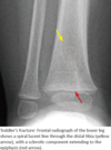
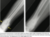
- Toddler’s fracture is a nondisplaced spiral fracture through the tibial** **metadiaphysis, caused by a rotational force to the leg.
- Toddler’s fracture may be clinically difficult to diagnose.
- Radiograph shows a hairline spiral lucency through the distal tibia.
- Nondisplaced fractures of the cuboid and calcaneus may appear similar, with a faint sclerotic band sometimes the only sign of a fracture.
Buckle Fractures
What are they?
Subtle imaging appearance?
How should the cortex normally appear?
- Buckle fractures are unique to the pediatric skeleton, representing a buckling of cortex rather than a true break.
- Buckle fractures can be subtle on imaging and sometimes are only detected by careful inspection of the cortex. Normally, the cortex should be so smooth that a virtual marble can roll down the cortex without bouncing off. Any focal cortical irregularity in a child should raise concern for a buckle fracture.

Pelvic Apophyseal Avulsion Injuries
What is an apophysis?
How many pelvic apophyses are there? how about proximal femoral apophysis?
When do they ossify and when do they fuse?
Why are they susceptible to avulsion? These injuries are most commonly seen in what population and why?
What do acute injuries look like? How about subacute?
Contrast lesser trochanter fractures in adolescents vs adults.
What are all the apophysis and what muscle insert and originate from each one?
- An apophysis is a growth plate that does not contribute to longitudinal growth.
- There are five pelvic apophyses and two proximal femoral apophyses, which arise in puberty and fuse by the third decade. The apophyses are the weakest link of the myotendinous unit. Pelvic apophyses close relatively late in skeletal development and are therefore susceptible to avulsion. Apophyseal avulsion fractures occur most commonly in athletic adolescents, who have strong muscles and open apophyses.
- Acute injuries appear as an avulsed bone fragment. Subacute avulsions are more complex appearing, as the donor site may undergo mixed lytic and sclerotic change in an attempted reparative response.
- Avulsion injuries may be subtle as they are Salter-Harris I equivalent fractures. There is often minimal or no displacement of the otherwise normal-appearing apophyseal ossification.
- Although a lesser trochanter avulsion may occur in an athletic adolescent due to avulsion of the iliopsoas insertion, in an adult a lesser trochanteric fracture is suspicious for a pathologic fracture.

Overview of Imaging for Child Abuse
If there is clinical or radiographic concern for child abuse what imaging needs to be done?
Bone scintigraphy is more sensitive for what type of fractures? What are the shortcomings of bone scintigraphy?
How do you estimate the age of fractures?
- If there is a clinical or radiographic concern for child abuse, a complete skeletal survey using high-resolution bone technique (for children under 2 years of age) should be performed, including frontal views of all long bones, rib views with obliques, skull, pelvis, hands/feet, and entire spine. The images are reviewed by the radiologist while the child is still in the department.
- Bone scintigraphy is more sensitive for posterior rib fractures, but has higher radiation dose than radiography, is insensitive for skull fractures, and cannot evaluate fracture morphology for age__.
- The age of fractures is estimated by the presence of callus formation. In general, a fracture is less than two weeks old if there is no callus, and at least one week old if there is callus.
- These estimates are rough guidelines. A younger child will form callus and heal more quickly.
What are the highly specific fractures for non-accidental trauma?
Are they the most common fractures?
- These fractures are highly specific for child abuse but are not always seen. If any of heset fractures are seen, immediate concern must be raised for child abuse.
-
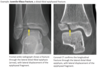
Classic metaphyseal lesion (also called metaphyseal corner fracture or bucket handle fracture) is highly specific for abuse, and is a circumferential fracture through the peripheral spongiosa bone of the distal metaphysis, most commonly around the knee or ankle. A classic metaphyseal lesion is thought to be due to violent shaking.
- The classic metaphyseal lesion typically heals quickly (within 10 days without callus formation, so prompt radiography is essential for diagnosis.
-
Posterior rib fractures are also highly specific for abuse but may be very difficult to diagnose when acute. A repeat chest radiograph with bone technique can be performed in the acute setting, or follow-up radiograph after one week to view interval callus formation.
- Unlike in adults, rib fractures are not typically seen after CPR in children. In the rare case of rib fractures from CPR, fractures tend to be anterior and lateral, not posterior.
- Scapula fracture.
- Sternum fracture.
- Spinous process fracture.
-
Classic metaphyseal lesion (also called metaphyseal corner fracture or bucket handle fracture) is highly specific for abuse, and is a circumferential fracture through the peripheral spongiosa bone of the distal metaphysis, most commonly around the knee or ankle. A classic metaphyseal lesion is thought to be due to violent shaking.
What are the suspicious (but no highly specific) fractures that suggest non-accidental trauma?
- These fractures should raise suspicion for child abuse, but are not highly specific.
- More than one fracture.
- Fracture out of proportion to the history.
- Digital fracture in infants.
- Long bone fracture in non-ambulatory child.
- Complex skull fracture.
What are the nonspecific (but frequently seen) fractures suggestive of non-accidental trauma?
- These fractures are commonly seen, both in the settng of child abuse and accidental trauma, but are not specific for child abuse when seen in isolation.
- Linear skull fracture.
- Long bone fracture in an ambulatory child.
Overview of Skeletal Dysplasia
What are bone dysplasias?
What are several terms used to describe abnormal limb growth?
- Bone dysplasias are characterized by impairment of normal growth of bone yielding an abnormal skeleton. The impairment can involve growth slowing down, speeding up, or can cause the bones to grow in an unusual manner.
- Several terms used to describe abnormal limb growth include:
- Rhizomelia: Proximal limb shortening (e.g., humerus is too short).
- Mesomelia: middle limb shortening (e.g., radius is too short).
- Acromelia: distal limb shortening (e.g., hand or wrist is too short).
- Micromelia: Entire limb is shortened.
- Amelia: Limb is absent.
Achondroplasia
What is it?
Affected individuals have what kind of intelligence?
What are the key radiographic findings?
- Achondroplasia is the most common cause of dwarfism.
- Affected individuals have normal intelligence.
- Key radiographic findings include the abnormal morphology of the lumbosacral spine and pelvis:
- Narrowing of interpedicular distances in the lower spine causes spinal stenosis that is compensated for by a characteristic lumbar lordosis as the child learns to walk.
- Posterior scalloping of vertebral bodies.
- Characteristic tombstone iliac wings.
- Flat acetabula, with short femoral necks.
- Other findings include frontal bossing of the skull.

Thanatophoic Dysplasia
What is it?
Inheritance pattern?
Key radiographic findings?
- Thanatophoric is most common lethal skeletal dysplasia.
- It is inherited in an autosomal dominant manner.
- Key radiographic findings are focused on the vertebral bodies and femur:
- Flattening of vertebral bodies (platyspondyly), causing H-shaped vertebral bodies with diffuse narrowing of interpedicular distance.
- Curved telephone receiver femurs.
Osteogenesis Imperfecta
What is it? Which subtype is lethal?
Key radiographic findings?
DDx for multiple fxs?
Contrast DDx to OI
- Osteogenesis imperfecta (OI) represents a varied spectrum of disorders due to abnormal type I collagen.
- OI type II is lethal.
- Key radiographic findings include multiple fractures in the ribs (causing characteristic accordion ribs), vertebral bodies, and long bones. Additional findings include:
- Bowed long bones.
- Osteopenia.
- Wormian bones in the skull, which are secondary to non-coalesced ossification centers in the skull.
- The differential diagnosis for multiple fractures includes OI, rickets and child abuse. of these three, OI is the only entity to cause antenatal fractures.
- With rickets, one would not expect antenatal injury. Fractures and osteopenia may be present.
- With child abuse, one would not expect osteopenia or antenatal injury. Fractures may be present.
Asphyxiating thoracic dystrophy (Jeune syndrome)
What is it? What happens to the lungs?
In addition to main finding, what other bony findings are seen?
- Asphyxiating thoracic dystrophy is an autosomal recessive disorder of a congenitally small thorax causing respiratory distress.
- In addition to the small thorax and associated pulmonary dysplasia, bony findings include:
- Short ribs that are bulbous anteriorly.
- High-riding handlebar clavicle.
- Trident acetabulum.
Cleidocranial Dysostosis
What is it?
Affected patients have what kind of intelligence?
What is this strongly associated with?
Wormian bones are seen in what other entities?
Additional radiographic findings?
- Cleidocranial dysostosis is a skeletal dysplasia characterized primarily by abnormalities in the clavicles.
- Affected patients are of normal intelligence but tend to be short.
- The key radiographic finding is complete or partial absence of the clavicles.
- Cleidocranial dysostosis is strongly associated with wormian bones in the skull.
- Wormian bones are nonspecific, and are also seen in hypothyroidism, osteogenesis imperfecta, healing rickets, and down syndrome.
- Additional radiographic findings of cleidocranial dysostosis include:
- Delayed ossification of the skull (neonatal).
- Widened pubic symphysis (typically not seen until later in life).

Stippled Epiphysis
What skeletal dysplasias and metabolic disease can cause this radiographic finding?
- Several skeletal dysplasias and metabolic bone diseases can cause stippling of the epiphyses, including:
- Chondrodysplasia punctata, a short-limbed dwarfism (usually rhizomelic).
- Multiple epiphyseal dysplasia (Fairbank disease), a mildly short-limbed, autosomal dominant skeletal dysplasia that usually manifests in late childhood or adolescence.
- Hypothyroidism.
- Complications of maternal warfarin use in a newborn.
Enchondromatosis
What are these characterized by?
What are these syndromes associated with?
What are the two syndromes? What’s the difference between the two?
- The enchondromatoses are characterized by multiple intra-osseous benign cartilaginous tumors in an asymmetric distribution.
- These syndromes are associated with an increased risk of malignant transformation to chondrosarcoma (Maffucci > Ollier).
- Ollier disease is an enchondromatosis without associated abnormalities.
- Maffucci syndrome is an enchondromatosis with venous malformations, which cause phleboliths that are evident on radiography.
Multiple Hereditary Exostosis (Osteochondromatosis)
What is this?
Complications?
Contrast to enchondromatosis
- Multiple hereditary exostoses is an autosomal dominant disorder of multiple benign osteochondromas (also called exostoses) growing from the metaphyses of long bones.
- Complications include pain, deformity, and malignant transformation into low-grade chondrosarcoma (between 5 and 25% of cases).
- In contrast to enchondromatosis, skeletal involvement is usually symmetric bilaterally.
Mucopolysaccharidosis
What are these? What are three examples?
Key radiographic findings?
Hurlers syndrome features what?
Morquio syndrome features what? Other associations with Morquio?
- Mucopolysaccharidoses are a group of lysosomal storage disorders including Hurlers, Morquio, and Hunters. These disorders share common radiographic findings and are typically distinguished clinically and biochemically.
- Key radiographic findings include anterior vertebral body beaking, thickened ribs, and undertubulated bones. Other findings include:
-
Madelung deformity of the wrists (wedge-shaped proximal carpal row, with radius shifted towards ulna).
- Mnemonic: Mucopolysaccharidosis - Madelung
- Thickened calvarium with a J-shaped sella.
-
Madelung deformity of the wrists (wedge-shaped proximal carpal row, with radius shifted towards ulna).
- Hurlers: Hurlers features anterior beaking of the vertebral bodies, primarily inferiorly.
- Morquio: Morquio also features anterior beaking of the middle portion of the vertebral body
- Mnemonic: Morquio/Middle.
- Morquio is associated with spinal stenosis and atlantoaxial instability.
Septic Hip Arthritis
Why is this so important to diagnose? What should be done for any pediatric hip effusion?
Primary DDx consideration?
Less common cause of hip effusion?
Septic hip is usually secondary to what? What is the most common etiology?
Plain film findings?
Test of choice?
- Infection of the synovium and joint space is an orthopedic emergency as joint destruction and growth arrest may occur without prompt washout and antibiotics.
- Any hip effusion must be urgently aspirated to evaluate for septic arthritis.
- The primary differential consideration for septic arthritis is aseptic toxic synovitis, which is a self-limited noninfectious diagnosis of exclusion.
- Less common causes of hip effusion and limp include hemarthrosis in trauma or hemophilia (history is usually evident).
- Septic arthritis is usually secondary to hematogenously seeded metaphyseal osteomyelitis that breaks through the periosteum to infect the joint capsule. S. aureus is the most common cause.
- Plain film findings are not sensitive or reliable for evaluation of effusion, but include:
- Displacement or distortion of gluteal or psoas fat planes. The gluteus medius and minimus insert on the greater trochanter. The psoas inserts on the lesser trochanter.
- Widening of the teardrop distance (space between the lateral margin of the pelvic teardrop and the medial margin of the femoral head). The teardrop is the antero-inferior acetabulum.
- Ultrasound is the imaging modality of choice. Both sides should be compared for symmetry.

Slipped Capital Femoral Epiphysis
What type of fracture is this?
In what population is this seen?
What are the radiographic findings?
What view is best to evaluate SCFE?
What should always be done in these cases?
- Slipped capital femoral epiphysis - SCFE is a Salter I fracture of the proximal femoral epiphysis (displacement of the epiphysis from the metaphysis) seen in obese preadolescents (most affected children are between 10 and 16 years old).
- SCFE typically affects children slightly older than those with Legg-Calvé-Perthes disease.
- Initial findings on an AP view can be subtle, including the asymmetric widening of the proximal femoral growth plate and lack of intersection of Klein’s line with the femoral head.
- Although Klein’s line is drawn on the AP view, the physeal widening and displacement is best evaluated on the frog-leg lateral projection.
- The contralateral hip should always be carefully evaluated. SCFE may be bilateral but is usually asymmetric.

Legg–Calvé–Perthes (LCP) Disease
What is this disease?
Etiology? In what population does it occur?
Is this usually unilateral or bilateral? What should be considered in bilateral cases?
Findings in early LCP?
Findings in late LCP?
- LCP disease is avascular necrosis of the capital femoral epiphysis ossification center.
- LCP is of unknown etiology and typically occurs in children between the ages of 4 and 8 years.
- LCP is usually unilateral. A systemic cause should be sought when bilateral, such as sickle cell disease or steroids.
- Imaging findings are dependent on stage and chronicity.
- Early LCP can be subtle, and often multiple modalities are used for initial diagnosis.
- Late LCP shows secondary signs of osteonecrosis. The femoral head becomes flattened and distorted, with secondary changes of osteoarthritis. Bone scan of late LCP typically shows increased uptake due to attempted repair.

Osteosarcoma
Prevalence? Which subtype represents 75% of all osteosarcoma?
Most common location?
Characteristic radiographic appearance?
- Osteosarcoma is the most common primary pediatric bone tumor. There are over 10 types of osteosarcoma, with the most common subtype the conventional intramedullary type, representing 75% of all osteosarcomas.
- Conventional osteosarcoma is seen most commonly about the knee in the distal femur or proximal tibia.
- The characteristic radiographic appearance of conventional osteosarcoma is a destructive lesion often invading the cortex, with an extensive osteoid matrix.
Ewing Sarcoma
Prevalence? What is it?
Most common location? Second most common?
The most common site of metastasis?
Characteristic radiologic appearance?
- Ewing sarcoma is the second most common primary pediatric bone tumor. It is an aggressive, small round blue cell tumor of neuroectodermal differentiation.
- Ewing sarcoma most commonly arises from the femoral diaphysis, followed by the flat bones of the pelvis. Ewing may also develop in the tibia, humerus, and ribs.
- The lung is the most common site of metastasis.
- The characteristic radiographic appearance is a permeative lesion in the medullary cavity with a wide zone of transition and associated aggressive lamellated onion- skinning or spiculated periosteal reaction.
- Ewing often causes a soft-tissue mass, which can be difficult to see by radiography due to lack of ossification.
Osseus Langerhans Cell Histiocytosis
What is it?
What are Langerhans cells? What characteristic findings are seen histopathologically?
What are the clinical subtybes of LCH? Describe them.
What is the most important factor to determine in this disease?
Which subtype is relatively common? How may it present? What age group does it affect?
Appearance?
What DDx should be considered in lytic lesions in any patient < 30 yo.
- Langerhans cell histiocytosis - LCH is an abnormal proliferation of Langerhans cells with a variety of clinical manifestations, ranging from an isolated lytic bony lesion to fulminant systemic disease.
- Langerhans cells are dendritic cells (histiocytes) that normally live in the epidermis and lymph nodes, where they act as antigen-presenting cells.
- Histopathologically, Birbeck bodies are seen on electron microscopy within the histiocytes.
- Clinical subtypes of LCH have varied presentations.
- Eosinophilic granuloma (Osseous LCH) features skeletal involvement only. It may be mono- or polyostotic. Despite the name, the involvement of eosinophils is variable.
- Hand-Schüller-Christian (multifocal, unisystem) is a clinical triad of pituitary hypophysitis (causing diabetes insipidus), exophthalmos, and lytic bone lesions (typically affecting the skull).
- Letterer-Siwe (multifocal, multisystem) is a fulminant disease with multisystem involvement, primarily seen in young infants and toddlers under age 2. Prognosis is poor.
- Pulmonary LCH (PLCH) is a disease of adult smokers, discussed in the thoracic imaging section. Osseous LCH is occasionally seen in conjunction with PLCH.
- In clinical practice, the most important factor is thought to be determination of unifocal or multifocal disease rather than the classification above.
- The eosinophilic granuloma variant of LCH is relatively common and may present clinically with pain, tenderness, and fever, often mimicking osteomyelitis. Children between ages 5 and 15 are typically affected.
- The appearance of the affected bone depends on the bone involved:
- Skull: Beveled edge lytic lesion.
- Flat bones: (e.g., pelvis): Hole within a hole lytic lesion.
- Long bone: (diaphysis most common, but may occur anywhere including rarely the epiphysis): Permeative destruction, with later lytic lesion and faint rim of sclerosis.
- Spine: Complete vertebral body collapse (vertebra plana), as pictured.
- Maxilla: Floating teeth.
- Along with infection, eosinophilic granuloma should be considered in the differential diagnosis in any patient under 30 with a lytic lesion anywhere.

Pediatric Osteomyelitis
Early osteomyelitis can require what kind of radiologic exams?
How does osteo occur in children? What is the most common site of initial healing?
Can infection cross the physis in infants? How about in children? Why?
Most common organism? How about in sickle cell dz?
How does the initial hematogenous infection appear on imaging and how does it progress?
How long does it take for findings to be evident on radiography? What is the initial finding? Later findings?
What is more sensitive for the detection of early osteo?
- The initial findings of osteomyelitis are often subtle on radiography and may require a combination of plain radiographs, MRI, and scintigraphy for accurate early diagnosis.
- Osteomyelitis in children is most commonly hematogenous, with the metaphyseal marrow the most common site of initial seeding.
- In infants, the epiphysis receives blood supply from transphyseal vessels arising from metaphysis__, so infection can cross the physis. In older children, capillaries do not cross the physis, so transphyseal extension of a metaphyseal infection is uncommon.
- The most common organism implicated in hematogenous osteomyelitis is S. aureus, with Salmonella seen in sickle cell patients.
- The imaging findings on radiography, MRI, and scintigraphy follow the anatomic progression of infection. The initial hematogenous infection is intramedullary and then subsequently spreads through the cortex and uplifts the periosteum. The pediatric periosteum is only loosely adherent to bone, and the purulent infection is able to dramatically uplift the periosteum, causing a prominent periosteal reaction.
- It usually takes 10-15 days for findings to become evident on radiography. The initial finding may be focal osteopenia due to hyperemia. Subsequently, a lucent, often aggressive-appearing medullary lesion can erode through the cortex and cause a periosteal reaction. MRI and scintigraphy are much more sensitive for the detection of early osteomyelitis.

Spinal Osteomyelitis/Diskitis
Compare diskitis/osteomyelitis in children vs adults.
Typical clinical history of discitis?
Most common location of discitis?
Less common location of discitis in pre-teen children?
Initial radiographic findings? When should you get an MRI?
MRI appearance?
- Isolated discitis is unique to children, due to the presence of blood vessels directly feeding the intervertebral disk. In adults, the infection can spread through the disk, but in children infection begins in the disk__.
- A typical clinical history of discitis is a young child up to 4 years old with preceding upper respiratory tract infection and back pain or refusal to sit.
- Discitis most commonly occurs in the lumbar spine in young children.
- Less commonly, discitis may occur in pre-teen children in the thoracic spine.
- The initial radiographic findings are disk space narrowing and vertebral end plate irregularity. These findings may be subtle. Plain films may also be normal. Because persistent back pain is never normal in children, further evaluation with MRI is recommended if clinical suspicion for discitis is high.
- MRI shows narrowing of the disk space, with bone marrow edema of two adjacent vertebral bodies. The affected vertebral bodies may enhance, and enhancement and edema may extend into the soft tissues and epidural space.

Chronic Recurrent Multifocal Osteomyelitis
What is it?
Where does it tend to occur?
Clinical outcomes of CRMO?
What is the key imaging findings?
Compare to infectious osteomyelitis
What syndrome is CRMO associated with?
- Chronic recurrent multifocal osteomyelitis - CRMO is a nonpyogenic, inflammatory disorder that can mimic osteomyelitis.
- It tends to occur in lower extremity long bones.
- CRMO is typically a self-limited diagnosis. A biopsy may need to be performed to exclude pyogenic infection.
- The key imaging finding is migratory lytic and sclerotic lesions in time and space. CRMO may be indistinguishable from pyogenic osteomyelitis on radiographs and MRI. Unlike infectious osteomyelitis, CRMO does not feature soft tissue abscess, bony sequestra, or fistula.
- CRMO is associated with SAPHO syndrome, characterized by:
- Synovitis
- Acne
- Pustulosis
- Hyperostosis
- Osteitis
What is the SAPHO syndrome?
Radiographic appearance? Most common location of involvement?
- SAPHO syndrome, characterized by:
- Synovitis: Anterior chest wall, unilateral sacroiliitis
- Acne: Hydradenitis suppurativa; acne conglobata
- Pustulosis: Palmoplantar pustulosis (50%)
- Hyperostosis: Enthesopathy, sclerosis
- Osteitis: Inflammatory changes, pain
- Radiographic appearance:
- Sternoclavicular joint: most common location of involvement, with osteitis and hyperostosis.
- Sacroiliitis osteosclerosis of vertebral bodies
- Long bones: metaphyseal osteosclerosis and osteolysis.
Leukemia
Prevalence?
Bony changes can be seen in what percent of cases?
What is one of the most distinctive radiographic feature of leukemia? Etiology for this finding? DDx for this finding?
What are other osseous findings?
- Leukemia is the most common pediatric malignancy, with bony changes seen in >50% of patients.
- Imaging findings can be varied. One of the most distinctive radiographic features of leukemia is the metaphyseal lucent band.
- The etiology of the lucent bands is controversial, thought to be possibly due to malnutrition or vitamin deficiency in young children. The differential diagnosis of metaphyseal lucent bands includes:
- Leukemia, lymphoma.
- Severe illness.
- TORCH infections.
- Scurvy.
- Other osseous findings of leukemia include generalized osteopenia or permeative lytic lesions.

Rickets
What is it?
What is it characterized by?
Radiographic appearance?
Other findings?
What is oncogenic rickets?
- Rickets is a metabolic bone disorder caused by inadequate vitamin D or abnormal vitamin D metabolism.
- It is characterized by abnormal calcification at the zone of provisional calcification, leading to abnormal physeal development.
- Radiographs show expansion, fraying, and cupping of the long bone metaphyses, often associated with adjacent metaphyseal periosteal new bone.
- Other findings of rickets include bowing of the legs, osteopenia, and fractures. The classic rachitic rosary represents a_nterior cupping of the ribs with_ widening of the rib epiphyseal cartilage. The skull may become demineralized.
- Oncogenic rickets is a variant of rickets seen with hemangiopericytoma or nonossifying fibroma due to tumor metabolites that cause deranged vitamin D metabolism.
Syphilis
What is it caused by? How can it be transmitted and what can it cause to bones?
What is the Wimberger sign? What is the Wimberger Rim sign?
What is finding on radiographs that also has many other causes?
- Syphilis is caused by the spirochete Treponema pallidum.
- Syphilis can be transmitted in utero through the placenta to cause congenital syphilitic osteomyelitis.
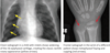
- The Wimberger sign is the destructive erosion of the medial aspect of the proximal tibial metaphysis. The Wimberger sign is not to be confused with the Wimberger ring sign of scurvy, which is increased density of the ossification centers.
- Syphilis is one of many causes of symmetric periosteal reaction in a child.

Juvenile Idiopathic Arthritis (JIA) / Juvenile Rheumatoid Arthritis (JRA) / Juvenile Chronic Arthritis (JCA)
What is it? How long and how old does patient need to be to diagnose this?
Rheumatoid factor lab work?
Most common subtype? Gender predilection? Regardless of subtype, which joint is most commonly involved?
Imaging appearance? Later changes?
What does chronic disease lead to? Contrast to adults? The most common site of this late complication?
What does it mimic in the cervical spine? Contrast this to JIA.
What happens to bone growth and why?
What is Still’s disease and does it have a gender predilection?
- Juvenile idiopathic arthritis - JIA is the most common chronic arthropathy of childhood. I_t is a systemic idiopathic disease defined as being present for more than 6 weeks in a patient younger than 16 years old_.
- JIA is usually rheumatoid-factor negative.
- The most common subtype of JIA is pauciarticular disease, typically affecting young girls. Regardless of subtype, the knee is the most commonly affected joint, but any joint can be affected.
- The imaging appearance reflects the underlying pathophysiology of synovitis, as the earliest radiographic changes are joint effusion, soft tissue swelling, and osteopenia.
- Later changes include periostitis and erosions.
- Chronic disease leads to eventual joint ankylosis, which is in contrast to adult rheumatoid arthritis, where ankylosis is rare. The most common sites of ankylosis are in the wrist, at the carpometacarpal joints, and the cervical spine.
- Cervical spine ankylosis in a child may appear similar to Kilppel-Feil syndrome. In contrast to KF, JIA does not feature segmentation anomalies.
- Accelerated skeletal maturation and accelerated bone growth are caused by synovitis and hyperemia, which lead to premature fusion of the physes and/or abnormal bone growth.
- Still disease is an acute systemic subtype of JIA affecting children under the age of five. Still disease presents with fever, anemia, leukocytosis, hepatosplenomegaly, and polyarthritis. Boys and girls are affected equally in Still disease.
Developmental Dysplasia of the Hip (DDH)
What is it? What happens in an abnormally shallow acetabular angle?
Increased incidence in what children? Do they get screened?
US is performed when? Why is imaging delayed?
What is the alpha angle? The bony portion of the acetabular roof should cover at least what % of the cartilaginous femoral head?
What measurements suggest DDH?
When do you start doing radiographs to diagnose this, and how do you diagnose it?
- Developmental dysplasia of the hip - DDH is abnormal development of the femoral head-acetabular relationship. An abnormally shallow acetabular angle results in uncovering of the femoral head by the acetabulum.
- There is an increased incidence of DDH in breech births, so all breech births are typically screened by ultrasound.
- Ultrasound is performed a few weeks after birth. Imaging is delayed because of the effects of perinatal maternal hormones on neonatal ligamentous laxity.
- The alpha angle is the angle formed by the bony ilium and acetabular roof, obtained from the coronal hip ultrasound. A normal alpha angle is greater than 60 degrees. The bony portion of the acetabular roof should cover at least 50% of the cartilaginous femoral head.
- An alpha angle less than 60 degrees or less than 50% coverage of the femoral head by acetabulum is abnormal, suggesting DDH.
- After femoral head ossification (typically after 6 months of age) radiographs are performed.
- On pelvic radiography, the ossified femoral head should normally sit in the inner lower quadrant formed by the intersection of the Hilgenreiner and Perkins lines.
Torticollis
What causes it? What does it result in?
Clinical outcome?
Ultrasound appearance?
- Torticollis is caused by fibromatosis coli (idiopathic sternocleidomastoid enlargement) resulting in an inability to fully straighten the neck.
- It usually resolves with physical therapy.
What are the causes of periosteal reaction in an infant or child?
- Physiologic periosteal reaction of the newborn
- Prostaglandin therapy
- Infectious
- Neoplastic (these are typically aggressive rxns)
- Trauma
- Metabolic
- Syndromic
(pretty much the broad differential for most things)
Examples of Osteochondrosis
- Blount’s disease - proximal tibial metaphysis
- Madelung deformity - medial sloping of distal radius
- Panner disease - caPitellum
- Little league elbow - medial epicondyle
- Leg-Calvé-Perthes disease - capital femoral epiphysis
- Keinbock disease - carpal lunate
- Osgood-Schlatter disease - tibial tuberosity
- Freiberg infarction - second metatarsal head
- Kohler disease - navicular bone
- Sever disease - calcaneal apophysitis
- Scheuermann disease - avascular necrosis of multiple thoracic
vertebral bodies leading to multiple compression fractures
Periosteal Reaction in an Infant or Child
What is periosteum?
What can cause irritation to the periosteum?
What are other commonly used synonyms?
Simply put, what is a periosteal reaction?
What may produce an aggressive appearance periosteal reaction?
- The periosteum is a thin membrane covering the entire surface of the bone excluding those areas covered by cartilage. It contributes to bone production and remodeling. Irritation of the periosteum, which can be due to trauma, infection, inflammation, neoplasm, or metabolic disturbance, can cause periosteal reaction.
- The terms “periosteal reaction,” “periostitis,” and “periosteal new bone formation” are commonly used as synonyms. Periosteal reaction is simply a nonspecific response to injury resulting in periosteal new bone production.
- Periosteal reaction may also be associated with tumors or infection, typically producing an aggressive appearance in these disorders.
Blount Disease
What is it?
What does it eventually lead to?
What is the infantile form thought to be due to?
Distribution?
When does the adolescent form occur and what is the distribution?
- Blount disease is an osteochondrosis of the proximal tibial metaphysis, causing tibia varus (bowing and internal rotation), which eventually leads to progressive deformity, gait deviations, and leg-length discrepancy.
- The infantile form of Blount disease is thought to be due to early walking and obesity and is typically bilateral.
- The adolescent form is seen in children over 6 years old and is usually unilateral.

Osteochondrosis
What is it?
What were they thought to be due to?
What is the current understanding?
More common in males or females?
- The osteochondroses are a heterogeneous group of disorders that were at one point thought to be initiated by avascular necrosis. The current understanding is that most of these disorders are secondary to trauma. While many are associated with avascular necrosis, AVN is not thought to be the initiating factor.
- Most are more common in males.
Sever Disease
What is this?
What imaging appearance is nonspecific?
Which finding may be more associated with pain?
- Sever disease is calcaneal apophysitis (rather than an osteochondrosis) and is a cause of heel pain. Sclerosis of the calcaneal apophysis is nonspecific and can be seen in asymptomatic children, although greater fragmentation of the apophysis may be associated with pain.
Tarsal Coalition
What is it?
What three types of coalitions are there?
What are the two most common tarsal coalitions?
How and when does it clinically present?
- Tarsal coalition is an abnormal joining of two normally separate bones in the tarsus.
- The three types of coalition are fibrous, cartilaginous, and osseous.
- The two most common coalitions (together making up 90% of all tarsal coalitions) are talocalcaneal and calcaneonavicular.
- Tarsal coalition clinically presents as foot pain in the child as the coalition begins to ossify.
Carpal Coalition
What is it?
Clinical presentation?
Types of coalitions?
Most common coalition?
Second most common?
- Carpal coalition is an abnormal joining of carpal bones.
- Carpal coalition is usually asymptomatic but may be a cause of wrist pain.
- Like tarsal coalition, the coalition may be fibrous, cartilaginous, or osseous.
- Lunotriquetral coalition is the most common carpal coalition.
- Capitohamate is second most common carpal coalition.

Physiologic Periosteal Reaction of the Newborn
When does it occur and why?
What does it appear like and where is it most commonly seen?
- Physiologic periosteal reaction occurs in infants 1-4 months old, thought to be due to rapid bone growth and loosely adherent periosteum.
- The periosteal reaction is smooth and thin, and usually only involves the lateral or medial aspect of the long bones.
What medication could cause a periosteal reaction in infants?
What is this medicine used for?
Why does the this medicine cuase periosteal reaction?
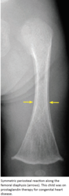
- Prostaglandin therapy
- Neonates on prostaglandin therapy for congenital heart disease (to maintain ductal patency) may have periosteal reaction.
- Prostaglandins are thought to decrease osteoclast bone resorption.

Periosteal Reaction Caused by Infection
What kind of periosteal reaction does this cause?
Name an infectious etiology that causes this
- Subperiosteal spread of pyogenic infection elevates the periosteum and irritates it, usually causing an aggressive periosteal reaction.
- Congenital syphilis causes periosteal reaction.
What malignancies can cause a periosteal reaction and what kind of reaction do these cause?
- Multiple malignancies, including leukemia, neuroblastoma, Ewing sarcoma, and osteosarcoma can produce an aggressive periosteal reaction.
What are metabolic causes of periosteal reaction?
What is the Wimberger Rim sign?
- Rickets, scurvy (which is also characterized by epiphyseal sclerosis - the Wimberger Rim sign), and hypervitaminosis A may all cause periosteal reaction.
What is a rare classic metabolic cause of periosteal reaction in children?
What bones are classically affected?
- Multiple syndromes may cause periostitis.
- Caffey disease (infantile cortical hyperostosis) is a rare inflammatory disease causing periostitis of the mandible, scapula, and clavicle.
