MOST COMMON QUESTIONS Flashcards
What are the 3 zones in target lesions of Erythema multiforme?
- central dusky purpura
- Elevated edematous pale ring
- surrounding macular erythema

compare AD ans seborrheic dermatitis based on their site involvement
AD involves the cheeks in infants (spares the central face), spares the diaper area
Seborrheic dermatitis can involve the diaper area, central face and scalp
first line treatment of vitiligo for pediatric and facial involvement
topical corticosteroids
most common clinical pattern of psoriatic arthritis
oligoarthritis with swelling and tenosynovitis of 1 or a few hand joints (70%)
nail groove can be seen in what condition
Myxoid cyst AKA synovial and digital mucous cyst

infants with seborrheic dermatitis with cradle cap were found to have impaired function of this enzyme which is required for the metabolism of essential fatty acids
delta - 6 - desaturase
Cellulitis etiology
S. Aureus/ Strep pyogenes
When is varicella most infectious
1 - 2 days before eruption of lesions
in cutaneous porphyrias, the photosensitivity is caused by absorption of UV radiation in what band of the UV spectrum by the porphyrins?
Soret Band (400 - 410 nm)
6th disease
Roseola infantum / Exantum subitum
common cause of sudden, unexplained high fever in young children between 6 and 36 months of age. Prodromal fever is usually high and may be accompanied by convulsions and lymphadenopathy. Suddenly, on about the fourth day, the fever drops. Coincident with the decrease in temperature, a morbilliform erythema of discrete, rose-colored macules appears on the neck, trunk, and buttocks and sometimes on the face and extremities. Often, there is a blanched halo around the lesions. The eruption may also be papular or rarely even vesicular. The mucous membranes are spared. Complete resolution of the eruption occurs in 1–2 days

deep folliculitis due to a cutaneous dermatophyte infection

Majocchi Granuloma
Majocchi granuloma is due to disruption of infected hair follicles so that hair shafts and fungi penetrate into the dermis and subcutaneous tissue. It presents with an irregular red, scaly plaque in which there are follicular papules, pustules and nodules. It is usually found on one lower leg.

Varnish-like crust
bullous impetigo
DOC for dermatophyte onychomycosis
Terbinafine
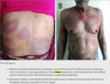
exaggerated skin injury occurring after minor trauma such as bump, bruise, needle stick injury. A more severe injury, such as a surgical procedure, can result in persistent ulceration in a patient with pathergy. It typically occurs in patients with Behcet disease.
Pathergy
n-serrated pattern of immunofluorescence in DIF means?
the split is above the basal lamina
n-serrated pattern is typically found in the most common sAIBD (Subepidermal Autoimmune Bullous Disease) bullous pemphigoid.

etiologic agent for non-inflammatory tinea capitis
All microsporum and trichophyton except T. concentricum
The underlying cause of TTP is a congenital or acquired deficiency of the vWF-cleaving protease, _________
ADAMTS13
vWF is secreted by the endothelial cell in long multimers, which should be cleaved into monomers by ADAMTS13 and released into the circulation. Instead, multimers circulate and extend from the surface of the endothelial cells in the microvasculature. Platelets adhere to these multimers and the surface of the endothelial cell, leading to microvascular thrombosis.
Herald / Mother patch
salmon-colored
Pityriasis rosea

golden period in detecting early signs of nerve function impairment
12 months or less
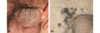
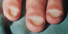
horizontal indentations, or ridges, that develop across the nails
Beau’s Lines
can be seen in psoriasis

give some differentials of leprosy
- tinea corporis
- psoriasis
- pityriasis rosea
- DLE
- granuloma annulare
- lupus vulgaris
- tuberculosis verruca cutis
- neurofibromatosis
- leukemia cutis
- xanthomas
- pityriasis versicolor
- pityriasis alba
- post-inflammatory hypopigmentation
- birthmarks
- vitiligo
- scleroderma
Subepidermal Immunobullous Diseases
- bullous pemphigoid (BP),
- epidermolysis bullosa acquisita (EBA),
- cicatricial pemphigoid (CP),
- pemphigoid gestationis (PG),
- linear IgA dermatosis (LAD),
- dermatitis herpetiformis (DH), and
- bullous systemic lupus erythematosus (BLSE)
azoles are active against what organisms
Yeasts > dermatophytes
hallmark of ACD
Itch
relative contraindications to prednisolone therapy in leprosy
- HTN
- DM
- Pregnancy
- mature cataracts
- glaucoma
- less than 15 y/o
- more than 60 y/o
- lack of cooperation
etiologic agent of total dystrophic pnychomycosis
Candida sp.
causative agent for black dot tinea
T. tonsurans and T. violaceum
Treatment for Impetigo contagiosa
- oral or topical antibiotics for 7 - 10 days
- Cloxacillin
- Children: 25 - 50 mkday in 4 divided doses
- Adults: 500 mg QID
- 1st generation cephalosporins like cephalexin
- Children: 25 - 50 mkday in 4 divided doses
- Adults: 500 mg QID
- Mupirocin ointment BID
- Cloxacillin
characterized by adherence of the distal portion of the nail bed to the ventral surface of the nail plate

Pterygium Inversum Unguis
It results from the extension of the zone of the nail bed that normally contributes to the formation of the nail plate. This eventually leads to a more ventral and distal extension of the hyponychium.
general management of AD
- skin hydration with moisturizers
- avoid overbathing
- avoid irritating or aggravating factors
- may give oral sedating antihistamines particularly at night
- short courses of topical steroids
- saline compresses or oral antibiotics for acute weeping eczematous dermatitis
Neutrophils exit from the tips of a subset of dermal capillaries (the “squirting papillae”), leading to their accumulation in the overlying parakeratotic stratum corneum

Munro’s microabscess
Present in psoriasis
Chronic Cutaneous LE

common histologic denominator in all forms of pemphigus
acantholysis, lysis of the intercellular adhesive junctions between neighboring squamous epithelial cells that results in the rounding up of detached cells
inflammatory manifestations of tinea capitis
Kerion
Favus
Telogen shed may be estimated by what test?
Pull Test
grasping 40 hairs firmly between thumb and forefinger, followed by a slow pull that causes minimal discomfort to the patient A count of more than 4–6 club hairs is abnormal
Classification of cutaneous Manifestations of lupus erythematosus
trench fever
B. quintana
give a differential diagnosis for guttate psoriasis
pityriasis rosea
acquired, depigmented dermatosis caused by repeated exposure to chemicals
Leukoderma
Pemphigus vulgaris has autoantibodies directed to what?
Desmoglein 1 and 3
* presents with generalized flaccid blisters with ulcers in the oral mucosa; many erosions because the flaccid bullae easily ruptures

Treatment of moderate acne

causative agent of tinea versicolor
Malassezia globosa
difference of verruca from calluses?
verruca has no dermatoglyphics (fingerprints)
SCC in situ of the glans penis or prepuce
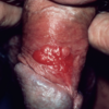
ERYTHROPLASIA OF QUEYRAT
* caused by high-risk HPV types (16, 18, 31, 35)
* single or multiple, fixed, well-circumscribed, erythematous, moist, velvety or smooth, red-surfaced plaques on the glans penis (Fig. 29.26).
what do you check when there is cellulitis of the lower extremity
Look at the foot for tenia pedis infection
Differentiate BCC from SCC
BCC - face especially the NOSE is the more common site of predilection (dorsum of the hands in SCC); rodent ulcer; (+) telangiectasia; (+) friability
SCC is more associated with chronic long term sun exposure; metastasis is more common; may have necrotic border

doubling time og M. leprae
12 days
5th Disease
Erythema Infectiosum
What is the lovibond angle in nail clubbing?
180o or more

characterized by an acute eczematous eruption triggered by purulent discharge from a primary infected site
Infectious eczematoid dermatitis (IED)
dermatitis that develops on the area macerated from the discharge from an infected ulcer or sinus
locations frequently involved in adult AD
Hands and wrists
class 2 potent steroids
Fluocinonide 0.05% cream/gel/ointment
mometasone furoate 0.1% ointment
Hutchinson Triad of Congenital Syphilis
- changes in the incisor teeth (hutchinson teeth - malformation of the central upper incisors that appear in the secondary or permanent teeth
- opacities of the cornea
- eighth cranial nerve deafness
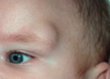
this indicates the involvement of the nasociliary nerve presenting as herpes zoster vesicle present on the tip or side of the nose.
hutchinson’s sign

Porphyria cutanea tarda is deficient in what enzyme?
uroporphyrinogen decarboxylase (UROD)
give some Differentials for exfoliative dermatitis
- psoriasis
- atopic dermatitis
- eczema
- allergic contact dermatitis
- irritant contact dermatitis
Chancroid
Haemophilus ducreyi
Chancroid (soft chancre) is an infectious, ulcerative STD caused by the gram-negative bacillus Haemophilus ducreyi (the Ducrey bacillus) One or more tender ulcers on the genitalia and painful inguinal adenitis that may suppurate, are characteristic of the disease.
Bullous impetigo etiology
Staphylococcus aureus
The angle formed by the dorsal surface of the distal phalanx and the nail plate

Lovibond angle
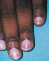
Black or brown pigmentation of the normal nail plate

Melanonychia
bacillary angiomatosis
bartonella hensellae
bartonella quintana
Individual melanocytes in the “buckshot” scatter throughout the epidermis are typical of?
Superficial Spreading melanoma
general mechanism for Type 1 reactions in leprosy
Enhanced cell-mediated immune response
*generally ocuur in borderline leprosy (BT, BB, BL)
*occur at existing skin lesions
etiology of EM
Adult: HSV 1 > 2
Children: Mycoplasma pneumoniae
Maculae cerulae is seen in whart disorder
Pediculosis Pubis
Maculae cerulae are blue-gray macules, which are actually a discoloration of the skin due to the insect’s bite. They are due to altered blood pigments of the infested humans or the excretion products of the louse’s salivary glands

Casal Necklace

Pellagra (Vitamin B3 Deficiency)
*photosensitive eruption
* occurs symmetrically on the face, neck and upper chest (casal necklace), extensor hands and backs of the hands
Possible side effect of dapsone
hemolytic anemia
Holster sign
dermatomyositis

KOH finding of dermatophytes
Long Septated hyphae
most common type of xamthomas
Xanthelasma/ Xamthelasma palpebrarum

incubation period of measles
8 - 12 days
Gottron Papules

Dermatomyositis
Gottron papules refer to a violaceous hue located at the dorsal-lateral interphalangeal (IP) and/or metacarpophalangeal (MCP) joints.
“split ends”

Trichoptilosis
most serious complication of varicella
Varicella Pneumonia
causative agent of granuloma inguinale
Klebsiella granulomatis
give differentials for tinea pedis
Atopic dermatitis - never on the plantar arch (can involve plantar arch in tinea)
Allergic contact dermatitis - usually symmetrical (in tinea, may involve one hand and both feet)
Treatment for herpes zoster
Acyclovir 800 mg 1 tab q4 5x/day for 7 days
San joaquin valley fever
Coccidioidomycosis
onychomycosis that is HIV related
proximal subungal onychomycosis
joints affected in psoriasis
DIP, PIP joints
spares the MCP and MTP joints
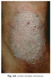
exaggerated skin markings; criss-cross pattern
Lichen Simplex Chronicus
Wickham striae

Lichen Planus
The usual presentation of the disease is classical lichen planus. Symptoms can range from none (uncommon) to intense itch.
Papules and polygonal plaques are shiny, flat-topped and firm on palpation.
The plaques are crossed by fine white lines called Wickham striae.
Hypertrophic lichen planus can be scaly.
Atrophic lichen planus is a rare annular variant with an atrophic centre.
Bullous lichen planus is rare.
Size ranges from pinpoint to larger than a centimetre.
Distribution may be scattered, clustered, linear, annular or actinic (sun-exposed sites such as face, neck and backs of the hands).
Location can be anywhere, but most often front of the wrists, lower back, and ankles.
Colour depends on the patient’s skin type. New papules and plaques often have a purple or violet hue, except on palms and soles where they are yellowish-brown.
Plaques resolve after some months to leave greyish-brown post-inflammatory macules that can take a year or longer to fade.
Chicken wire pattern of intercellular IgG in DIF
Pemphigus vulgaris
Give evidence of recent Strep infection for Scarlet fever
Increased Antistreptolysin O or DNAse B titer
HPV type of external genital warts
HPV 6 and 11
KOH finding of tinea versicolor
short hyphae with spores
(spaghetti and meatballs)
color of pityriasis versicolor in wood’s lamp
yellowish to yellowish-green fluorescence
aspirin is contraindicated in varicella because?
may produce Reye’s syndrome (encephalopathy with liver damage)
what is the normal lovibond angle?
160o
differentials for molluscum contagiosum
- verruca
- syringoma - benign sweat gland tumor usually on the face around the eyes
- sebaceous hyperplasia - sebaceous gland hypertrophy usually on seborrheic areas of the face
- basal cell carcinoma
- condyloma acuminata
- epithelial inclusion cyst
Multiple facial mollusca - disseminated invasive fungal infection like cryptococcosis, histoplasmosis, coccidioidomycosis and penicillosis
(Tinea Corporis) –due to corticosteroid therapy
Tinea Incognito
fungal skin infection when the clinical appearance has been altered by inappropriate treatment, usually a topical steroid cream. The result is that the original infection slowly extends.

Darier Sign
Urticaria Pigmentosa, Mastocytoma, Mastocytosis
refers to the urtication and erythematous halo that are produced in response to the rubbing or scratching of these lesions. This is due to mast cell degranulation induced by physical stimulation

number of hair normally shed daily
100 - 150 strands daily
concentric blanching of erythematous skin near periphery of healing psoriatic plaque
Woronoff ring
- it is often the first sign that the patient’s psoriasis is responding to phototherapy.
- ring-like hypopigmentation zone around regressing psoriasis lesions.

what increases and decreases absorpption of itraconazole
food INCREASES absorption
antacids and gastric acid suppressors DECREASES absorption
areas of hair loss with absence of follicular ostia
Citcatricial / Scarring alopecia

common disorder of the hair follicles that clinically gives the impression of blackheads (Fig. 33.30), but the follicles are filled with funnel-shaped, horny plugs within which are bundles of vellus hairs. The hairs are round at their proximal ends and shredded distally.
Trichostasis spinulosa results from retention of telogen hairs, which are derived from a single hair matrix. It is primarily caused by a hyperkeratosis of the follicular infundibulum, which leads to a partial obstruction of the follicular orifice and thus does not permit shedding of small telogen hairs.
Extension of discolouration into the skin surrounding the nail
Hutchinson sign
- clinical clue to subungal melanoma
- involvement of the nail plate + periungual skin

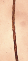
how can you differentiate anagen and telogen hair?

- Anagen hair has a pigmented bulb and is surrounded by a gelatinous root sheath (left);
- Telogen hair has a nonpigmented bulb and lacks a root sheath. (right)
Squamous cell carcinoma in situ

Bowen Disease
BD may be found on any part of the body as an erythematous, slightly scaly and crusted, noninfiltrated patch from a few millimeters to many centimeters in diameter (Fig. 29.24). The lesion is sharply defined. The scale may be pronounced enough for the lesion to be mistaken for psoriasis, or the plaque may have a stuck-on appearance and be mistaken for a broad, sessile seborrheic keratosis

Ugly duckling sign
Melanoma
- one mole among may that sticks out and looks different

how do you differentiate pediculosis corporis from scabies?
pediculosis corporis spares the hands and feet
Bullae are often arranged in rosettes or an annular array, the so-called string of pearls configuration

Childhood Linear IgA Disease (Chronic Bullous Disease of Childhood)
etiology of Erysipelas
Group A strep
how can you view burrows of scabies

via india ink or gentian violet

Buttonhole sign

With application of gentle pressure, a neurofibroma will easily invaginate into the subcutis
Neurofibromas can be invaginated with the tip of index finger back into the subcutis and again reappear after release of pressure. [14] Other condition where one can find positive buttonhole sign are anetoderma (macular atrophy - localized loss of elastoc tissue resulting in herniation of subcutaneous tissue) and dermatofibroma.
produces non-inflammatory type of Tinea pedis
T. rubrum - produces dry erythema with scaling
most common type of melanoma
Superficial Spreading melanoma

most common internal cause of pruritus
CKD
Hepatitis associated with polyarteritis nodosa (PAN)
Hepatitis B
most common causes of mortality in SJS
- sepsis
- electrolyte imbalance
secondary lesions of syphilis
Condyloma lata
- also check lesions on the palms and soles

most common type of melanoma in asians/ darker population
Acral lentiginous melanoma - specific type of melanoma that appears on the palms of the hands, the soles of the feet, or under the nails. The word “acral” means “extremity” in Greek and refers to the occurrence of this type of melanoma on the extremities (hands and feet).

Differentiate Hypertrichosis form hirsutism
Hypertrichosis is an overgrowth of hair not localized to the androgen-dependent areas of the skin.
Hirsutism is an excess of terminal hair growth in women in a pattern more typical of men.
drug of choice for bullous systemic LE
Dapsone
areas spared in scabies in adults
face, scalp, mucosa
most common complication of herpes zoster
post-herpetic neuralgia
delicate scaling in pityriasis versicolor can be elicited by what maneuver
Grattinage maneuver
https://www.youtube.com/watch?v=0cAJpDJT95g
scaling is dust-like/furfuraceous, and in some macules, may only become apparent after a combination of light scaping and scratching
average incubation period of leprosy
2 - 5 years
It usually begins in adolescence, appearing first as minute, round, skin-colored or hyperpigmented macules or papules that develop singly or in sparse numbers on the malar regions or on the cheeks below the eyes.
DERMATOSIS PAPULOSA NIGRA
causative agent of lyme disease
Borrelia burgdorferi - transmitted via bit of an ixodes tick
Forchheimer sign

German measles (Rubella)
*exanthem of pinhead-sized red macules or petechiae on the soft palate and uvula
3 major triggers of dyshidrotic eczema
stresss, atopy, contactants
presents as SUBCORNEAL blister formation
Pemphigus foliaceus
% BSA of SJS
<10% BSA
u-serrated pattern of immunofluorescence in DIF means?
The split is in the sub-lamina densa
Presence of the u-serrated pattern indicates the sAIBD subtype epidermolysis bullosa acquisita (EBA)

give some differentials for psoriasis
- fungal infections
- seborrheic dermatitis
- pityriasis rosea
- lichen planus - chiefly affects flexor surfaces of the forearms, wrists, shins and ankles; may have very fine scales; scalp less frequently involved; nails may have pterygium
- psoriasiform subset of subacute cutaneous LE - located usually on the upper trunk, arms as well as signs of photsensitivity
- chronic contact dermatitis and dyshidrotic eczema
usually results from hair shaft fracture
Anagen Effluvium
It is frequently seen following the administration of cancer chemotherapeutic agents, such as the antimetabolites, alkylating agents, and mitotic inhibitors. These agents result in temporary shutdown of the hair matrix with resultant tapering of the shaft (Pohl-Pinkus constrictions).
percent of KOH used for dermatophyte infection
10% KOH
junction between palmoplantar and glabrous skin
Wallace line
causative agent of pediculosis capitis
Pediculus humanus var. capitis
describe childhood AD
- less exudative, drier and more PAPULAR lesions
- classic locations are the antecubital and the popliteal fossa, flexor wrists, eyelids, face and around the neck
- lesions are 2-4 mm excoriated papules and lichenified plaques, slightly scaly
adverse effect of cyclosporine
HTN and nephrotoxicity
differentials for tinea corporis
- contact dermatitis - history of exposure to contactant; absence of central clearing and advancing border
- atopic dermatitis - absence of advancing borders and central clearing plaques; found on flexural areas in adults and extensors in infants
- psoriasis - erythematous plaques with silvery white scales; (+) koebner phenomenon and auspitz sign
- pityriasis rosea
- pityriasis alba
- tinea versicolor
- erythema migrans
- subacute LE
- cutaneous T cell lymphoma
differentials for tinea pedis
- pitted keratolysis - bacterial infection producinf pits on the stratum corneun; positive foot odor; negative KOH
- P. aeruginosa webspace infection - presence of gram negative bacteria on gram stain and culture; negative KOH
- Erythrasma - coral red fluorescence on wood’s light; negative KOH
- Contact dermatitis - exposure to contactants; negative KOH
- Dyshidrotic eczema - minute tapioca-like vesicles on the lateral sides of the toes and soles; negative KOH
- Psoriasis
Nail pitting
*caused by parakeratosis from the proximal matrix
psoriasis
cause of black dot tinea
*this occurs due to the fungi invading the follicle (endothrix)
T. tonsurans
neonatal varicella may occur if the mother develops infection when?
5 days before and 2 days after delivery
etiology of pityriasis rosea
HHV 6 & 7
%BSA of TEN
>30% BSA
HPV types of plantar warts (Verruca plantaris)
HPV 1, 2, 27, 57
Thrombotic thrombocytopenic Pentad
- Microangiopathic Hemolytic anemia - coomb’s negative
- Thrombocytopenic purpura - (+) platelet aggregation in the microvasculature
- Neurologic abnormalities
- Fever
- Renal Disease
incubation period of rubella
14 - 17 days
most common cause of inflammatory type of tinea pedis
T. mentagrophytes
primary lesion of syphilis
chancre

cardinal signs of Leprosy
- hypopigmented or reddish skin lesion with definite loss of sensation
- skin smear positive for AFB
- enlargement plus tenderness of the peripheral nerves
exclamation point hairs
Alopecia Areata

Cat scratch disease
Bartonella henselae
SAUCER RIGHT SIDE UP APPEARANCE in leprosy
Tuberculoid leprosy
Saucer right side up: sharpy defined and elevated border that slopes down to a flattened atrophic center

nephritogenic strains of Streptococcus
Type 49, 55, 57, 60 and strain M - Type 2
Treatment for Paucibacillary Leprosy
Dapsone, Rifampicin
clues to correct diagnosis of alopecia areata
- history of periodic regrowth,
- nail pitting (Fig. 33.2), and the
- presence of tapered fractures or “exclamation point” hairs.
what causes hypopigmentation in pityriasis versicolor
due to the metabolites PITYRIACITRIN which absorbs UV light ans AZELAIC ACID which inhibits tyrosinase enzyme
site of injury in the epidermis of miliaria rubra
prickle cell layer (stratum spinosum)
ash-leaf macules

tuberous sclerosis
(+) anti-Jo-1 antibody
Dermatomyositis
unilateral eye swelling after nighttime encounter with typanosoma cruzi
Romana’s sign (Reduviid bites)

type of miliaria that is ALWAYS preceded by some other dematitis (contact derm, lichen simplex xhronicus, intertrigo)
Miliaria pustulosa
Osler Nodes

Infective endocarditis
*PAINFUL, erythematous nodule with pale center on the fingertips
*from septic emboli leading to clogging of distal vessles

Impetigo contagiosa etiology
S. aureus, Group A strep;
Group B strep in newborns
give a differential for dermatitis herpetiformis
Scabies
* DH can be seen on the nape, scapula, extensors, buttocks
severity-of-illness score that estimates the risk of death in TEN
SCORTEN
HSV 2 reactivates most commonly in what ganglia
Sacral ganglia (S2 - S4)
spares the folds of the inguinal area
diaper dermatitis
conditions that are (+) for pathergy test

- Behçet disease,
- pyoderma gangrenosum,
- Sweet syndrome, and
- bowel-associated dermatosis–arthritis syndrome.
HPV type of pigmented warts
HPV 4, 60, 65
Give conditions that presents with Koebner phenomenon (Isomorphic Response)
Psoriasis
Vitiligo
Verruca plana
Lichen planus
Molluscum contagiosum

Granuloma inguinale
The disease begins as single or multiple subcutaneous nodules, which erode through the skin to produce clean, sharply defined lesions, which are usually painless (Fig. 14.36A).
More than 80% of cases demonstrate hypertrophic, vegetative granulation tissue, which is soft, has a beefy-red appearance, and bleeds readily. Approximately 10% of cases have ulcerative lesions with overhanging edges and a dry or moist floor (Fig. 14.36B).
Treatment for scabies
5% Permethrin lotion - apply from the neck down. leave on overnight for 8-10 hours and rinse off in the morning. Use once a week for 2 weeks
*we repeat after 1 week because we wait for the eggs to hatch again
Most common type of BCC

Nodular/Classic Basal Cell Carcinoma
Nodular BCC is composed of one or a few small, waxy, semitranslucent nodules forming around a central depression that may or may not be ulcerated, crusted, and bleeding. The edge of larger lesions has a characteristic rolled border. Telangiectases course through the lesion. Bleeding on slight injury is a common sign. As growth progresses, crusting appears over a central erosion or ulcer, and when the crust is knocked or picked off, bleeding occurs, and the ulcer becomes apparent. This ulcer is characterized by chronicity and gradual enlargement over time The lesions are asymptomatic, and bleeding is the only difficulty encountered. The lesions are most frequently found on the face

etiologic agent of tinea pedis
T. rubrum, T. interdigitale and E. flocossum
Differential diagnosis for intertrigo
- tinea cruris
- candidal intertrigo
- inverse psoriasis
- erythrasma
6 Types of Vitiligo
- Localized/Focal
- Segmental
- Generalized - most common
- Universal
- Acrofacial - distal fingers and facial orifices
- Mucosal

Bullous Pemphigoid has IgG Ab against what?
Bullous Pemphigoid Antigen 1(BPAG 1 or BP 230)
and
Bullous Pemphigoid Antigen 2 (BPAG 2 or BP 180 or Type XVII collagen)
*presents as TENSE blisters and it is VERY PRURITIC

MOA of acitretin in psoriasis
antiproliferative and enhances keratinocyte differentiation
usually cause the non-inflammatory type of tinea pedis
T. rubrum
periodic idiopathic shedding of the nail beginning at its proximal end
The temporary arrest of the function of the nail matrix may cause onychomadesis.
what is targeted by the antibodies in Epidermolysis Bullosa Acquisita?
Type VII Collagen, which is a major component of the anchoring fibrils

KOH finding of Pityriasis versicolor
short, thich hyphae with numerous round yeast cells (spaghetti and meatballs)
3 day measles
german measles
Calabar swelling
Loa loa infection
The first sign is often painful, localized, subcutaneous, nonpitting edema called Calabar or fugitive swelling
This is a sign to be elicited in case of secondary syphilis and cutaneous vasculitis, where there is deep dermal tenderness on pressing the lesion (e.g., papular lesions of syphilis) with a pinhead.
Buschke-Ollendorff Sign
Groove sign or Dry riverbed sign
*follows the course of the underlying vessels

Eosinophilic fasciitis
differentials for total dystrophic onychomycosis
- alopecia areata - twenty nail dystrophy; no hyphae on KOH
- lichen planus - may also present as 20 nail dystrophy or dorsal pterygium (scarring process leads to fusion of the proximal nail fold to the nail bed)
- Nail psoriasis - may present as onychodystrophy but with associated psoriatic lesions in the body
Staphylococcal Scalded skin syndrome is caused by what toxins?
Exfoliative toxins Type A and B
Pastia Lines are accentuated in what disease

This is accentuated in SCARLET FEVER
*In body folds, especially the armpits and elbows, fragile blood vessels (capillaries) can rupture and cause classic red streaks called Pastia lines.
*accebtuation of the skin folds & linear petechial eruption
*usually on the antecubital and axillary folds
“bloodhound-like” facies

Cutis Laxa (Generalized elastolysis)
treatment of psoriasis that is safe in children, pregnant and lactating women
Broad band and narrowband UVB (311nm)
satellite pustules; intertriginous area
Candida
most common type of psoriasis
plaque type/ psoriasis vulgaris
pruritus may persist for several weeks after treatment after treatment due to the body’s response to dead mites
post-scabetic pruritus
lesions of bullous pemphigoid appear as tense bullae, filled with clear fluid, on normal or erythematous skin (Fig. 23–12, B). Bullous pemphigoid is characterized by _____________. Early lesions show a perivascular infiltrate of lymphocytes and variable numbers of eosinophils, occasional neutrophils, superficial dermal edema, and associated basal cell layer vacuolization. The vacuolated basal cell layer eventually gives rise to a fluid-filled blister (Fig. 23–12, C). The blister roof consists of fullthickness epidermis with intact intercellular junctions, a key distinction from the blisters seen in pemphigus.
subepidermal nonacantholytic blisters
Differential diagnosis for vitiligo
- morphea
- lichen sclerosus
- Pityriasis alba - fine scale, s slightly papular, and is poorly defined.
- Tinea versicolor favors the center back and chest and has a fine scale; yeast and hyphal forms are demonstrable with potassium hydroxide (KOH) examination
- severe chronic actinic dermatitis - may develop vitiligo-like depigmentation.
- Chemical leukoderma may closely resemble vitiligo
Oil spot
Psoriasis
*yellow areas of subungal parakeratosis from the distal matrix

most common type of porphyria
Porphyria Cutanea Tarda
most common benign tumor of the skin
seborrheic keratosis
- stuck on greasy appearance

incubation period of varicella
10 - 23 days
heliotrope rash
dermatomyositis

indications for systemic corticosteroid use in leprosy
- Severe Type I and Type II reaction
- Silent neuritis
- inflamed skin patch over a major nerve trunk like cheeks, elboows and knee
melanoma with the highest risk of metastasis
Nodular melanoma

class 3 potent, upper midstrength steroids
betamethasone valerate 0.1% ointment
fluticasone propionate 0.005% ointment
general pathology of vitiligo
autoimmune destruction of melanoctyes
surface has the peau d’ orange appearance
erysipelas
most common epithelial precancerous lesion

Actinic / Solar Keratosis
represent in situ dysplasias resulting from sun exposure They are found chiefly on the chronically sun-exposed surfaces of the face (Fig. 29.11), ears, balding scalp, dorsal hands, and forearms. They are usually multiple, discrete, flat or elevated, verrucous or keratotic, red, pigmented, or skin colored. Usually, the surface is covered by an adherent scale, but sometimes it is smooth and shiny. On palpation, the surface is rough, like sandpaper, and at times lesions are more easily felt than seen. T
characterized by episodic, recurrent vasospasm of the fingers and toes resulting in white, blue, and red discoloration provoked by cold or stress
Raynaud phenomenon
* Pallor (white) - Cyanosis (blue) - Rubor (red) (PCR)
most effective scabicide but NEUROTOXIC for infants
Lindane
unable to close eyes due to paralysis of the zygomatic branch of the facial nerve; seen in leprosy
Lagophthalmos
Ectothrix dermatophytes that are positive for wood’s lamp
- Microsporum canis
- Microsporum audouinii
- Microsporum distortum
- Microsporum ferrugineum
incubation period of molluscum contagiosum
2 - 7 weeks
tender boggy plaques exuding pus; cause scarring and permanent alopecia in Tinea capitis
Kerion Celsii

White piedra is caused by

Trichosporon (Trichosporum) beigelii or Trichosporon inkin
etiologic agent of Tinea corporis
T. rubrum, mentagrophytes and tonsurans
necrotic keratinocytes are seen in
SJS
Also known as “bamboo hair,”this is caused by intussusception of the hair shaft at the zone where keratinization begins. The invagination is caused by softness of the cortex in the keratogenous zone.

Trichorrhexis Invaginata
The patient with bamboo hair will have nodose ball-and-socket deformities, with the socket forming the proximal and the ball part forming the distal portion of the node along the hair shaft. This type of hair is associated with Netherton syndrome (Fig. 33.24). Occasionally, only the proximal half of the abnormality is seen; this has been called “golf tee hairs.”

paucibacillary corresponds to what leprosy spectrum
Indeterminate and Tuberculoid leprosy (TT)
the most common dermatophyte worldwide
T. rubrum
HPV type of anogenital dysplasia
HPV 16 and 18
4 types of tinea pedis
- Chronic hyperkeratotis (moccasin) Type
- Interdigital Type
- Vesicobullous Type
- Acute ulcerative type
KOH finding of candida
pseudohyphae and spores

complication of diffuse leprosy characterized by severe necrotizing cutaneous lesions

Lucio phenomenon (Erythema Necroticans)
most common offending drug in FDE
NSAIDs
This form of BCC is the most common pattern seen in patients with human immunodeficiency virus (HIV) infection and BCC.

Superficial basal cell carcinoma
This type of BCC most frequently presents as a dry, psoriasiform, scaly lesion. It is usually a superficial flat growth, which in many cases exhibits little tendency to invade or ulcerate. The lesions enlarge very slowly and may be misdiagnosed as patches of eczema or psoriasis. They may grow to be 10–15 cm in diameter. Close examination of the edges of the lesion will show a threadlike raised border (Fig. 29.16). These erythematous plaques with telangiectasia may occasionally show atrophy or scarring.

test for blanchability
diascopy
breakfast, lunch, dinner lesions
Bedbug bites/ Cimicosis

bulla-spread phenomenon
Asboe-Hansen Sign
*elicited by pressure on an intact bulla, gently forcing the fluid to spread under the adjacent skin
Also known as “twisted hairs”.
Malformation of hair characterized by twisting of the hair shaft on its own axis. The hair shaft is segmentally thickened, and light and dark segments are seen. Scalp hair, eyebrows, and eyelashes may be affected. The hairs are brittle and easily broken.
Pili Torti
most common form of oral candidiasis
acute pseudomembranous candidiasis or thrush
- compressible, but not fluctuant, cystic mass from 0.5 cm to several centimeters in diameter (Fig. 29.51).
- The surface of the overlying skin is usually smooth and shiny from the upward pressure.
- These nodules are freely movable over underlying tissue and are attached to the normal skin above them by a comedolike central infundibular structure or punctum.
- The pasty contents of the cysts are formed mostly of macerated keratin, which has a cheesy consistency and pungent odor.

Epidermal Cyst (Epidermal Inclusion Cyst, Infundibular Cyst)
They frequently result from plugging of the follicular orifice, often in association with acne vulgaris. They may also occur by epidermal implantation.

most common causative agent of Ecthyma
Group A streptococcus
(next is S. aureus)
etiology of molluscum contagiosum
poxvirus
other term for white superficial onychomycosis
leukonychia trichophytica
most common etiologic agent for erysipelas
Group A Streptococcus
etiologic agent of bullous impetigo
S. aureus
allergen in cement
chromates
most common form of cutaneous lupus
Chronic cutaneous lupus erythematosus
- better prognosis
- usually not associated with systemic symptoms (SLE), unlike the Acute cutaneous LE where it is only seen in SYSTEMIC lupus erythematosus
EM can be confused with SJS coz they may also appear with target lesions, may be drug induced and may have the same areas of predilection. how can you differentiate them
(+) nikolsky for SJS
differentials for verruca vulgaris
- molluscum contagiosum - umbilicated surface
- syringoma - benign sweat gland tumor on the face
- seborrheic keratosis - stuck-on hyperkeratotic, pigmented papules and plaques
- acrochordon - skin tags; skin colored, soft, exophytic papule
- callus
- corn
- actinic keratosis
- keratoacanthoma
- SCCIS
- invasive SCC
*
oral antifungals are always warranted in
nails and hair fungal infection
Differentials for tinea manuum
- contact dermatitis - hx of contactant; negative KOH
- dyshidrotic eczema - tapioca-like vesicles on the lateral sides of the finders and palms; negative KOH
- pityriasis rubra pilaris - islands of normal skin on the trunk and extremities with RED-ORANGE desquamation, keratoderma of the palms and soles; negative KOH smear
- atopic dermatitis
- lichen simplex chronicus
- ACD/ICD
- psoriasis vulgaris
Types of Melanoma
- Superficial spreading melanoma
- Nodular melanoma
- Acral lentiginous melanoma
- Lentigo maligna
causative agent of black piedra

Piedraia hortai
Non-blanchable
purpura - due to extravasated RBC
characterize infantile AD
- usually begins as a pruritic eczema of the cheeks
- intraepidermal vesicles are present in the erythematous patches which results to moist crusted areas
- it spreads to the scalp, foreheadm neck, wrists and extensors
- it spares the buttocks and diaper area
- resolves at the end of the second year
Criteria of Sjogren / Sicca Syndrome
2 of major 3 criteria:
- xerophthalmia
- xerostomia
- an associated autoimmune, rheumatic or lymphoproliferative disorder
first reading of patch testing is done when
48 hours
repeat reading at day 4 or 5
Gottron sign

Dermatomyositis
* pink to reddish-purple atrophic or scaling eruption over the knuckles, knees or elbows
treatment for erythrasma
- Localized disease
- Topical erythromycin/clindamycin solution BID x 4 - 6 weeks
- Topical azole creams BID x 2 weeks
- Larger lesions
- Clarythromycin 1g single dose
- Erythromycin 250 mg QID x 2 weeks
- prophylaxis
- benzoyl peroxide bar while showering
- minimize skin occlusion and moisture
- control of hyperhydrosis with aluminum chloride
fungi are best demonstrated with what stains
Periodic Acid Schiff stain (PAS) and methenamine silver stains
differentials for AD
- seborrheic dermatitis - usually presents as greasy, scally, yellowish patches on areas where the sebaceous glands are most active; it may affect the GROIN and GLUTEAL AREA which is atypical in AD
- Contact dermatitis - history of prior contactant and presents as sharply demarcated eczematous plaques localized on the exposed area
- nummular eczema - presents as discoid or coin-shaped vesicular papules predominantly on the extremities; may also coexist with AD
- Scabies - extremely pruritic excoriated papules predominantly found in the web spaces, buttocks, axillae, groin, periumbiliccal area; pruritus more intense at night; (+) also for other contacts at home
- Psoriasis - thick plaques with wilvery white scales; positive koebnerizationa nd auspitz sign
- Dermatophytosis
- early stage of mycosis fungoides
type of folliculitis associated with long term antibiotics
Gram (-) folliculitis
presents with hair at varying lengths
Treatment of type 2 reaction in leprosy
Thalidomide
oral drug of choice for psoriatic arthritis
Methotrexate
General mechanism of alopecia areata
Alopecia areata generally presents as an anagen effluvium, with an inflammatory insult to the hair matrix resulting in tapering of the hair shaft and fracture of anagen hairs. As the hair miniaturizes or converts from anagen to telogen, the remaining lower portion of the hair rises above the level of the scalp, producing the exclamation point hair.
HSV 1 reactivates most commonly in what ganglia
Trigeminal ganglia
characteristic rolled border
Basal Cell carcinoma

a chronic skin disease characterized by small follicular papules that coalesce into salmon colored pink scaling patches, and often, solid confluent yellow-orange palmoplantar hyperkeratosis. The papules are the most important diagnostic feature, being more or less acuminate (pointy), salmon colored to reddish brown, about pinhead sized, and topped by a central horny plug

Ptyriasis Rubra pilaris

result from local anomalies in embryonic development and occur along embryonic closure zones. On the face, they occur above the lateral end of the eyebrow (external angular dermoid) (Fig. 29.54), at the nasal root, along the midline of the forehead, over the mastoid process, on the floor of the mouth, and anywhere along the midline of the scalp from the frontal to the occipital region.
Cutaneous dermoid cyst / congenital inclusion dermoid cyst
They are nonpulsatile, firm, and cystic, and they do not transilluminate. A punctum or opening to the skin surface may be present, but dermoid cysts are not usually attached to the overlying skin. A tuft of hair may project from a pit, signifying the presence of an underlying sinus or cyst.

treatment for pityriasis versicolor
- Topicals
- 1 - 2% Ketoconazole shampoo. applied on the affected areas and left in contact for 10 minutes before rinsing. Repeat daily for 2 weeks
- Selenium sulfide shampoo
- topical azoles and allylamines. applied BID
- ciclopirox olamine
- nystatin
- 50% propylene glycol in water
- zinc pyrithione
- salicylic acid preparation
- Oral
- ketoconazole 200 mg/ day x 7-10 days
- fluconazole 400 mg single dose or once a week for 2 - 3 weeks if necessary
- itraconazole 200 mg/day x 5-7 days
Shagreen plaque (collagenoma)

Tuberous sclerosis
presents with doll’s hair–like bundling of follicular units
Tufted Folliculitis
griseofulvin is active against
dermatophytes
give examples of imidazoles (Azoles)
ketoconazole
miconazole
econazole
clotrimazole
common etiology of tinea capitis
Trichophyton tonsurans and microsporum canis
differentials for tinea barbae
- sycosis barbae - papules and pustules pierced in the center by a hair which is loose and easily extracted after suppuration has occured; a form of folliculitis caused by S. aureus
- Contact dermatitis - history of exposure to a contactant; positive patch testing (ACD)
- Herpes simplex infection - group vesicles or vesiculopustular lesions; positive multinucleated epidermal cells on Tzanck smear; positive serologic tests
- S. aureus folliculitis
- furuncle
- carbuncle
- acne vulgaris
- rosacea
- pseudofolliculitis
an ENDOTHRIX type of infection presenting as patches of alopecia with associated infected broken-off hairs near the surface of the scalp
Black dot Tinea capitis
Koplik Spots

pathognomonic for MEASLES
* nearest to the lower molars; white papules on an erythematous base
type of scabies that can be seen in immunocompromised or debilitated patients
Crusted/ Norwegian/ Hyperkeratotic Scabies

treatment of erysipelas
Systemic antibiotic for 10 days
- Penicillin VK
- adults: 250 - 500 mg PO QID
- 25 - 50 mkday PO in 4 divided doses
- IV Penicillin
- adults: 600 000 to 2 000 000 U/kg in 4 divided doses
- 50 000 - 250 000 U/kg in 4 divided doses
- Erythromycin
- Adults: 250 - 500 mg PO QID
- Children: 30 - 50 mkday PO QID
Pellagra
Vitamin B3 (Niacin) deficiency
lesion in tertiary syphilis
Gumma

CREST syndrome
Calcinosis cutis
Raynaud’s phenomenon
Esophageal dysmotility
Sclerodactyly
Telangiectasia

major complication of Type 1 reactional leprosy
Nerve damage
usually cause the inflammatory type of tinea pedis
T. mentagrophytes
give examples of allylamines
terbinafine
butenafine
naftifine
butcher’s warts are caused by what type of HPV
HPV 7
*they are cauliflower like lesions seen on hands of meat handlers
differentials for oral candidiasis
- oral hairy leukoplakia - white plaques NOT easily scraped off; negative KOH
- Condyloma acuminatum - whitish papillomatous papules or verrucous plaques on the oral mucosa
- geographic tongue - map-like reddish and whitish areas on the tongue
- hairy tongue - whitish plaques on tongue due to elongation of the filliform papillae
- lichen planus - usually reticulated or lacelike pattern; may also be atrophic, papular, erosive bullous or plaque-like
- bite irritation - irregularly- shaped whitish plaques; history of biting/trauma to affected areas
most common portal of entry of cellulitis
tinea pedis
Paucibacillary leprosy
(-) bacilli on smears and biopsy
5 or less lesions
premalignant lesion of SCC
actinic keratosis
Multiple discrete symmetrical hyperpigmented macules with collarette of scales on both palms

Biett Sign
Presence of hyperpigmented macules or papules with collarette of scales - Biett’s sign on the palms and soles is considered to be a strong indicator of secondary syphilis and clinically distinguishes it from other papulosquamous conditions affecting the palms and soles.
etiology of cellulitis
S. aureus, Group A strep (S. pyogenes)
Examples of subepidermal bullous diseases
Bullous pemphigoid, Bullous insect bites and bullous lupus erythematosus
- blisters are TENSE
- (-) Nikolsky and Asboe hansen’s sign
- there is disruption of the HEMIDESMOSOME: pathology is deeper
Bacterial infection with Raised indurated border
Erysipelas
class 7 least potent steroid
hydrocortisone 1% cream
Gas gangrene
Clostridium species
hallmark of leukocytoclastic vasculitis
palpable purpura

Shawl sign
dermatomyositis
3 D’s of Pellagra
Diarrhea
Dementia
Dermatitis
the substance that makes the wood’s light fluorescence of erythrasma coral red in color
Porphyrin
In pemphigus foliaceus, acantholysis selectively involves the superficial epidermis at the level of the____________. Variable superficial dermal infiltrates comprised of lymphocytes, macrophages, and eosinophils accompany all forms of pemphigus.
stratum granulosum (Fig. 23–11, B).
Janeway lesion
Infective endocarditis
NON-TENDER, angular, hemorrhagic lesion on the palms and soles

3 cardinal signs of leprosy
- hypopigmented or reddish skin lesions with definite sensory loss
- damage to the peripheral nerves as demonstrated by their thickening and loss of sensation and weakness of the muscles of the hands, feet and/or face
- positive skin smear for AFB
Carpet tack sign
Discoid Lupus Erythematosus
Follicular keratosis, or plugs of keratin within hair follicles, is noted when the surface scale is removed, for example with tape (carpet-tack sign)
what type of desmoglein is present in the mucosa?
Desmoglein 3
*so pemphigus vulgaris will have lesions on the mucosa because there is anti-Dsg3 abtibodies ;
* on the otherhand, pemphigus foliaceus has anti-Dsg1 so no mucosal lesions)
causative agent of CHRONIC paronychia
Candida albicans
end of the thumb is widened and flattened, the nail plate is flattened as well, and the distal phalanx is abnormally short

Racquet Nails (Nail en Raquette)
type of onychomycosis assiciated with paronychia and onychodystrophy
Candidal Onychomycosis
pathognomonic for scabies
Burrows, scybala

gold standart treatment for systemic candidiasis
amphotericin B
diagnosis?

Palmoplantar Keratoderma
thickening of the skin on the palms of the hands and soles of the feet
differentiate purpura vs petechiae vs ecchymosis
Purpura is the term used to describe extravasation of blood into the skin or mucous membranes. It presents as distinctive, brownish red or purplish macules a few millimeters to many centimeters in diameter.
Petechiae are superficial, pinhead-sized (<3 mm), round, hemorrhagic macules, bright red at first, then brownish or rust colored. They are most often seen in dependent areas, occur in crops, regress over days, and usually imply a disorder of platelets rather than of coagulation factors, which typically give rise to ecchymoses or hematomas rather than petechiae.
Ecchymoses are commonly known as bruises. These extravasations signify a deeper, more extensive interstitial hemorrhage that forms a flat, irregularly shaped, blue-purple patch. Such patches gradually turn yellow and finally fade away.
DIF finding of GRANULAR DEPOSITS OF IGA localized at the tips of the DERMAL PAPILLAE
Dermatitis Herpetiformis
This rare malformation is characterized by the presence of bifurcated or multiple divided hair matrices and papillae, giving rise to the formation of multiple hair shafts within the individual follicles

Pili Multigemini
involvement of the facial and auditory nerves bt the varicella zoster virus
Ramsay hunt Syndrome

- reactivation of the varicella zoster virus (VZV) in the geniculate ganglion of cranial nerve VII, which supplies the facial nerve.
- characterised by unilateral facial weakness and painful blisters — either in the ear canal on the same side as the facial palsy or inside the mouth.
WHO recommended prednisolone regimen for adults
40 mg/day - 1st and 2nd week
30mg/day - 3rg and 4th week
20 mg/day - 5th and 6th week
15 mg/day - 7th and 8th week
10 mg/day - 9th and 10th week
5 mg/day - 11th and 12th week
*given in the morning after a full meal
most common cause of pyogenic skin and soft tissue infection
Staphylococcus aureus
vagabond’s disease
Pediculosis corporis
KOH finding of tinea
long septated hyphae
KOH finding of Malassezia furfur

Short nonseptated hyphae and cluster of spores

an allergic rash caused by an inflammatory fungal infection (tinea) at a distant site.
a fungal infection on one area of the body can cause an allergic skin eruption to appear on another area of the body that is not infected
dermatophytid or id reaction
Scrapings taken from the areas that have the dermatophyte infection show the fungus, but scrapings taken from the areas that have the dermatophytid reaction do not. This combination of findings indicates that the second (separate) eruption is a dermatophytid reaction.
differentials for dyshidrotic eczema
- contact dermatitis - more eczematous and prominent on the dorsal aspects of the hands and feet; exposure to contactant
- Drug eruptions - hx of drug intake; lesions are located predominantly on the palms and less likely on the lateral fingers
- pustular psoriasis of the palms and soles
- Tinea manuum or pedis
- scabies
deep dermatophytic folliculitis presenting as papulupustules or nodules
Majocchi Granuloma
3 groups primarily affected by molluscum
young children
sexually active
immunocompromised
single or multiple white transverse bands in 1919 as a sign of inorganic arsenic poisoning

Mees lines
* disturbance is on the NAIL PLATE
differentials for pityriasis versicolor
- seborrheic dermatitis - erythematous patches with yellowish greasy scales; in PV, the scales are fine/furfuraceous
- pityriasis rosea - herald patch followed by daughter lesions with collarette scales; Christmas tree pattern on the back and negative KOH
- pityriasis alba - hypopigmented macules or patches on the head and neck; commonly seen in children and negative KOH
- leprosy - hypopigmented, hypoesthetic lesions, may have nerve enlargement, negative KOH
- secondary syphilis - faint pink, slightly scaly, less than 1 cm, irregularly round or oval lesions, distributed on the nape, trunk and flexors; (+) palms and soles involvement; positive serologic tests
- vitiligo - depigmented macules or patches, negative KOH
- postinflammatory hypopigmentation
superficial saucer shaped ulcer

Ecthyma
caused by S. aureus or GAS
DOC for granuloma inguinale
Azithromycin 1g once a week for 3 weeks
definition of disseminated herpes zoster
more than 25 lesions outside the affected dermatome or more than 2 dermatomes involved
PASH syndrome
- Pyoderma gangrenosum
- Acne conglobata
- Suppurative Hidradenitis
Shoulder pad sign

Pathognomonic for AL Amyloidosis
* prominent deltoid muscles as a result of deposition of amyloid in the muscles
give a differential for condyloma lata
condyloma acuminata (anogenital warts)
- caused by HPV 6 and 11
measles vaccine can be given up to how many days after the exposure
5 days after exposure
South american blastomycosis
Paracoccidioidomycosis
diagnosis?
Koilonychia (Spoon Nails)
* thin and concave, with the edges everted so that if a drop of water were placed on the nail, it would not run off
Koilonychia may result from faulty iron metabolism and is one of the signs of Plummer-Vinson syndrome, as well as of hemochromatosis.
(+) Lepromin Test
Tuberculoid leprosy (TT)
Ingrown nail

Onychocryptosis / unguis incarnatus
It occurs chiefly on the great toes, where there is an excessive lateral nail growth into the nailfold, leading to this painful inflammatory condition. The lateral margin of the nail acts as a foreign body and may cause exuberant granulation tissue.
most common autoimmune association with vitiligo
Autoimmune thyroid disease
four Hs of Scurvy
Hemorrhagic signs
Hyperkeratosis of the hair follicles
Hypochondriasis
Hematologic abnormalities
Most common causative agent of Majocchi granuloma
Trichophyton rubrum
primary predisposing factor for pyogenic paronychia
separation of the eponychium (cuticle) from the nail plate
perioral, perinasal and periorbital pallor seen in atopic dermatitis
Headlight sign (Atopic Dermatitis)
The “headlight sign,” which refers to the presence of facial dermatitis that dramatically spares the nose
dermoscopy findings of seborrheic keratosis
Comedo-like cyst

presents as SUPRABASAL blister formation
Pemphigus vulgaris

Hallmark of ICD
Pain and burning
treatment of choice for pustular psoriasis
Acitretin
Treatment of choice for melasma
Hydroquinone
- but may produce hyperpigmentation if used for a long time
HPV types that cause Verruca vulgaris (common wart)
HPV 1, 2, 4, 27, 57, 63
Basal cell carcinoma is associated with mutations in what gene
PTCH1 gene
DDx of erythema toxicum neonatorum

- miliaria
- bacterial folliculitis
- neonatal herpes
- scabies
until when are varicella patients infectious?
1 - 2 days before and 4 - 5 days after appearance of the exanthem
an ECTOTHRIX form of tinea capitis characterized by scaly patches of circular alopecia which are dull gray due to the presence of arthrospores surrounding the affected hair
Gray patch Tinea capitis
sudden eruption of multiple seborrheic keratosis
Leser-trelat Sign
- may indicate adenocarcinoma of the GI tract

diagnosis?

Keratolysis exfoliativa AKA exfoliative keratolysis, dyshidrosis lamellosa sicca, and focal palmar peeling.
common skin condition in which there is focal peeling of the palms and less often the soles

etiologic agent of Majocchi granuloma
T. rubrum
Sulfur granules
* fine, delicate branching filaments

Actinomycosis
The characteristic lesion of actinomycosis is an indurated area of multiple, small, communicating abscesses surrounded by granulation tissue. Lesions tend to form sinus tracts that communicate to the skin and drain a purulent discharge containing “sulfur” granules (rounded or spherical, usually yellowish, and ≤ 1 mm in diameter). Sulfur granules, which do not contain sulfur, are so named because of their yellow appearance; they consist of a tangled mass of the branching filaments of Actinomyces.
differentials for tinea cruris
- erythrasma - erythematous, scaly patch with coral red fluorescence on wood’s lamp
- candidal intertrigo - erythematous plaque with satellite papules and pustules
- Intertriginous/ inverse psoriasis
- Tinea versicolor
shows dry, wrinkled surface and furfuraceous thin scales
Pityriasis/ Tinea versicolor
Treatment of Severe acne

what supplement should you avoid in Porphyria cutanea tarda?
IRON, because this inhibits the UROD enzyme
Wood’s light finding of Erythrasma
Coral-red fluorescence due to porphyrin
Signs of Melanoma (ABCDE)
A - Asymmetry
B - Border Irregularity
C - Color variability
D - Diameter >6mm
E - Evolving: lesionis changing in size, shape or shade of color

give the spectrum of Lepsosy
- Early and indeterminate
- Tuberculoid Leprosy (TT)
- Borderline tuberculoid (BT)
- Borderline borderline (BB)
- Borderline lepromatous (BL)
- Lepromatous Leprosy (LL)
Bacterial infection that may have concomitant diarrhea with greenish stools
bullous impetigo
type of psoriasis that may be provoked by withdrawal of systemic corticosteroids
Generalized pustular psoriasis (von Zumbusch Psoriasis)

medical term for stretch marks
Striae Distensae
causative agent of lymphogranuloma venereum

Chlamydia trachomatis
characterized by suppurative inguinal adenitis with matted lymph nodes, inguinal bubo with secondary ulceration, and constitutional symptoms. After an incubation period of 3–20 days, a primary lesion consisting of a 2–3 mm herpetiform vesicle or erosion develops on the glans penis, prepuce, or coronal sulcus, or at the meatus. In MSM, the lesion may be in the rectum. In women, it occurs on the vulva, vagina, or cervix. The lesion is painless and soon becomes a shallow ulceration.
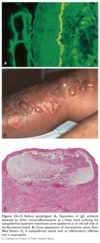
tiny black dots in verruca vulgaris respresents what
thrombosed dillated capillaries
Mycetoma/ Madura foot/ Maduromycosis etiology

Mycetoma, also known as Madura foot and maduromycosis, is a chronic, granulomatous, subcutaneous, inflammatory disease caused by filamentous bacteria (actinomycetoma) or true fungi (eumycetoma). The organisms enter the skin by traumatic inoculation. Both forms of mycetoma present as a triad of progressive subcutaneous swelling with sinus tracts that discharge grains (Fig. 15.29).
Mycetoma is divided into actinomycetoma, produced by bacteria, and eumycetoma, produced by fungi Actinomycetomas are caused by Nocardia asteroides, N. brasiliensis, N. caviae, N. otitidiscaviarum, Actinomadura madurae, Actinomadura pelletieri, Actinomyces israelii (the major cause of lumpy jaw), and Streptomyces somaliensis. Eumycetomas are caused by fungi, including pigmented fungi such as Madurella spp., and hyaline fungi such as Pseudallescheria.
Reiter syndrome

narrow, white transverse bands occurring in pairs as a sign of chronic hypoalbuminemia.

Muehrcke Lines
the disturbance appears to be in the nail bed
congenital varicella and congenital varicella results when maternal infection occurs during
first 20 weeks AOG
Katayama fever / urticarial fever
Schistosomiasis
Katayama fever is a manifestation of acute schistosomiasis. Typical features include fever, an urticarial rash, enlarged liver and spleen, and bronchospasm. The precise pathogenesis of Katayama fever is unknown, but it is thought to be an immune complex phenomenon, initiated by eggs laid by maturing schistosomes.
describe the course of measles
predrome of fever, malaise, conjunctivitis URTI that may last 4 days. Rash may appear 1 - 7 days after prodrome. erythematous macules and papules that spreads quickly over the face down to the trunk and extremities (cephalocaudal and centrifugal).
what is auspitz sign
removal of scales discloses bleeding points
differentiate Trichophyton tonsurans and microsporum canis based on wood’s light fluorescence
Trichophyton tonsurans (-) fluorescence - endothrix
Microsporum canis (+) fluorescence - ectothrix
a form of rhinitis which is the most frequent and often the 1st specific finding of early congenital syphilis
Snuffles
General mechanism for Type 2 reaction in leprosy
Immune complex-mediated vasculitis, with most of hte complexes forming locally rather than circulating
*there is in situ generation of membrane attack complex
*occur in lepromatous patients (BL, LL)
*(+) systemic symptoms
*do not occur at sites of existing lesions
Treatment of type 1 reaction in leprosy
Steroids
In pemphigus vulgaris, acantholysis selectively involves the layer of cells immediately above the basal cell layer, giving rise to a ___________.
suprabasal acantholytic blister (Fig. 23–10, B)

Bullous systemic lupus erythematosus (SLE) and EBA demonstrate clinical and histologic overlap. How can you differentiate them?
EBA: skin fragility, predilection for traumatized areas, and healing with scars and milia.
bullous SLE: sun-exposed skin is involved by preference, and the patient has a diagnosis of SLE established by American College of Rheumatology criteria; bullous SLE patients usually have a dramatic response to dapsone.
show the proximal portion of the nail white and the distal half red, pink, or brown, with a sharp line of demarcation between the two halves
Half and Half Nails
. About 76% of hemodialysis patients and 56% of renal transplant patients have at least one type of nail abnormality. Half and half nails are the most common, affecting 20% of hemodialysis patients.
Subacute Cutaneous LE

causative agent of erythrasma
Corynebacterium minutissimum
pruritic tapioca-like deep seated vesicles over the lateral surface of the fingers
Dyshidrotic eczema / pompholyx

Disorder that is exacerbated by wheat-rich foods
Dermatitis Herpetiformis

parameters of SCORTEN

Also known as fetid sweat, osmidrosis, and malodorous sweating,
Bromhidrosis
allylamines are active against
Dermatophytes > yeast
PAPA syndrome
Pyogenic sterile arthritis
Pyoderma gangrenosum
Acne
Treatment for pediculosis capitis
1% Permethrin Shampoo - apply to dry hair and scalp in sections. Leave on and occlude for 10 - 15 mins
*appying to dry hair lessens the dilution of the medication
allergen in rubber
carbamates
most common neurologic complications of varicella
cerebellar ataxia and encephalitis
also known as circumscribed neurodermatitis
Lichen simplex chronicus
distribution of pityriasis rosea
langer lines/ long axis runs parallel to the line of cleavage
“stork bite”

Nevus simplex / nevus flammeus
*pink-red macule on the posterior midline between the occipital protruberance and the tip of the spine of the 5th cervical vertebra
one of ketoconazole’s side effect
hepatotoxicity
HPV types that cause Verruca plana (flat warts)
HPV Type 3, 10, 28, 41
broken off hairs, comma hair

Tinea capitis
3 types of nevus
Junctional Nevus - dermoepidermal junction; dark
Compound Nevus - Junction + DERMIS
Intradermal Nevus - DERMIS; depigmented / skin-colored
*superficial - darker + flatter
+deeper - lighter + more elevated
“angel’s kiss”

Nevus Simplex / nevus flammeus
*located on the glabella
Flag sign

Kwashiorkor
The hair is hypopigmented, varying in color from a reddish yellow to gray or even white The hair is dry and lusterless; curly hair becomes soft and straight; and marked scaling (crackled hair) is seen. Especially striking is the “flag sign,” affecting long, normally dark hair. The hair grown during periods of poor nutrition is pale, so that alternating bands of pale and dark hair can be seen along a single strand, indicating alternating periods of good and poor nutrition.
chronic inflammatory disease characterized by minute, shiny, flat-topped, pale, discrete, uniform papules, rarely larger than 1-2 mm.

Lichen nitidus
Kala-azar/ Black fever
Visceral leishmaniasis
blanchable
Erythema - due to vasodilation
What medication whould you avoid in treating Tinea
avoid POTENT TOPICAL STEROIDS because this may cause widespread tinea and fungal folliculitis
Sign that refers to involvement of the external division of the nasociliary branch of CN V1 (ophthalmic division) that presents with vesicles on the side and tip of the nose
Hutchinson’s Sign
seen in Herpes zoster

what areas are rarely involved in tinea cruris
penoscrotal folds, scrotum and penis
describe the course of varicella
prodrome of low grade fever, malaise and headache followed by appearance of exanthem 2-3 days later. Lesions begin on the head (face and scalp) spreading inferiorly to the trunk. there is relative sparing of the extremities, palms and soles. There is rapid progression of lesions ( 8 -12 hours) from rose colored macules to papules, vesicles, pustules and crusts.
How can you differentiate SSSS from SJS?
SJS/TEN - (+) mucosal involvement; separates at the DEJ
SSSS - SPARES mucous membranes, palms and soles; separates at the granular layer
give differentials for subacute cutaneous LE
Tinea
Psoriasis
patches produce powdery scales when scratched
furfuraceous/branny scales of tinea versicolor
microsporum metabolite that causes the (+) fluorescence in wood’s light

Pteridine
What type of cell is present in the blood smear of TTP and is considered as morphologic hallmark of the disease?
Schistocytes

causative agent of Tinea pedis
T. mentagrophytes (interdigitale)
causative agent of Yaws
T. pallidum subs. pertenue
lesions following the lines of cleavage
pityriasis rosea
surface glycolipid unique to the leprosy bacillus
Phenolic glycolipid 1 (PGL-1)
extension of a blister to adjacent unblistered skin when pressure is put on top of the blister
Asboe-hansen sign
The affected hair shafts fracture easily and may have small white nodes arranged at irregular intervals. These nodes are the sites of fraying of the hair cortex. The splitting into strands produces a microscopic appearance suggestive of a pair of brooms stuck together end to end by their bristles. The hairs soon break at these nodes

Trichorrhexis Nodosa
two peaks in age of onset of leprosy
10 - 20 years and 30 to 60 years
allergen in preservatives
parabens
Neutrophils accumulate in the stratum spinosum in psoriasis
Pustules of kogoj
SAPHO Syndrome
Synovitis
Acne
Pustulosis
Hyperostosis
Osteitis
Swiss cheese appearance in leprosy
Borderline borderline leprosy (BB)
Follicular occlusion Triad
- Dissecting cellulitis of the scalp
- Hidradenitis suppurativa
- Acne conglobata
most common fungal infection
Tinea pedis
MC caused by T.rubrum
primary lesion of scabies
papulovesicles
used as treatment for nodular scabies
Ivermectin
rapidly growing papule that enlarges from a 1-mm macule or papule to as large as 25 mm in 3–8 weeks. When fully developed, it is a hemispheric, dome-shaped, skin-colored nodule that has a smooth crater filled with a central keratin plug

Keratoacanthoma
Treatment for multibacillary leprosy
Dapsone, Rifampicin, Clofazimine
treatment options for zoster-associated pain (Postherpetic neuralgia)
- gabapentin 300 mg TID; Pregabalin
- Tricyclic antidepressants such as doxepin 10 - 100 mg PO at bed time
- capsaicin cream every 4 hours
*
- capsaicin cream every 4 hours
an idiopathic disease of collagen necrobiosis

Granuloma Annulare / Necrobiotic papulosis
* lesions are erythematous to violaceous, thinly bordered plaques/papules that slowly spread peripherally while undergoing central involution
*lack of scales may differentiate this to other annular scaling lesions like tinea

Milian’s ear sign
Erysipelas
Ear involvement is called Milian’s ear sign, and it is a feature distinguishing erysipelas from cellulitis, because the pinna has no deeper dermis and subcutaneous tissue (2). Bilateral ear erysipelas is rare, but it should be considered in the differential diagnosis when encountering patients presenting with red ears.
The lesions of dermatitis herpetiformis are bilateral, symmetric, and grouped and preferentially involve the extensor surfaces, elbows, knees, upper back, and buttocks (Fig. 2313, B). Initially, neutrophils accumulate selectively at the tips of dermal papillae, forming small microabscesses (Fig. 23–13, C). The basal cells overlying these microabscesses show vacuolization and focal dermoepidermal separation that ultimately coalesce to form a true _____________
subepidermal blister
B, Lesions consist of intact and eroded (usually scratched) erythematous blisters, often grouped (seen here on elbows and arms).
C, The blisters are associated with basal cell layer injury, initially caused by accumulation of neutrophils (microabscesses) at the tips of dermal papillae.

hanging curtain sign
Pityriasis Rosea

This finding is present in all patients with Hemolytic Uremic Syndrome and is the hallmark of the disease
Renal insufficiency
Erythema migrans

Lyme disease
*there is gradual expansion on the redness around the papule; the advancing border is usually slightly raised, warm, red to bluish red and free of any scale
causative agent of pediculosis corporis (body louse)
Pediculosis humanus var. corporis (body louse)
umbilibated pustule
vaccinia virus
type of psoriasis that occurs usually after an infection
Guttate psoriasis

Kligman’s Formula
- Hydroquinone + tretinoin
- Topical corticosteroid
*most effective in treating melasma
also known as “beaded hairs,”
It is characterized by dryness, fragility, and sparseness of the scalp hair
Monilethrix
special stain used in histopathologic specimens of hansen’s disease
Fite faraco stain
can be used for both candidal and dermatophyte onychomycosis
Itraconazole (triazole)
concave, sulfuryellow crusts arranged around loose wiry hair, atrophic scarring results to smooth, glossy, paper-white patch
superficial dermatophyte infection usually caused by T schoenleinii and characterized by scutula (cupshaped crust)

multiple yellowish papules, sometimes with a central dell, with overlying telangiectasia

Sebaceous hyperplasia
*benign enlargement of ht esebaceous glands

conditions positive for auspitz sign
seborrheic dermatitis
psoriasis
give examples of Class 1 superpotent steroids
Betamethasone dipropionate 0.05% ointment
Clobetasol propionate 0.05% ointment
describe the course of scarlet fever
- affects primarily children, who develop the eruption 24–48 hours after onset of pharyngeal symptoms
- tonsils are red, edematous, and covered with exudate.
- tongue has a white coating through which reddened, hypertrophied papillae project, giving the so-called white strawberry tongue appearance (as opposed to the red strawberry tongue of Kawasaki that lacks an exudate).
- By the fourth or fifth day the coating disappears, the tongue is bright red, and the red strawberry tongue remains.
- The cutaneous eruption begins on the neck then spreads to the trunk and finally the extremities (Fig. 14.15).
- Within the widespread erythema are 1–2 mm papules, which give the skin a rough, sandpaper quality.
- There is accentuation over the skinfolds, and a linear petechial eruption, called Pastia lines, is often present in the antecubital and axillary folds.
- There is facial flushing and circumoral pallor.
- A branny desquamation occurs as the eruption fades, with peeling of the palms and soles taking place about 2 weeks after the acute illness.
considered cutaneous markers of diabetes mellitus
acrochordon
infection wchich involves the superficial dermal lymphatics
Erysipelas
collarette scaling

Pityriasis rosea
gold standard in the diagnosis of ACD
patch test
antibodies with high specificity for ACTIVE NEPHRITIS is SLE
anti-dsDNA and Anti-C1q antibody
Differential diagnosis of sun esposed areas
- Subacute cutaneous Lupus erythematosus
- Phototoxicity
- Photoallergy
etiologic agent of Tinea imbricata
T. concentricum
major complication of herpes zoster
post-herpetic neuralgia
MOA of ALLYLAMINES (Terbinafine, Naftifine, Butenafine)

what type of hypersensitivity reaction are Pemphigus vulgaris and foliaceus?
antibody-mediated (type II) hypersensitivity reactions
*pathogenic antibodies are IgG autoantibodies that bind to intercellular desmosomal proteins (desmoglein types 1 and 3) of skin and mucous membranes. The antibodies disrupt the intercellular adhesive function of the desmosomes and may activate intercellular proteases as well. The distribution of these proteins within the epidermis determines the location of the lesions. By direct immunofluorescence study, lesional sites show a characteristic fishnet-like pattern of intercellular IgG deposits (Fig. 23–9). As in other autoimmune diseases, pemphigus shows linkage with particular HLA alleles.
A technique used to differentiate pemphigoid group from other Subepidermal immunobullous disease
Salt Split technique
Useful in determining the site of deposition of autoantibodies
Punch biopsy samples were incubated in 5 mL of NaCl (1 mol/L) at 4ºC for 24 h. The epidermis was then teased from the dermis with the use of a fine forceps. The specimens were then processed in the same manner and treated with IgG and C3 conjugates as in DIF.
This technique involves artificially splitting the skin at the level of lamina lucida by incubating it in 1 M solution of sodium chloride for 24 h.
By DIF testing of the patient’s salt-split-skin biopsy, EBA will manifest IgG deposition only on the dermal side of the split, whereas the majority of BP patients will have IgG bound only to the epidermal side or to both sides.
etiology of kerion
T. mentagrophytes, verrucosum, tonsurans and violaceum
“BOTE” (Beginning of the end) Sign

Molluscum contagiosum
It has described a phenomenon called “beginning of the end” (BOTE) sign which refers to clinical erythema and swelling of an MC skin lesion when the regression phase begins (Figure 2). This phenomenon is likely due to an immune response towards the MC infection rather than a bacterial superinfection.
Inflamed MC is characterized by erythema and swelling of the individual lesions, sometimes with pustulation or fluctuance. It occurs in 20% of children with MC and usually heralds the resolution of disease. This has been named the “BOTE” sign (beginning of the end) and can be helpful for parents who are concerned about the redness and pain to understand that the infection may be close to resolution.
inclusion bodies of molluscum contagiosum

Henderson Paterson bodies

diagnosis?

Pterygium caused by lichen planus
(+) satellite lesions around macules or plaques in leprosy
Borderline Tuberculoid (BT)
Acrodermatitis Enteropathica

Zinc deficiency
The dermatitis found in all forms of zinc deficiency is pustular and bullous, but the lesions are often flaccid leading to the clinical appearance of crusting. There is an acral and a periorificial distribution. On the face, in the groin, and in other flexors there is a patchy, red, dry scaling with exudation and crusting (Fig. 22.7). Angular cheilitis and stomatitis may be present.

give the doses of MDT in multibacillary leprosy
- Rifampin 600 mg and Clofazimine 300 mg once a month
- Dapsone 100 mg and Clofazimine 50 mg daily
for 12 months
Drug of choice for Chancroid
Azithromycin 1g or Ceftriaxone 250 mg IM single dose
shortes phase of the hair cycle
catagen
hallmark of myelofibrosis
extramedullary hematopoiesis
appearance of lesions at various stage of evolution
Varicella

Drug of choice for Herpes simplex
Acyclovir 200 mg 1 tab q4 5x/day for 7 days
give differentials for verruca plantaris
Callus - exaggerated skin markings; (+) dermatoglyphics
Corn - hyperkeratotic papule with a central core
give examples of triazoles (Azoles)
itraconazole
fluconazole
give differentials for tinea capitis
- alopecia areata - affected areas are bald, smooth skin and shiny without inflammation and scales
- seborrheic dermatitis - involved areas are covered by fine, whitish dry or greasy yellowish scales
- psoriasis - well demarcated areas of erythema with silvery white scales, MAY EXTEND BEYOND THE HAIRLINE
- lichen simplex chronicus - accentuated skin markings usually localized to the inferior occipital scalp; negative KOH and fungal culture
-
Trichotillomania - stubs of broken hair due to hair pulling; no inflammation and scaling
*
abnormal result in hair pull test
~ 40 strands of hair are pulled and a count of 4-6 is considered abnormal
Etiology of hand-foot-and-mouth disease
Coxsackievirus A16
Dimple Sign

Dermatofibroma
refers to the downward movement of a dermatofibroma when it is pinched
Samitz Sign
Dermatomyositis
*cuticular overgrowth with an irregular frayed appearance
* Dystrophic and ragged cuticles (circles)
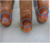
what enzyme is inhibited by azoles?
lanosterol 14a-demethylase - which is an essential enzyme for ergosterol (component of the fungal cell membrane) synthesis
Why can’t we give tetracycline and isotretinoin at the same time?
May caue pseudotumor cerebri AKA idiopathic intracranial hypertension (benign intracranial swelling)
Leprosy
Countable, irregular-shaped ASYMMETRICAL
Borderline Borderline (BB)
Leprosy
Too many to count lesions; SYMMETRICAL
Borderline lepromatous
etiologic agent of favus
T. schoenleinii
etiolodic agent of seborrheic dermatitis
Malassezia ovalis (Pityrosporum ovale)
HHV-8
Kaposi Sarcoma
transverse furrows that begin in the matrix and progress distally as the nail grows

Beau Lines
They are ascribed to the temporary arrest of function of the nail matrix.
wood’s lamp finding of favic (T. schoenleinii)
Bluish white or dull gray-green fluorescence
Treatment for mild acne

Touton giant cells containing hemosiderin is pathognomonic to what disease

Dermatofibroma (Fibrous Histiocytoma)
etiologic agent of gray patch tinea capitis
Microsporum species such as M. audouinii
etiologic agent for tinea cruris
T. rubrum, mentagrophytes and E. floccossum
PASS Syndrome
Pyoderma gangrenosum
Acne conglobata
Suppurative Hidradenitis
Axial Spondyloarthritis
What sign is this?

Schamroth sign is observed when the normally appearing diamond-shaped window that is formed by placing the dorsal surfaces of opposite terminal phalanges together disappears. Seen in clubbing fingers
MOA of Vitamin D (Calcipotriol, Calcitriol) in psoriasis
controls keratinocyte proliferation and promotes differentiation
differentials for Tinea faciei
- Seborrheic dermatitis - usually yellowish. greasy scales on the nasolabial folds, glabella, preauricular, retroauricular ares; absence of central clearing and raised borders
- contact dermatitis - history of exposure to contactenat. no central clearing and negative for KOH
- Pityriasis alba - hypopigmented patch with fuzzy border and slight scaling; absence of advancing border and central clearing
- erythema migrans
- lupus erythematosus
- Polymorphic light eruption
- phototoxic drug eruption
- lymphocytic infiltrate
suppurative inflammation of subcutaneous tissue
cellulitis
Vesicles or vesiculopustule with erythematous base and surrounding halo that enlarges over days and crusts - superficial saucer-shaped ulcers with raw base with an indurated ulcer margin; granulating base which extend deep into dermis
Ecthyma
features that will differentiate bullous pemphigoid to other immunobullous diseases
- subepidermal separation at the DEJ,
- an inflammatory cell infiltrate that tends to be rich in eosinophils, and
- antibodies directed against two hemidesmosomal antigens, BP230 and BP180.
How do you do pathergy test
It is done by injecting 0.1 mL of normal saline solution into the skin or by simply pricking the skin with a sterile needle. A pustule appears at the site within 24 hours. If results are negative, the test should be repeated at two to five points before results are accepted.
Cutaneous larva migrans / creeping eruption

Hookworm
differentiate erysipelas and cellulitis based on borders
- Erysipelas - with well-defined border
- cellulitis - ill-defined border
allergen in hair dyes
paraphenylenediamine
most common type of fungal infection of the nail plate
distal subungual onychomycosis

represent remnants of the inner root sheath

Hair Casts (Pseudonits)
They often occur in great numbers and may mimic nits in the scalp. Whereas nits are firmly cemented to the hair shaft, however, hair casts slide freely along the shaft.
explain itch scratch cycle
–pruritus leads to scratch and scratching causes secondary changes that causes itching
seen in atopic dermatitis and lichen simplex chronicus
Splitting of the distal nail plate into layers at the free edge is a very common problem among women and in 30% of newborns. It represents a dyshesion of the layers of keratin, likely as a result of dehydration.

Onychoschizia
Double upper lip sign

Ascher Syndrome
Ascher syndrome consists of progressive enlargement of the upper lip and blepharochalasis (eyelid skin becomes lax and falls in redundant folds over the lid margins)
Mechanic’s Hand

Dermatomyositis
Mechanic’s Hands is a descriptive term for the changes in the hands of patients with dermatomyositis and polymyositis. Mechanic’s hand is usually associated with anti-synthetase autoantibodies, including the myositis-specific anti-Jo1 autoantibodies.
Characterized by hyperkeratosis, scaling, fissuring, hyperpigmentation over the fingertips, sides of the thumb and fingers and occasional involvement of the palms
single most important prognostic factor for melanoma
Presence or absence of melanoma in regional lymph nodes
drugs used for candidal onychomycosis
Azoles (itraconazole, fluconazole)
Precaution: liver disease
type of hypersensitivity reaction of ACD
Type IV
type of psoriasis that may occur after an acute infection, such as STREPTOCOCCAL PHARYNGITIS
GUTTATE PSORIASIS

leprosy patients are no longer infectious after how many months of treatment
after 1 month of treatment
Winterbotton sign
Trypanosomiasis
swelling of lymph nodes (lymphadenopathy) along the posterior cervical lymph node chain, associated with the early phase of African trypanosomiasis (African sleeping sickness), a disease caused by the parasites Trypanosoma brucei rhodesiense and Trypanosoma brucei gambiense.

absolute contraindications to prednisolone treatment in leprosy
- peptic ulcer
- psychosis/depression
- acute or chronic bacterial infection
etiologic agent of dandruff
Pityriasis sicca
Crazy pavement skin, crackled skin, mosaic skin, enamel paint, plaky paint

Kwashiorkor
most commonly affected site of tinea pedis
3rd toe web
compare seborrheic dermatitis and psoriasis
Psoriasis is (+) auspitz sign, more erythematous and GOES BEYOND the hairline.
On the otherhand, another differential is Tinea capitits. this does not go beyond the hairline.
sensitization period of scabies
2 - 4 weeks after infection
longitudinal, red-brown haemorrhage under a nail and looks like a wood splinter.

Splinter hemorrhages
Splinter haemorrhages are present in 15–33% of patients with infective endocarditis in association with Osler nodes and Janeway lesions [3]. They may be due to septic emboli in the small vessels of the nail bed and the increased fragility of the vessel walls in sepsis [2,3].
Splinter haemorrhages are common signs of psoriatic nail disease and nail disease due to lichen planus [3]. They can also be associated with a tumour.
Splinter haemorrhages may be due to microemboli or injury to vessel walls associated with vasculiti
characterized by dehydrated skin showing redness, dry scaling and fine crackling that may resemble crackled porcelain or the fissures in a dried lake

Asteatotic dermatitis/ winter itch/ eczema craquele/ asteatotic eczema
smears of pus from pustular psoriasis will reveal
neutrophils with no organisms
larva currens
Strongyloides stercoralis
Intestinal infections with Strongyloides stercoralis may be associated with a perianal larva migrans syndrome called larva currens because of the rapidity of larval migration (currens means “running” or “racing”). Larva currens is an autoinfection caused by penetration of the perianal skin by infectious larvae as they are excreted in the feces.
causative agent of scabies
sarcoptes scabiei
DOC for all stages of syphilis
Penicillin
KOH finding of candida
budding yeast and pseudohyphae
Pemphigus foliaceus has antibodies that target what?
Desmoglein 1
causative agent of sporotrichosis
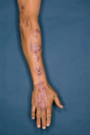
Sporothrix schenckii
* nodules develop along the draining lymphatics
causative agent of pediculosis pubis
Phthirus pubis (pubic or crab louse)
Examples of intraepidermal bullous dermatitis
Pemphigus vulgaris, SJS, SSSS, Erythema multiforme
- blisters are FLACCID, (+) Nikolsky sign and (+) Asboe-hansen’s sign
Triad of Scabies
- circle of hebra
- nocturnal itch
- (+) contact people at home
thinning of eyebrows seen ion atopic dermatitis

Hertoghe’s sign (Atopic Dermatitis)
