Module Workshops Flashcards
Describe the locations and functional associations of the spinal cord grey and white matter
Grey matter: Contains the cell bodies of neurones
White matter: Contains the myelinated neuronal processes

Outline the changes observed in transverse sections of the spinal cord at different regions
The beginning of the spinal cord is marked by the emergence of the first pair of spinal nerves.
Thoracic region: Increased white matter seen due to the large number of nerves exiting the spinal cord in this region
From T1-L2 a lateral horn is seen - indicates the cell bodies of sympathetic neurones.
Cervical region: The dorsal column medial lemiscus pathway has 2 elevations. Only 1 is seen in the lumbar region (as gracilias cuneatus is for the upper limb)
Outline the function and route of the main descending and ascending tracts
Outline the positions of the ascending and descending tracts within the spinal cord

Describe the arterial supply to the spinal cord.
Clinical link: Outline the potential consequences of blockage to these vessels and how this may be induced
Great radicular artery (Artery of Adamkiewicz): Reinforces blood supply to the distal parts of the spinal cord
Causes of spinal ischaemia: Vertebral fracture; vascultic disease; external compression
Potential consequences
Anterior spinal artery: Loss of descending pathways hence loss of voluntary and involuntary motor movements. Loss of some ascending pathways (spinothalamic and spinocerebellar) leading to loss of pain and temperature sensation alongside reduced co-ordination. Loss of spinal reflexes
Posterior spinal artery: Loss of the dorsal column medial lemniscal pathway, causing loss of fine touch, vibration and unconscious proprioception sensory input.

Describe the autonomic outflow from the CNS
Sympathetic: T1 - L2
- Cell bodies are seen in the lateral horn of the gray matter
- Travel in the sympathetic chain.
3 possible routes:
- Synapse within the paravertebral ganglia. Re-enter the spinal nerve via the gray rami communicantes. Travel to the periphery
- Exits the ganglia without synapsing. Travel with splanchnic nerves to distal ganglia where they then synapse. Post-ganglionic fibres then innervate visceral structures.
- Travel up or down the sympathetic chain.
- Fibres travel from the spinal nerve to the sympathetic chain (paravertebral ganglion) via the white rami communicantes. They are able to re-enter the spinal nerve from the sympathetic chain via the grey rami communicantes.
- Ganglion situated around the body. Tend to closer to the CNS than those to which the parasympathetic fibres travel
Parasympathetic: Cranial nerves (CN V, VII and IX) and S2-S4
- Travel to ganglia
Outline the route taken by the sympathetic nerve supply to the head and neck
Clinical link: Explain Horner’s syndrome and the potential lesion sites
Originate from T1 - T6
Sympathetic fibres travelling to the head and neck exit the spinal nerve and travel to the sympathetic chain via the white rami communicantes.
There are 3 ganglia of the chain related to the head and neck, which are in turn related to specific arteries:
Superior (internal carotid artery); middle (inferior thryoid artery) and inferior (vertebral and subclavian arteries) cerival ganglia.
Sympathetic fibres synapse at these ganglia with post-ganglionic structures continuing to supply the head and neck. These post-ganglionic fibres ‘hitch-a-ride’ with their assoicated arterial vessels.
Horner’s syndrome: Results from loss of sympathetic input to the head. Characterised by ptosis, anyhydrosis and miosis.
Ptosis: Eyelid drooping due to loss of superior tarsal muscle innervation
Miosis: Pupillary contriction due to loss of dilator pupillae innervation
Causes: Spinal cord lesion, traumatic injury, Pancoast tumour (apex of the lung),
Identify and describe the major parts of the eyeball
* Central retinal artery and vein travel in the optic nerve
* Fovea centralis: A area condensed with photoreceptor cells, seen at the centre of the macula. Allows for high visual acuity
* Optic disc: Devoid of photoreceptor cells. Allows the passage of the optic nerve and blood vessels.
* Cornea = a transparent continuation of the sclera
* Conjuctiva: Mucous membrane covering of the eyelid and eye. Maintains moisture
* Ciliary muscles and associated suspensory ligaments: Adjust the convexity of the lens
Describe the movements of the eye and their associated muscles
Clinical link: Describe the clinical testing for these muscles
Clinical testing: Superior/inferior obliques and rectus muscles swap
State and explain the resting position and appearance of the eye with a given injury to CN III, IV or VI
Describe the visual pathway
Describe the different visual field defects that may result due to damage to the visual pathway

* Macula is spared due to its dual blood supply from PICA and the middle cerebral artery

Orbit: Identify component bones, anatomical relations and contents
Outline the innervation of the lacrimal gland and the drainage of secretions
Innervation:
Sensory from lacrimal nerve, a branch of Va
Parasympathetic from greater petrosal (CN VII) → maxillary (Vb) → zygomatic nerve. Stimulates fluid production.
Sympathetic orginate from the superior cervical ganglion and then follow the same route as parasympathetic fibres. Inhibit fluid production.
Drainage: To the nasal cavity via the lacrimal lake → lacrimal sac → nasolacrimal canal
Describe the corneal reflex
Afferent: Nasocoliary branch of the ophthalmic branch of the trigeminal nerve (Va)
Efferent: Temporal branch of the facial nerve (VII)
Elicited by using a wisp of cotton wool to touch the cornea.
Describe the pupillary reflex
***DOES NOT INVOLVE THE PRIMARY VISUAL CORTEX OR THE LGN (THALAMUS)***
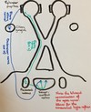
Describe the accommodation reflex
Describe the role and relative importance of the cornea and lens in the focusing power of the eye
- The cornea provides the majority of the eyes focussing power, through the refraction of the incidence of light
- The cornea is a continuation of the sclera and is avascular.
- The lens is also able to refract light and may do so to varying degrees through adjustment of its convexity. The lens provides ‘fine tuning’ in the refraction of light.
Describe astigmatism
Resultant of an ‘abnormal’ morphology of the eye which leads to refractive error. The cornea forms a more oval shape (rugby ball), instead of round. This changes the path of light meaning that the visual image is not focussed sharply. This causes blurred vision, headaches and eye strain. In young children a high astigmatism may cause lazy eye.
Treatment: Glasses, contact lenses, laser eye surgery
Describe the mechanism by which the shape of the lens is altered
The shape of the lens is altered by the ciliary muscles (a component of the ciliary body).
Contraction of the ciliary muscles relaxes the suspensory ligaments. The lens increases in convexity, becomes more round and decreases in size. This allows greater refractive power.
Describe the structural abnormalities which lead to myopia and heterometropia (hyperopia)
Myopia:
- Short sightedness
- The distance between the lens and the retina increased causing light to be focussed in front of the retina
- This abnormality can be corrected using a convex/minus lens
Heterometropia:
- Long sightedness
- The distance between the lens and the retina is decreased causing light to be focussed behind the retina
- This abnormality can be corrected using a concave/plus lens
Describe the effect of aging on the lens and how this leads to presbyopia
With the age the lens becomes thickened and hardened, reducing its flexibility/elasticity.
Presbyopia: The increasing long sightenedness associated with aging. Resultant of changes in the elasticity of the lens
Identify the bones of the skull, foramina and sutures
Outline the structures passing through the foramen of the skull and potential consequences of fracture to their component bones or compression of their contents
Pontomedullary junction
Cerebellomedullary angle
Pontomedullary junction: Seen on the ventral surface. Defined by the angle of the lower border of the pons and the upper border of the medulla.
- Origin site of abducens (VI) nerve
Cerebellopontine angle: Formed by the junction of the pons, medulla and cerebellum
- Cerebellar flocculus, ventricular choroid plexus and emerging CNs VII and VIII surround the lateral apertures of the fourth ventricle
- Origin site of facial (VII - motor), nervus intermedius (sensory and autonomic fibres of VII) and the vestibulocochlear nerve (VIII) NOTE: The glossopharyngeal nerve (CN IX) arises from the posteriorlateral sulcus (alongside X and XI)
-
Clinical link:
- Often the site of intracranial growths, with initial signs linked to the impingement of CN VIII
-
Vestibular schwannomas are the most common growth in the area. They are benign and slow growing but complications arise due to their compression of nearby structures.
- Symptoms: Ipsilateral hearing loss, tinnitus and dysequilibrium (due to CN VIII compression); nystagmus (due to cerebellar flocculus compression). Untreated large tumours will lead to compression of nearby structures, CN V and CN VII (loss of facial sensation, facial expression, reduced corneal reflex)
Identify the lobes of the brain and the principle features of cerebral hemispheres
List the main features and function of the brain stem
Describe the structure and function of the ventricular system, relate the postion of of the system to parts of the brain and brainstem
Clinical link: Be able to deduce the potential outcomes of a swelling/ tumour in a given part of the system
Lateral ventricles from both hemispheres → intraventricle foramen → third ventricle → cerebral aqueduct → fourth ventricle → 2 x lateral and 1 medial apertures
Function of the ventricular system
The choroid plexus of the system produces CSF, through filtration of blood. CSF acts to provide nutritional support to the cerebrum. It also acts as a protective mechanism, through cushioning the tissue.
Anatomical relations:
The lateral ventricles are seen superior to the thalamus whilst the third ventricle is medial to the thalamus.
The fourth ventricle is closely related to the pons, with the floor of the fourth ventricle seen on the dorsal surface.
The lateral aperture is seen at the pontomedullary junction.
Potential outcomes of obstruction
Describe the main functions of the thalamus and be able to identify its key anatomical relations
Functions:
- A relay station for the distribution of sensory information with nearly all sensory information passing through the thalamus, allowing thalamic nuclei to send sensory fibres to the cortex.
- EXCEPTION: Olfaction (via CN I) does not pass through the thalamus. This sensory information is sent to the temporal lobe. Linked to embryological origin (from telecephalon)
- Relays information from the ipsilateral cerebral cortex to the outside world via cortico-fugal neurons
Key anatomical relations:
- Lateral to the 3rd ventricle
- Medial to the internal capsule
- Posterior to the caudate nucleus
- Superior to the sub- and hypothalamus
- Rostral to the midbrain

Outline the structure and functions of the following neurones:
- Pseudounipolar
- Bipolar
Pseudounipolar: A sensory neurone of the peripheral nervous system. The axon is split into 2 branches, with one running to the periphery and the other to the spinal cord.
Bipolar: Has 2 extensions, 1 axon and one dendrite. Act as specialised sensory neurones for the transmission of special sensory information - smell, sight, taste, hearing and vestibular system e.g. the ganglia of the vestibulocochlear nerve and the bipolar cell of the retina.

Deduce the functional consequences of damage to a given area of the thalamus or area damaged from a given set of symptoms
Periphery → thalamus = CONTRALATERAL
Thalamus → cerebral cortex = IPSILATERAL
Left thalamic region lesion → Symptoms on the contralateral of the body, loss of sensation to the right side of the body
State the thalamic nuclei and outline their functions
State the visual defect that would be experienced following a lesion at the sites indicated
Complete the sensory receptor table


Outline the structure of the Dural venous sinuses
Draw diagram - state location
This sinuses are formed at the sites of separation between the periosteal and meningeal Dura matter. The venous blood contained within the sinuses eventually drains into the internal jugular vein.

State the key anatomical sites of the tongue
Draw diagram

Outline the sensory and special sensory supply to the tongue

State the processes associated with the TMJ and the process responsible for articulation at the joint

Outline what would be observed in the case of a unilateral and a bilateral lesion to the nerves supplying the muscles of mastification
Unilateral: Deviation of the jaw towards the side of the lesion
Bilateral: Dropping of the jaw
Outline Bell’s Palsy
A unilateral palsy of the facial nerve which fully evolves over a period of 72 hours - important aspect for Ddx.
Temporary weakness or lack of facial movement results.
Likely to be viral aetiology.
Usually self resolves within 9 weeks.
Outline Trigeminal Neuralgia
Resultant of irritation of a branch of the Trigeminal nerve, most commonly caused by compresson of the nerve by a vascular loop (superior cerebellar artery).
Compression of the nerve leads to severe unilateral pain (lasting seconds to minutes) which may be worsened by exposure to cold.
DO NOT MISS - Temporal/Giant cell arteritis: Arteritis. Inflammation of the external branches of the carotid artery (most commonly). The most common serious consequence is irreversible blindness due to optic nerve ischaemia.
Identify and describe the lobes of the brain and the functional associations of each lobe
Describe association, commissural and projection fibres
Association: Fibres which connects different regions of the same hemisphere
Commissual: Fibres which connect the right and left hemispheres
e.g. corpus callosum
Projection: Fibres from travel between the cerebrum and the brainstem and/or spinal cord
e.g. Corona radiata
Identify the main features of cerebral hemispheres on transverse and coronal sections

Outline the contributors and components of the vertebrobasilar arterial system and circle of Willis
Describe the parts of the brain supplied by branches of the vertebrobasilar arterial system
Draw transverse and coronal sections
Outline the consequences of loss/blockage of blood supply to areas of the brain
Identify the main anatomical features of the oral cavity and submandibular region
Describe the anatomy of the tongue and outline its sensory, motor and taste nerve supply
Describe the sensory and motor innervation of the face
Clinical link: Outline the functional consequences of loss of innervation to the muscles of facial expression
Sensory innervation: The main divisions of the trigeminal nerve (Opthalmic, maxillary and mandibular)
Motor innervation: Terminal branches of the facial nerve (temporal, zygomatic, buccal, medial mandibular and cervical)
Functional consequences of loss:
- Unable to close the eyelids (orbicularis oculi) except through relaxation of the superior tarsal and levator palpebrae
- Unable to raise the eyebrows, smile or puff out of the cheeks
- Unable to keep food/drink within the mouth whilst eating - no ‘compression’ of the cheeks by buccinator
- Loss of orbicular oris leads to inabilty to suck or to spit (important if unable to swallow)
Describe the examination findings of a unilateral or bilateral lesion of CN XII
Unilateral lesion: Contralateral paralysis of the lower face, with no paralysis of the forehead
- Due to the bilateral innervation of the upper face/forehead
- The same presentation would be observed in an UMN lesion
Bilateral lesion: Paralysis of both the upper and lower face
Outline the presentation of an UMN and LMN lesion of CN VII, explain why the presentation is different (referring to neural pathways)
UMN: Forehead sparing, due to bilateral innervation of the forehead, with contralateral paralysis of the lower face.
LMN: Ipsilateral forehead and lower face paralysis

Identify and describe the muscle groups and nerves responsible for chewing, swallowing and speaking
Describe the basic anatomy, location, relations and innervation of the salivary glands
Identify the main features and describe the basic functions of the brain stem
Identify the cranial nerves originating from different levels of the brain stem
Describe the structure and basic function of the cochlea
Draw diagram

Describe the pathway of fibres from the cochlea to the primary auditory cortex
Draw pathway

Define sensorineural and conductive deafness. Outline possible causes of each
Sensorineural: A defect within the inner ear leading to inability to convert vibrations from the middle ear into neuronal impulses
- Ototoxicity
- Cochlear pathology: Presbycusis, meningitis, labrinthyitis
- Acoustic neuroma
- Exposure to loud noises e.g. explosions
Conductive: A defect within the conductive pathways of the ear, within the middle ear, leading to an inabilty to pass vibrations along the auditory ossicles to the oval window.
- Damage to the tympanic membrane
- Arthritis (synovial joints of the auditory ossicles)
- External auditory canal obstruction
- Otitis media
Describe the location and basis structure of the vestibular apparatus and describe the function of its static and dynamic parts
Location: Within the petrous part of the temporal bone
Structure:
- Formed from bony and membranous components/labyrinth.
- The bony vestibule contains the membranous saccule and utricle - which form the static part of the vestibular system
- The bony semicircular canals contain membranous structures - forming the dynamic part of the vestibular system
- Perilymph is contained within the bony structures whilst endolymph is seen within the membranous structures
- The membranes of the vestibular system are formed by sensorineural epithelium
- Macula - Movement is detected by deflection of hair cells, caused by the movement of otolith crystals
Function: Detection of movement
Static component
- Detects linear movement of the head
- Saccule detects movement within the vertical plane
- Utricle detects movement within the horixontal plane
Dynamic component
- Semicircular canals detect rotational movements of the head
Anterior - Yes movements
Lateral - No movements
Posterior - Ear to shoulder movements
Describe how the dynamic labyrinth can bring about automatic eye movement, specifically horizontal conjugate gaze only
Draw pathway
☆ The MLF (medial longitudinal fasciculus) is crucial for conjugate eye movement - lesions lead to loss of conjugate movement (intranuclear opthalmoplegia - seen in MS)

Outline the potential range of symptoms associated with damage/disease of the vestibular system and explain the basic mechanism surrounding each
Explain the role of the frontal eye fields in eye movements and explain the consequence of a unilateral lesion to the frontal eye field
Role of the frontal eye field:
- Conjugate eye movement
- Saccadic eye movement
- Allows for the tracking of moving objects - over powers the VOR
Consequence of a unilateral lesion:
- Deviation of the eyes towards the side of the lesion
- Frontal eye field → PPRF → Abducens nucleus → Oculomotor nucleus
*Contrast to PPRF lesion - Eyes deviates away from the side of the lesion
Define the term nystagmus; explain the vestibular cause of horizontal nystagmus and the examination findings in a patient with vestibular system/nucleus damage
Nystagmus: An unconscious repetitive movement of the eyes
Vestibular causes: Changes in the firing rate of the semicircular canals (irritative/damaging) such that turning of the head is mimicked. This leads to unnecessary triggering of the VOR. Saccades are generated to correct this movement, leading to nystagmus.
Examination findings:
- Nystagmus (fast phase) towards the ear with the highest firing rate
Describe the cold caloric test and outline possible findings relating to brainstem and cerebral function
Cold - Direction of nystagmus: Opposite to ear in which cold air/water is introduced
Hot - Direction of nystagmus: To the same side as the ear in which cold air/water is introduced
Scenarios:
- Cold air introduced into right ear. Eyes drift to the right and then slowly back to the midline.
Comatosed patient - no PPRF activity
- Cold air introduced into the right ear. Eyes remain in the midline.
Brain stem death
Identify and describe the basic anatomy of the cerebellum
Describe the function of the cerebellum and its clinical importance
Function: Integral in the learning of skilled motor tasks (require planning); coordination of muscular activity (creates smooth movements); muscular control
NOT involved in the initiation of motor movements
☆ The cerebellum itself has no motor pathways - it influences movement via its connections with other motor pathways e.g. the rubrospinal pathway
Clinical importance:
Damage to the cerebellum leads to ataxia and a distinctive set of symptoms - can be mimicked by excessive alcohol intake.
Cerebellar lesion ⇒ IPSILATERAL symptoms
Describe the functional roles of the cerebellum
Spinocerebellum/paleocerebellum: Maintenance of posture and muscle tone
Damage → Inabilty to sit unsupported, hypotonia
Neocerebellum/cerebrocerebellum: Control of musculature, including speed, power and trajectory. Ensures smooth movements.
Damage → Jerky movements, intention tremor
Vestibulocerebellum: Balance and coordination
Damage → Nystagmus
Define tonsillar herniation and explain its consequences and potential causes
Tonsillar herniation: The inferior descent of the cerebellar tonsils below the foramen magnum.
Chiari malformation (structural defects in the base of the skull and cerebellum) - type I
Potential causes: Secondary to the presence of raised intracranial mass or pressure
- Hydrocephalus
- Brain tumour
- Haemorrhage
- Stroke
Consequences:
- Compression of respiratory and cardiac centres (of the pons and medulla) against the clivus, leading to respiratory/CV depression
- Impulse headaches - prompted by coughing or straining