Mediastinum Flashcards
Mediastinum
- central region of the thoracic cavity
- contains the heart and great vessels

Transverse Thoracic Plane
- division of the mediastinum into superior/inferior portions by horizontal plane that passes through:
Anterior - Sternal Angle
Posterior - disc between T4-T5
Inferior Divisions of the Mediastinum
- anterior, middle, posterior divisions of the pericardium
Borders of the Mediastinum
Superior - superior thoracic aperture
Inferior - diaphragm
Lateral - Pleural cavities and lungs
Anterior - sternum
Posterior - thoracic vertebrae

Pericardium
- pericardial sac - the membrane that surrounds the heart
Layers - outer fibrous layer and inner serous layer
- superior limit - transverse thoracic plane

Fibrous Pericardium
- tough outer layer of the pericardium that does not stretch
- fused to the diaphragm and continuous with the tunica adventitia of the great vessels

Serous Pericardium
Visceral Serous Layer - applied to the surface of the heart (forms outer layer of heart wall and can be called epicardium in that context)
Parietal Serous Layer - lines the internal surface of the fibrous pericardium
- both are continuous near the origins of the great vessels (i.e. fist in a balloon)

Pericardial Cavity
- potential space between the visceral and parietal serous pericardium

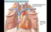
Great Vessels
- large arteries and veins connected to the heart
Includes - SVC, IVC, Ascending Aorta, Pulmonary Trunk, Pulmonary Veins
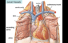
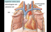
Superior Vena Cava
- large vein that receives venous drainage from the head, upper extremeties, and thorax
- drains into the RA
- convergence of the Left and Right Brachiocephalic Veins (Right Brachiocephalic Vein formed by convergence of Right Subclavian Vein and Right Internal Jugular Vein)
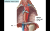
Inferior Vena Cava
- large vein that receives venous drainage from the lower half of the body (abdomen, pelvis, lower extremities)
- drains into the RA
- enters thoracic cavity by traveling through an opening in the diaphragm at T8
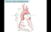
Aorta
- largest artery in the body
- arises from LV and immediately gives off Right and Left Coronary Arteries
In thorax, has 3 parts:
Ascending Aorta (ends at transverse thoracic plane)
Aortic Arch (begins and ends the transverse thoracic plane
Descending Aorta (aka thoracic aorta - begins at the transverse thoracic plane)
In abdomen: Abdominal Aorta - descends and bifurcates at L4 into common iliac arteries

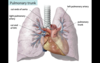
Pulmonary Trunk
- aka Main Pulmonary Artery
- outflow tract from RV
- bifurcates into right and left pulmonary arteries
Right Pulmonary Artery - travels posterior to ascending aorta and SVC towards Right Lung
Left Pulmonary Artery - travels anterior to the thoracic aorta
Pulmonary Veins
- four veins carrying oxygenated blood from the lungs to the LA

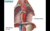
Thymus
- lymphoid organ involved in dev’t of immune system (T-cells)
- primarily active during childhood, undergoes involution during puberty and mostly replaced by fat
Location - posterior to the sternum, anterior to the great vessels and pericardium
Brachiocephalic Trunk
- the first branch of the aortic arch
- gives rise to the Right Sublcavian Artery and the Right Common Carotid Artery
- supplies upper right quadrant of the body
Left Common Carotid Artery
- second branch of the aortic arch
- supplies head and neck region
Left Subclavian Artery
- third branch of the aortic arch
- supplies the left upper quadrant of the body
Aortic Arch
- peak of the aorta bw ascending and descending aorta (above the transverse thoracic plane)
Three branches:
- Brachiocephalic Trunk
- Left Common Carotid Artery
- Left Subclavian Artery
* Remember you “ABCs” (Aorta, Brachicephalic, Carotid, Subclavian)*

Epicardium
- visceral serous layer of the pericardium that forms the outer layer of the heart
Ascending Aorta
- gives rise to the coronary arteries
- lies below the transverse thoracic plane

Descending Aorta
- aka Thoracic Aorta
- begins at the transverse thoracic plane
- descends anterolateral to the left of the vertebral column
- passes posterior to the diaphragm and becomes abdominal aorta from T12-L4 until it bifurcates
Ligamentum Arteriosum
- remnant of the ductus arteriosis (channel bw the pulmonary trunk and aortic arch that allowed blood to bipass lungs in fetus)
- fibrous after closure of ductus arteriosis at birth
- location between aortic arch and pulmonary vessels called “Autopulmonary window” by radiologists

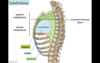
Trachea
- posterior to the great vessels in the midline
- bifurcates into right and left main bronchi at T4 vertebral level

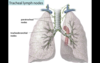
Tracheobronchial Lymph Nodes
- lymph nodes surrounding the trachea at the tracheal bifurcation
- receive lymphatic drainage from lungs and other thoracic viscera

Paratracheal Lymph Nodes
- lymph nodes surrounding the lateral aspect of the trachea above the bifurcation
- receive lymphatic drainage from lungs and other thoracic viscera
Esophagus
- located posterior to the trachea
- continuous with the pharynx superiorly and the stomach inferiorly
- exits thorax/enters the abdoment through the diaphragm at T10 vertebral level through the Esophageal Hiatus
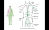
Azygous Venous System
- receives drainage from the thoracic wall (i.e. posterior intercostal veins) and viscera (i.e. esophageal and mediastinal veins)
Travels - anterolateral to the right of the vertebral column
- hemiazygos and accessory hemiazygos travel anterolateral to the left of the vertebral column - drain into azygous via branches across the midline)

Azygous Drainage
Superiorly - SVC
Inferiorly - IVC in the abdomen
**Azygous serves as a collateral channel if the IVC becomes blocked

Thoracic Duct
- largest lymphatic vessel in the body
- begins in upper abdomen as a dilation called Cisterna Chyli
- in mediastinum travels on the anterior aspect of the vertebral column (to the right of descending aorta and left of azygous vein)
- passes through the posterior diaphram into the mediastinum alongside the aorta
Termination: merges with venous system at junction of Left Internal Jugular Vein and Left Subclavian Vein
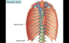
Cisterna Chyli
- dilation/beginning of the thoracic duct in the abdomen
Thoracic Duct Sources
- entire lower half of the body and upper left quadrant of the body
- returns fluid to the venous system
(RUQ drains into Right Lymphatic Duct)
Relationship between azygous, thoracic duct, and descending aorta
From right to left
Azygous Vein → Thoracic Duct → Descending/Thoracic Aorta

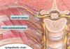
Sympathetic Trunks
(Sympathetic Chain)
- extend from the base of the skull to the coccyx
- travel lateral to the vertebral column
- various branches leave the sympathetic chain ganglia throughout the thorax

White and Gray Rami Communicantes
- connect the sympathetic trunks to the spinal nerves
- emerge from the posterolateral parts of the ganglia
Cardiac Nerves
- emerge from the anteromedial parts of the cervical and upper thoracic sympathetic ganglia
- contain post-ganglionic sympathetic neurons that innervate the viscera of the thorax by traveling through visceral plexuses

Visceral Plexuses
- contains visceral nerves in the thorax (i.e. cardiac nerves) that travel through the plexus to get to their targets
Three major subdivisions:
Cardiac Plexus - superficial and deep aspects of the aortic arch
Pulmonary Plexus - surrounds the trachial bifurcation and primary bronchi
Esophageal Plexus - surrounds the esophagus

Thoracic Splenchnic Nerves
- emerge from the anteromedial parts of the T5-T12 sympathetic ganglia
- contain pre-ganglionic sympathetic neurons that innervate viscera in the abdominal and pelvic cavities
**just pass through thorax**
Three Nerves:
Greater Splanchnic (T5-T9)
Lesser Splanchnic (T10-T11)
Least Splanchnic (T12)

Structures Passing Through Diaphragm
Inferior Vena Cava (T8)
Esophagus (T10)
Aorta (T12)
- sympathetic chains pass through with aorta
“I 8 10 E A 12” - I ate 10 eggs at 12

Phrenic Nerves
(Origination)
- arise from C3-C5 ventral rami

Phrenic Nerves
(Travel)
- enter mediastinum through superior thoracic aperture
- anterior to the roots of the lungs and descend to the diaphragm along the lateral aspect of the pericardium

Phrenic Nerves
(Actions)
- provide somatic motor fibers to the diaphragm
- convey somatic sensations from the central part of the diaphragm, fibrous and parietal serous pericardium, mediastinal pleura, and the central part of the diaphragmatic pleura

Pericardiacophrenic Vessels
Travel - with phrenic nerve
Provide - blood to the pericardium and diaphragm
Vagus Nerves
(Origination)
- arise from the brainstem
Travel - in neck near the coratid arteries and posterior to the root of the lung
Enter - thorax via the superior thoracic aperture
- branch into cardiac plexus, pulmonary plexus, and esophageal plexus

Vagus Nerves
(Right, Left Travel)
Left Vagus Nerve
- in contact with aortic arch
- enters esophageal plexus on the anterior aspect of esophagus
Right Vagus Nerve
- travels along the trachea
- enters esophageal plexus on the posterior aspect of the esophagus
Both - travel posterior to the root of the lung
- regroup as Anterior and Posterior Vagal Trunks after plexuses before traveling through diaphragm with esophagus

Vagus Nerves
(Actions)
Parasympathetic - decrease HR, constrict bronchial tree, stimulate peristalsis and secretion of mucous from esophagus
Visceral Afferent - convey sensations of stretch from the lungs, pain from the heart, and participate in visceral reflexes

Phrenic vs. Vagus Nerve Travel
Phrenic Nerve - travels anterior to the root of the lung
Vagus Nerve - travels posterior to the root of the lung
Left Recurrent Laryngeal Nerve
(Origination)
- branch of the Vagus Nerve
- arises near the aortic arch
Left Reccurent Laryngeal Nerve
(Travel)
- through the aortopulmonary window posterior to the ligamentum arteriosum
- ascends back into the neck along the lateral aspect of the trachea

Right Recurrent Laryngeal Nerve
- travels around the right subclavian artery in the neck (i.e. not in the mediastinum)
Left Recurrent Laryngeal Nerve (Action)
- innervates portions of the larynx - somatic efferent fibers to muscles
- compression (i.e. by aortic aneurysm or mass) can produce hoarseness in the voice
Major Branches of Descending Aorta
Major Branches:
Bronchial Arteries - supply lungs
Esophageal Arteries - supply esophagus
Posterior Intercostal Arteries - supply chest wall

