Heart Flashcards
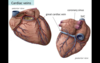
Coronary Sulcus
- groove on the external surface of the heart that marks the position of the interatrial septum
- conveys coronary vessels

Interventricular Sulcus
- groove on the external surface of the heart that marks the position of the interventricular septum
- conveys coronary vessels

Interatrial Septum
- internal partition between the right and left atria

Interventricular Septum
- internal partition between the right and left ventricles
- primarily made up of muscle with small area of membrane
- defects often located in membranous part of septum
Crista Terminalis
- a ridge that separates the smooth part (der. sinus venosus) from the rough part (der. primitive atrium) of the right atrium

Pectinate Muscles
- comb-like muscular ridges found on the wall of part of the right atrium and within the left auricle
Fossa Ovalis
- an oval-shaped depression on the interatrial septum of the right atrium that marks the location of the foramen ovale that was present in the fetus

Valve of Foramen Ovale
- piece of tissue on the interatrial septum of the left atrium that is a remnant of the primitive interatrial septum in the embryo - septum primum
- valve typically completely fused with interatrial wall (although occasionally incomplete fusion causing a small opening in the interatrial septum)
Tricuspid Valve
- valve between the right atrium and right ventricle
- aka Right Atrioventricular Valve

Pulmonary (Semilunar) Valve
- valve between the conus arteriosus and the pulmonary trunk
- consists of three cup-like cusps that have a central thickening called a nodule that is imp for valve closure
- passively opened by blood flow during systole
- during diastole, blood falls back down outflow tract and collects in sinuses of semilunar valve, causing nodules to meet and form an inverted pyramid - prevents cusps from descending further and keeps valve closed

Trabeculae Carnea
- muscular bundles on the walls of the ventricles
Papillary Muscles
- muscles attached to the atrioventricular valve cusps via chordae tendinae
- function to hold valve closed during systole preventing the valve cusps from flapping back into atria
- DO NOT contract to open the valve
- i.e. opening is passive, closing is active*
Moderator Band
- band of muscle containing conductive tissue connecting the interventricular septum to the anterior papillary muscle and the anterior wall of the right ventrical
- aka Septomarginal Trabecula
Conus Arteriosis
- the smooth outflow tract of the right ventricle leading to the pulmonary valve
- aka infundibulum
Bicuspid Valve
- aka Mitral Valve
- valve in the heart between the LA and LV
- aka Left Atrioventricular Valve

Aortic Valve
- aka Semilunar Valve
- the valve in the heart bw the LV and aorta
- has three cusps with a central nodule on each
**- opening for coronary arteries within two cusps
- passively opened by blood flow during systole
- passively closes during diastole (blood flowing back fills cusps and causes closure)

Auricles
- small ear-shaped appendages attached to the atria
- developmental remnants of primitive atria (no function)

Grooves on the external heart
- Coronary Sulcus - between the atria and ventricles
- Interventricular Sulcus - between the ventricles
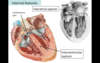
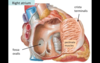
Right Atrium
Receives blood from: SVC, IVC, Coronary Sinus
Features:
- Crista terminalis
- Pectinate muscles
- Fossa ovalis
- Openings for SVC, IVC, Coronary Sinus

Right Ventricle
Receives blood from: RA through tricuspid valve
Features:
- Trabeculae carneae
- Papillary muscles
- Chordae tendineae
- Septomarginal Trabecula (moderator band)
- Conus arteriosis (infundibulum)
- Pulmonary (semilunar) valve

Left Atrium
Receives blood from: four pulmonary veins
Features:
- Pectinate muscles
- Valve of the Foramen Ovale
- Openings of the four Pulmonary Veins

Left Ventricle
Receives blood from: RA through the mitral valve
Features:
- Trabeculae carneae
- Chordae tendineae
- Papillary muscles
- Aortic (semilunar) valve

Coronary Arteries
- two openings in the aorta within cusps of the aortic semilunar valve
- blood flows into the arteries when blood flows back into the sinuses of the cusps (i.e. during diastole)
Heart Shape
- cone
Base: posterior, superior, right
- LA, small part of RA, prosimal parts of great veins (SVC, IVC, pulmonary veins)
Apex: anteroinferior, left
- LV
- lies deep to the 5th intercostal space
Anterior (Sternocostal) Surface
- portion of the heart facing anteriorly (posterior to sternum and costal cartilage)
- RV
Posterior Surface
- base of the heart
- LA, (part of RA attached to SVC and IVC)
Right (Pulmonary) Surface
- RA
- in contact with R lung
Left (pulmonary) surface
- LV, L auricle
- in contact with left lung
Inferior (diaphragmatic) Surface
- RV, LV
- in contact with the diaphragm
Right Border
- RA
- SVC
- IVC
Left Border
- LV
- Aortic Arch
- Left auricle
Inferior Border
- RV, LV
- border bw sternocostal and diaphragmatic surfaces
Apex
- located at 5th intercostal space
- located at midclavicular line
- LV
Blood Flow
IVC & SVC & Coronary Sinus → RA → Tricuspic Valve → RV → Infundibulum → Pulmonary Semilunar Valve → Pulmonary Trunk → Pulmonary Arteries → Lungs → Pulmonary Veins (4) → Left Atria → Mitral Valve → Left Ventrical → Aortic Semilunar Valve → Aorta → Body during systole, Coronary Arteries during diastole

Diastole & Systole
Diastole - ventricular relaxation (filling)
Systole - ventricular contraction (emptying)
Location of coronary arteries, veins, and branches
- in the grooves of the heart
- Coronary Sulcus and Interventricular Sulcus
Coronary Arteries
- two
- branch from the ascending aorta
Right Coronary Artery - travels in coronary sulcus
Left Coronary Artery - travels in interventricular sulcus

Right Coronary Artery
- emerges from ascending aorta near tip of right auricle
- travels along the coronary sulcus bw the RA and RV
- Three important branches:
- Sinoatrial Node Artery
- Marginal Artery
- Posterior Interventricular Artery

Left Coronary Artery
- emerges from the ascending aorta posterior to the pulmonary trunk
- bifurcates into two branches:
- Anterior Interventricular Artery (aka LAD - Left Anterior Descending) which travels along the anterior interventricular sulcus - mainly supplies LV
- Circumflex Artery - travels in the coronary sulcus of LH and terminates before reaching posterior interventricular sulcus

Pattern of Coronary Arteries
- small anastomoses, but not sufficient to nourish tissue if a major branch occluded
Widow Maker
- LAD from Left Coronary Artery (Anterior Interventricular Artery)
Cardiac Veins
- venous blood from heart tissue travels to the Coronary Sinus

Coronary Sinus
- where most cardiac veins terminate
- delivers blood to RA
- sac-like structure on posterior side of the heart

Great Cardiac Vein
- originates near the apex of the heart
- travels along the LAD in the anterior interventricular sulcus → enters cardiac sulcus and travels with circumflex artery to posterior side of heart → merges with coronary sinus
Middle Cardiac Vein
- travels in posterior interventricular sulcus with posterior interventricular artery → terminates in coronary sinus

Small Cardiac Vein
- travels with marginal branch of right coronary artery → enters coronary sinus near the IVC

Anterior Cardiac Veins
- small veins that transmit blood from the LV directly to the RA (i.e. not via the coronary sinus)
Cardiac Conduction System
- consists of specialized cardiac muscle cells that conduct electrical impulses
SA Node → AV Node → AV Bundle (of His) → AV Bundle Branches → Purkinje Fibers

Cardiac Conducting Cells Blood Supply
SA Node & AV Node - Right Coronary Artery
(SA Nodal Artery and Atrioventricular Nodal Artery)
AV Bundle and Branches - Left Coronary Artery
(Anterior Interventricular Artery)
Cardiac Plexus
- surrounds the aortic arch and the anterior surface of the tracheal bifurcation
- Efferent branches supply the heart
- Afferent neurons from the heart to the CNS

Sympathetic Innervation of the Heart
Cardiac Nerves
Functions - increase HR, increase contractility
Preganglionic Neurons
Cell body originates lateral horn in T1-T5 of spinal cord → axons enter sympathetic chain → travel up to cervical region (due to dev’t) or stay in thoracic region → synapse in cervical ganglia or thoracic ganglia (depending on where traveled)
Postganglionic Neurons
Leave sympathetic chain as cardiac nerves → enter cardiac plexus → travel to heart

Parasympathetic Innervation of the Heart
Vagus Nerve (CX - cranial nerve)
Function: decrease HR
Preganglionic Neurons
Cell bodies in brainstem → axons travel in vagus nerves to the thorax → vagus nerve gives off cardiac branches → enter cardiac plexus → Synapse with postganglionic parasympathetic neurons
Postganglionic neurons
Ganglia located in cardiac plexus or on the wall of the heart

Visceral Afferent Neurons of the Heart
Location - in the cardiac plexus
Purpose: pain and reflex response
- carry pain sensations from the heart (pain often from ischemia); Travel: with sympathetic cardiac nerves
- carry reflex from the heart (BP changes and chemical content of blood); Travel: with the cardiac branches of the vagus nerves

Referred Pain
- pain from the heart often felt in skin of the chest and left arm
- brain misinterpret the source of afferent neurons because enter the spinal cord together

Heart Block
- desynchronization of contraction of the atria with respect to the ventricles
Atrioventricular Valves
- Tricuspid and Mitral Valves
- Close due to papillary muscle contraction

Semilunar Valves
- Pulmonary Valve and Aortic Valve
- Close due to diastole - blood flow back from arteries collects in cusps and causes valves to close

Pulmonary Veins
- drain blood into LA

Circumflex Artery
- from Left Coronary Artery
- follows coronary sulcus to posterior side of heart

Posterior Interventricular Artery
- from Right Coronary Artery
- follows coronary Sulcus around to posterior of heart then goes down interventricular sulcus

