Lumps and Bumps Flashcards
what is a benign lesion and what are its characteristics?
abnormal growth of cells that lack the ability to invade neighboring tissues or metastasize uniform, smooth or papillary and can displace normal structures
what is a malignant lesion and what are its characteristics?
cancerous cells that invade and destroy body tissues irregular borders, vascularization/telangiectasia, ulceration and bleeding, alteration or normal architecture, loss of cilia and irregular pigmentary changes
what is a metastatic lesion and what are the characteristics?
cancerous cells that spread from a primary site to tissue not directly adjacent to the primary site spread via blood stream or lymph
what is seborrheic keratosis and what causes it?
benign pigmented cutaneous lesion - no malignant potential caused by proliferation of basal cells (immature keratinocytes)

who typically gets seborrheic keratosis and how does it present?
sun exposure, age, genetics onset in 30’s - more common in patients over 50 present in hair-bearing areas of skin (chest, face, back - not palms or soles)
what are the characteristics of a seborrheic keratosis?
usually solitary lesion 1-2cm, moveable hyperpigmented plaque, elevated with waxy surface and sharp demarcation
what is the treatment for a seborrheic keratosis?
obervation and cryotherapy, laser therapy or excision if they bother the patient
what syndrome is associated with seborrheic keratosis?
leser-trelat syndrome = sudden onset of multiple SK’s - usually onset of internal malignancy (stomach, liver, colon, pancreas cancers)
what is squamous papilloma?
benign hyperplasia of squamous epithelium flesh colored with cerebriform surface (pedunculated or sessile)

who typically presents with squamous papilloma?
middle-aged and elderly patients (gradual onset and slow growth)
what are some differential diagnoses for squamous papilloma?
basal cell carcinoma, seborrheic keratosis, and verruca vulgaris (* all benign tumors of the epidermis)
what is verruca vulgaris?
squamous papilloma caused by the human papilloma virus - can have concomitant conjunctivitis multiple lesions

who typically gets verruca vulgaris?
immunocompromised patients, children and young adults
what is the treatment for verruca vulgaris?
observation, complete surgical excision or cryotherapy - usually spontaneous resolution
what is molluscum contagiosum?
viral infection of the skin - multiple pearly flesh colored lesions with a small central crater

who typically gets molluscum contagiosum?
common in children suspect immunocompromised state if present in adults or severe bilateral involvement in children
what is the transmission of molluscum contagiosum?
pediatric = direct contact adult = STD
what is the treatment for molluscum contagiosum?
incision and expression, cryotherapy, excision or laser treatment - recurrence is rare after complete resolution
what condition is molluscum contagiosum associated with?
chronic follicular conjunctivitis
what is keratoacanthoma?
a pre-malignant tumor of the epidermis, develops on hair-bearing sun exposed skin (85% on face and 5% on eyelids)

who typically gets keratoacanthoma?
males > females and greater prevalence in immunosuppressed patients (s/p renal transplantations)
what are the characteristics of keratoacanthoma?
elevated margins with central crater, usually solitary lesion, rapid onset/growth, spontaneous regression
what is the histopathology for keratoacanthoma?
well differentiated squamous cells with keratin-containing center
what are some risk factors for keratoacanthoma?
skin color, UV radiation, trauma and genetics - may be a clinical variant of squamous cell carcinoma
what is the treatment for keratoacanthoma?
observation and complete removal via surgical excision
what is actinic keratosis?
most common pre-cancerous cutaneous lesion - caused by proliferation of atypical keratinocytes

who typically gets actinic keratosis?
light-skinned, F > M, mean age is 62, UV exposed skin
where is actinic keratosis usually located?
face, eyelids, dorsa of hands and bald areas on men
what can actinic keratosis progress to if untreated?
20% progression to squamous cell carcinoma
what does actinic keratosis look like?
multiple, erythrmatous, sessile plaques, 1-10mm, pink in color but can be pigmented, less distinct margins than seborrheic keratosis
when do you biopsy a actinic keratosis lesion?
when lesions appear indurated, painful, ulcerated, bleeding or hyperkeratotic lesions unresponsive to standard therapy
what are some treatment options for actinic keratosis?
destructive therapy - single lesions (cryotherapy, shave excision or surgical excision), topical medications - multiple lesions, photodynamic therapy and chemical peels
what is squamous cell carcinoma?
2nd most common eyelid malignancy (after basal cell carcinoma)

who typically gets squamous cell carcinoma?
fair-skin, 50-80 y/o, M > F, chronic UV exposure, many x-rays, severe sunburns
what are the 2 types of squamous cell carcinoma?
bowen’s disease and invasive SCC
what does squamous cell carcinoma look like?
broad spectrum of appearances: small red scaly patches, large ulcerated lesions, small nodular lesions, occurs more on upper eyelid, can be irritating or bleed
what is the course of squamous cell carcinoma?
aggressive = fast growing, 2-5% metastasize and more likely to recur
what is the treatment for squamous cell carcinoma?
mohs microsurgery or frozen section - radiotherapy, cryotherapy, intralesional chemotherapy and photodynamic therapy
what is basal cell carcinoma?
most common malignant tumor of the skin (90% of eyelid tumors)
what are the risk factors for basal cell carcinoma?
older age, light skin, sunlight exposure, prior irradiation, and immunosuppression F > M and 50-80 y/o
where are basal cell carcinoma’s usually located? what do they look like?
lower eyelid, head/neck region pearly, waxy, rolled, telangiectatic borders with central ulceration
what are the treatments for basal cell carcinoma?
small lesions = resection large lesions = mohs chemotherapy
what is the prognosis for basal cell carcinoma?
rarely metastasize, low mortality (intracranial invasion), locally invasive and destructive if left untreated
what is a melanocytic nevus?
darkly pigmented lesion containing modified melanocytes, may contain hair

how do you get a melanocytic nevus?
acquired or congenital (acquired - 5-15 y/o) and if multiple lesions = dysplastic nevus syndrome
what is oculodermal melanocytosis (nevus of ota)?
congenital pigmentation of periocular skin, uveal tract, sclera or ipsilateral meninges

who typically gets oculodermal melanocytosis?
rare in Caucasians - more common in Asian and African Americans
what does oculodermal melanocytosis look like?
flat lesion, tan-gray, follows V1 and V2 of CNV (bilateral in 10% of cases)
what is the treatment for oculodermal melanocytosis?
periodic DFE to r/o uveal melanoma
what is lentigo maligna (melanotic freckle of hutchinson)?
an acquired cutaneous pigmentation on exposed skin areas

who typically gets lentigo maligna?
middle-aged, elderly Caucasians
what does lentigo maligna look like?
flat, well-circumscribed, irregular, tan-brown macule, grows slowly
what are some differential diagnoses for lentigo maligna?
seborrheic keratosis, acquired melanocytic nevus, malignant melanoma
what is lentigo maligna associated with and what is the treatment?
primary acquired melanosis of conjunctiva wide surgical resection
what is primary malignant melanoma?
due to sun exposure in Caucasians, due to proliferation of atypical melanocytes, occurs mostly on lower eyelid (worse prognosis)

what are the clinical features of a primary malignant melanoma?
can extend into orbit via neural invasion, can metastasize after many years, advanced have ulcerative and nodular presentations
what is the management and prognosis for primary malignant melanoma?
management = surgical excision and eyelid reconstruction prognosis = nodular has worst, then thicker lesions then thin lesions
what can you use for early detection of melanomas?
A = asymmetry B = borders C = color D = diameter
how far into the skin do actinic keratosis, squamous cell carcinoma, basal cell carcinoma and melanomas go?
actinic keratosis = epidermis squamous cell carcinoma = dermis (though basal cell layer) basal cell carcinoma = dermis melanoma = all layers (through dermis)

what is sebaceous gland carcinoma?
solitary nodule or diffuse eyelid thickening origins = meibomian glands of upper tarsus, glands of zeis and caruncle

who typically gets sebaceous cell carcinoma?
2-7% of malignant eye tumors, most common in 5-7th decades and 70% female
what is the prognosis of sebaceous gland carcinoma?
83% mortality if upper and lower eyelids are involved
what are the clinical features of a sebaceous gland carcinoma?
aggressive local behavior, metastasizes to retinal lymph nodes and distant organs, masquerades as other benign lesions resulting in delay in diagnosis and treatment cardinal signs = madarosis, poliosis, thickening of lid margin more common in upper eyelid
what is an eyelid xanthelasma?
benign aggregation of lipid filled macrophages within the dermis - usually bilateral

what does an eyelid xanthelasma look like?
bilateral, single/multiple flat, yellow placoid lesion lesion that affects loose aspects of eyelids
what is a sebaceous cyst?
benign - due to occlusion of a sebaceous gland duct (most commonly in meibomian glands of upper tarsus) common on scalp and eyebrow

what is the treatment for a sebaceous cyst?
hot compresses
what is an eccrine hidrocystoma?
benign - ductal retention cyst of sweat glands (heat, humidity, perspiration can cause an enlargement)

what does an eccrine hidrocystoma look like and what is the treatment?
clear cystic translucent lesion near eyelid margin observation or excision
what is an apocrine hidrocystoma?
benign - retention cyst of apocrine gland (moll), usually solitary, common near medial canthus, bluish in color

what is an eyelid syringoma?
benign - sweat gland cyst, solitary or multiple, more pronounced on lower eyelid, very small

who typically gets eyelid syringoma?
young adult women, Asian patients
what is eyelid sweat gland carcinoma?
malignant - very uncommon tumors arise from epidermal cells of sweat glands of Moll, common in lower eyelid

who typically gets eyelid sweat gland carcinoma?
mean age = 63, 2:1 male to female ratio
what does an eyelid sweat gland carcinoma look like?
high content of mucin, slow growth, pink-blue elevated nodule, solid or cystic and near eyelid margin
what is a capillary hemangioma?
benign - strawberry birthmarks in infants (grows during first few months of life) one of the most common tumors in infancy

what is the pathology of capillary hemangioma?
proliferating benign endothelial cells and numerous small vascular channels
what are some complications of capillary hemangioma’s and what is the treatment?
amblyopia and strabismus topical beta blockers (timolol) - superficial lesions oral propanolol - deeper lesions intralesional steroid injection for lesions with amblyopgenic potential
what is an acquired hemangioma?
benign - common on trunk and extremities, moveable with skin, may bleed with trauma and common in elderly patients

what is a nevus flammeus?
benign - port wine hemangioma corresponds with cutaneous distribution of CN V present at birth and enlarges with time

what can occur if the upper lid is involved in a nevus flammeus?
a strong indicator for glaucoma development
what is the treatment for nevus flammeus?
laser photocoagulation
what is an eyelid lymphangioma?
benign - growth of lymph channels - can occur in various parts of the body occurs deep to dermis as dark blue and soft mass (can slowly enlarge)

what is an eyelid kaposi’s sarcoma?
malignant - multiple lesions usually benign in lower extremities and spreads to other parts of the skin and visceral organs

what does an eyelid kaposi’s sarcoma look like?
red, purple, brown, or blue, flat subcutaneous lesion can be diffuse, nodular or pedunculated initially has a smooth surface that becomes rough and crusty
who typically gets an eyelid kaposi’s sarcoma?
AIDS patients, immunosuppressed adults after renal transplantation
what is the treatment for eyelid kaposi’s sarcoma?
chemotherapy for extensive lesions, radiotherapy for small local lesions
what is an eyelid lymphoma?
can be benign, intermediate or malignant types = hodgkin’s vs. non-hodgkins and B-cell or T-cell

who typically gets eyelid lymphoma’s?
usually affects the elderly - suspect immunocompromised if younger patients (AIDS)
what is a metastatic neoplasm?
metastatic cancer of the eyelids - very uncommon usually solitary subcutaneous nodule resembling a chalazion
what is lymphangiectasia?
benign - dilation of lymphatic channels within conjunctiva bleeding can occur, solitary or multifocal, typically unilateral and sporadic

what is a conjunctival dermoid?
benign - tumorous malformation composed of tissue not typically present at involved site variably sized yellow-white limbal mass (usually infrotemporal)

what are the symptoms for a conjunctival dermoid?
small = asymptomatic large = irritation, astigmatism and inadequate eyelid closure
what is the treatment for a conjunctival dermoid?
surgical removal if causing amblyopia, astigmatism, dellen formation
what is a conjunctival dermolipoma?
benign - less well defined than a dermoid and usually located superotemporally yellow sessile lesion may contain bone, cartilage, and ectopic lacrimal gland

when does a conjunctival dermolipoma usually appear and what is the treatment?
1st or 2nd decade typically non-progressive and doesn’t require treatment
what is a conjunctival papilloma seen in childhood?
benign - virus induced (HPV types 6 or 11) usually larger and multiple, fleshy red appearance due to multiple vascular channels inferior fornix or bulbar conjunctiva

what is a conjunctival papilloma seen in adults?
benign - HPV type 7, unilateral and solitary, begins at limbus or bulbar conjunctiva, lighter pink color, low malignancy
what is racial melanosis?
benign - complexion related pigmentation, bilateral diffuse flat pigmentation of conjunctiva, most concentrated at limbus

what causes racial melanosis?
hyperpigmentation of basal cells of the conjunctival epithelium
what is primary acquired melanosis?
due to increase in melanocytes in basal layers of epithelium

what does primary acquired melanosis look like?
gradual onset in middle age typically unilateral, non-cystic patches on conjunctiva and peripheral cornea
what is the treatment for primary acquired melanosis?
biopsy and if suspicious - excision and mitomycin C
what is intraepithelial neoplasia?
benign squamous cell neoplasia of the surface epithelium - doesn’t metastasize but is pre-cancerous

what does intraepithelial neoplasia look like?
unilateral, fleshy, sessile lesion near limbus or interpalpebral fissue (can extend into corneal epithelium)
who typically gets an interepithelial neoplasia?
predisposing factors = sunlight or HPV immunosuppressed patients, middle-age/elderly
what is a squamous cell carcinoma?
CIN that has breached basement membrane of the epithelium and invaded underlying stroma wide variety of appearances = gelatinous, sessile, papillomatous large conjunctival feeder vessels are usually present

what is the management for squamous cell carcinoma?
complete surgical excision - topical mitomycin-C, 5-fluorouracil, cidofovir
what is conjunctival lymphoma?
malignant - diffuse, slightly elevated, redish/pink mass

where do conjunctival lymphomas usually present?
fornices or bulbar conjunctiva unilateral or bilateral
what is the treatment for conjunctival lymphoma’s?
excision of small lesions excision and chemotherapy for larger lesions
what is a malignant melanoma?
pigmented, fleshy, elevated, poorly defined conjunctival lesion, usually located on bulbar conjunctiva near limbus

who typically gets a malignant melanoma?
light skin, middle age/elderly, M = F
what is the management and mortality rate for malignant melanomas?
excision and mortality rate of 25%
what is an iris cyst?
benign - arise from iris pigment epithelium and located in inferotemporal quadrant

what can an iris cyst cause?
typically asymptomatic - can cause elevated IOP due to angle obstruction
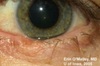
what is an iris melanoma?
pigmented or amelanotic, elevated with feeder vessels and usually there is a history of a nevus undergoing growth

what is orbital fat prolapse?
benign - protrusion of orbital fat through defect in Tenon’s capsule into conjunctival fornix soft yellow mass in superotemporal conjunctival fornix

who typically gets orbital fat prolapse?
very common in older patients and obese male patients
what are some differential diagnoses for orbital fat prolapse?
dermolipoma, lymphoma, and lacrimal gland tumor
what is a dermoid cyst?
benign - congenital cystic lesion most common cystic lesion of the orbit

what causes a dermoid cyst?
entrapped ectoderm at site of embryologic bony future
what does a dermoid cyst look like?
firm subcutaneous mass, non-movable as attached to bone, filled with hair, sebaceous glands and sweat glands
