Lecture 7 and 8: Diagnosis of Viral Diseases Flashcards
What are the purposes of viral disease diagnosis?
- Surveillance of viral diseases among certain population
- Identify the causative agent of certain suspected clinical cases of viral origin
- To monitor the progress of some viral diseases
- To monitor the antigenic/genetic variations of certain virus
- To help in the design of the right vaccine against the homologous circulating strains of viruses
What is the direct approach to viral diagnosis?
Identifying the virus or viral products (such as proteins,
nucleic acids) in clinical samples or after virus isolation
from clinical samples
What is the indirect approach to viral diagnosis?
Detecting an immunological response to the virus (detect antibodies)
What are the viral diagnosis strategies?
Detection of virus
Isolation of viruses
Detection of viral antigens
Detection of viral antibodies
Virus infectivity titration
Detection of viral genomes

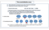
Ways specimens are evaluated.

What are the strategies to approach viral diagnostics?

What are the approach approach/ type of speciment in viral diagnostics? What notes are there about each way?

What is the rational behind viral diagnosis at the heard level/ individual animal level?
- Management of the animal or its prognosis is influenced by the diagnosis
- Rapid and accurate diagnosis of the causative virus can be the basis for establishing the management plan
(Biosecurity, vaccination, antimicrobial treatment)
What is the rational behind certification of freedom from specific viral life long infections or proof of vaccination (BLV, BVDV, EIAV) level?
-These certificates or vaccines are mandatory for animal travelling, participation in certain exhibition, or show for sale
-Artificial insemination, embryo transfer, and blood transfusion
-Male used for semen collection, female used for embryo transfer, blood donor animals should be screened for wide range of
viral infections (EAV, EIAV, BLV, etc)
-Zoonotic viruses: (RVfV, Rabies, WNV, EEEV, Hendra virus, etc)
-All these animals require screening and testing as well as veterinary care
What is the rational behind viral diagnosis at the State, country and International level?
- Test and removal programs for some viruses such as (MDV, EIAV, BHV-1, BVDV)
- Surveillance programs in support to enzootic diseases research control activates
- Surveillance programs in support to exotic diseases research control activates
IV- Prevention of new emerging and re-emerging diseases
?
What are the kinds of sample collections for these vaccines at the postmortem and antemortem level?

What should be considered during sample collection?
- Proper site of collection
- Right time
- Suitable volume/quantity
What should be considered during transportation of viral samples?
- Viral transport media (VTM)
- Antibiotic/antifungal cocktail
- Sterile containers
What should be considered for sample preservation?
- Proper preservation temperature
- Avoid freezing and thawing
What are the uses of light microscopy in diagnostic virology?
- To monitor the growth and multiplication of cell culture
- To monitor the viral infection in cell culture (CPE)
• Detection of viral inclusion bodies (TBDL)
- To study the histopathological changes of some viral infected tissues
- Immuno-histo-chemistry: Rabies virus antigen (brown dots)

What can be seen in this image?

In most other cases viruses are _______ in cell culture before they can be visualized by ____
isolated, EM
What is the virus surrounded by ? What are these substances? What happens when we stain these particles?
Virus is surrounded by electron dense (electron opaque) material
-2% uranyl acetate,
-1-3% sodium or
-potassium tungstate
Electrons are scattered from regions covered with stain thus creating a contrast which outlines viral structures
What are the advantages of EM in diagnostic virology?
- Can detect the virus in body secretions and execrations
- Do not require special reagents such as proteins standard, etc
- No cross reaction with other similar viruses
- Rapid technique
What are the disadvantages of diagnostic virology?
-Less sensitive than other tests: requires high
virus concentration
-The EM machine is expensive
-Requires expert personnel to do interpretation
What is true/ good about scanning electron microscopes?
- Lower resolution of tens of nm
- Shows only morphology of specimens.
- cheap
- Relatively safe.
What is some pros and cons to transmission electron microscopy?
- Higher resolution of 1 nm or less
- Shows multiple characteristics of objects such as crystalization, morphology, stress and much more.
- Specimen preparation requires thinning which is tiring and time consuming.
- Expensive
- Relatively harmful to human health


Transmission EM- SARS-CoV-2
Spike proteins are shown as protrusions from the surface of the virus and attach to the host cell receptors

EM - virus only

Virus + Abs
IEM (Immuno-electron microscopy)
What is important to remember about IEM?
IEM: addition of viral-specific antibodies will allow the concentration of virus particles thus can be seen under EM easily
What is seen in this image?

Common kits for sample collection
How are viral samples collected, transported, and handled/processed in a direct method?
• Nasal swabs (in transport media containing antibiotics)
• Tissue (homogenize), buffy coat cells and feces - make 10-20% solution in cell culture
medium – sonicate* homogenized tissues and buffy coat cells or freeze/thaw to lyse the cells (release the virus)
• Centrifuge (3,000X g) or filter (0.2 µ)
➢In most cases inoculation of sample (supernatant/ filtrate) onto cells of tissue culture –
incubate at 37˚C in a CO2 incubator
* Feces – no sonication
What are the methods of virus isolation?
Embryonated chicken egg innoculation (ECE)
Cell culture
Laboratory animals inoculation
What are the routes of inoculation for chicken eggs?
- Yolk sac
- Amniotic sac
- Allantoic Sac
- CAM
What are the types of cell culture used for virus isolation?
- Primary cell culture
- Secondary cell culture
- Established cell line
What are the routes of inoculation for laboratory animals?
- IM
- IV
- IP
- SC
- ID
I- Isolation of viruses by EC

I- Isolation of viruses by EC
Embryonated chicken egg inoculation (ECE)

Embryonated chicken egg inoculation (ECE)
What is the steps to the process of inoculating the Chorioallantoic Membrane (CAM) of a chicken egg?
**Quite difficult, but very common**
** Use older embryos (10 - 12 days)**
1. Drill hole at air space and then drill 2nd hold at top
2. Apply suction to 1st hole, then inoculate 0.1 ml of sample (to be tested) with syringe and needle in the 2nd hole.
3. Incubate & look for membrane edema or focal necrosis
4. Harvest the CAM
5. Preparation of artificial air sac
What is the method of harvesting inoculated materials?
• Open the inoculated eggs with sterile scissors
• Remove the egg shell and the egg shell membranes
• Pour the content of the egg into a clean, sterile petri dish
• Examine the inoculated embryos and the embryonic
membranes
What are the pathological changes of the virus on the ECE?
1-Curling: and dwarfing of embryos: IBV
2-Death: of the embryo: some viruses induce the death of the embryo such as NDV
3-Deformities: dwarfing, and hemorrhage of the embryo such as IBV
4-Hemorrhage: and thickening of the Chorioallantoic membranes as in case of Pox and Herpesviruses
5-Detection of hemagglutinin: in the egg fluids as in the case of NDV and AIV, which can be detected by the HA test.

What is the 7 pocket lesion? What does it refer to? What is seen in this image?

7-Pock lesions: circumscribed, rounded foci of necrosis on the surface of CAM: Varies in shape and size, Poxvirus
The term pock is designated to the white opaque necrotic foci resulting from virus
multiplication of the CAM as in the case of Pox and Herpesviruses
• These necrotic foci vary in size (large, medium, large), shape (circular, oval,
irregular). It may with raised or depressed centers and may be hemorrhagic in some
cases.
What is seen in this image?

Pock lesions
What is seen in this image?

These necrotic foci vary in size (large, medium, large), shape (circular, oval,
irregular). It may with raised or depressed centers and may be hemorrhagic in some
cases.
What are inclusion bodies?
• Viral components (most likely proteins) accumulated at the site of virus multiplication • They produced due to some replication/maturation process of viruses inside the infected cells • The position of the inclusion bodes depends on the site of viral assembly either in the nucleus or in the
cytoplasm
What are some examples of viruses with Intracytoplasmic IBs?
- Rabies: Negri bodies
- Small pox- Gaunielr bodies
- Fowl pox Bollinger bodies

What are some examples of viruses with Intracytoplasmic and intranuclear IBs?
- Canine distemper virus
- Measles

What are some examples of viruses with intranuclear IBs?
- Adenovirus
- Togavirus
- Herpesvirus

What kind of inclusion body is seen in this image?

Intracytoplasmic IB
What kind of inclusion body is seen in this image?

Intracytoplasmic IB
What kind of inclusion body is seen in this image?

Intracytoplasmic IB
What kind of inclusion body is seen in this image?

Intracytoplasmic and Intranuclear IB
What kind of inclusion body is seen in this image?

Intranuclear IB
What is seen in this image?

Giant cell: syncytia formation
What is a primary cell culture?
Cultured cells that are derrived directly from tissue ( often embryonic tissue)

What are the advantages of primary cell culture?
Advantages: cells have not been “modified” in any way
What are the disadvantages of primary cell culture?
limited lifespan of the culture potential contamination problems
What is an established cell lines?
- Specific cell types artificially maintained in the laboratory (ie, in vitro) for scientific purposes
- A population of cultured cells, of animal origin, undergone a change allowing the cells to grow indefinitely.

What are the advantages of an established cell line?
• Advantages cells grow indefinitely
What are the disadvantages of an established cell line?
• Disadvantage not all viruses replicate well in
cell lines
What is seen in this image?

methods of primary cell cultures
What are some examples of common cell culture media?

What are the steps to preparing primary kidney cell culture?

What are the causes of CPE?
1-Effect of virus on the cell membrane during attachment or release
2-Effect of the virus on the host cells DNA, RNA or proteins
3-Transformation of host cells by some viral genomes
4-Accumulation of the viral proteins and the new progeny viruses
What are some examples of CPE in cell culture?
1-Cell lysis: FMDV
2-Cell rounding: IBDV on Vero Cell line
3-Giant cells : Muti-nucleated cells: Syncytial formation: NDV,
Ex:Avian Reovirus
4-Transformation of cells: MDV, Avian Leucosis
5-Haemadsorption: Influenza, PI-3
True or False: Not all viruses cause CPE.
True.
What is seen in this image?

III-Virus isolation via lab animal inoculation
What factors affect hemagglutination test principles?
1-Temperature: Each virus require certain temp for HA
-NDV: 37 ºC -Enterovirus: 4 ºC
2- PH -NDV: PH (7.3) -Enterovirus PH(5.8)
3-Salt concentration: NaCl: important for binding of
virus & RBCs
4-Presence of non-specific inhibitors in sera


In the AGIDT or AGIP test, both soluble ______ and _____ interact in an aqueous solution to form a _____ (insoluble visible precipitate)
antigens, antibodies, lattice


What is radical immunodiffusion?
• Antigen solution diffuses into agar in which a specific antiserum has been incorporated, and a ring of
precipitate will form around the antigen well.
• The area of this ring is proportional to the amount of antigen in the well.
What is the principle of the complement fixation test (CFT)?
Principles: Complement that fixed by Ag/Ab reaction
thus, lysis of RBCs will not occur.
In the absence of a specific Ag/Ab reaction or absence of
either Ag or Ab, the complement is free and lyses the
RBCs

What are the principles of the fluorescent antibody techniques (FAT)?


What results do a qualitative ELISA give you?
Positive or negative results
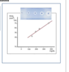
What results does an quantitative ELISA give you?
The optical density (OD) of the fluorescent dye is directly proportional to the concentration in the Ag or
Ab in the tested samples


What is seroconversion? What are the sample requirements?
- Seroconversion: development of detectable specific Abs to microorganisms in the blood serum as a result of infection or immunization
- Collection of 2 serum samples (Acute and convalescent) with 4 weeks apart
•If the convalescent sample is greater than the acute sample with t least 4 folds: Recent infection

What are the principles of polymerase chain reaction?
Principles: A reaction that uses the enzyme DNA polymerase to
catalyze the formation of more DNA strands from
an original one by the execution of repeated cycles of DNA
synthesis
What are the main steps of PCR amplification?
Main steps in PCR amplification:
1- Denaturing of ds- DNA template
2- Annealing of the primer
3- Extension of ds-DNA molecules
How do you visualize PCR products?
Gel electrophoresis
DNA gel
Sequencing
What are the advantages of PCR?
- Highly sensitive can detect one copy of the viral genome per sample
- Easy to set up
- Fast time (close to 2 hrs)
What are the disadvantages of PCR?
- Prone to contamination
- Requires high skilled personnel to conduct the experiment
- -Requires optimization of all parameteres in some cases
(annealing temp, primer design, etc) - -Difficulty in the interoperations os some positive results especially the latent infection with some viruses
What is virus quantification?
Virus quantification: counting the number of viruses in a specific volume to determine the virus concentration
What are the comparisons of some of the quantatative assays of viruses?
• I-Physical Quantitative assays
-Counting of viral particles by EM
II. Biological assays (TBS) later in the course
-HA assay -Plaque assay -TCID50
III. Quantitative Assas :
-Polymerase Chain Reaction (PCR) -qPCR and qRT-PCR
What is real time pcr? What does it allow us to measure?
Real-Time PCR a specialized technique that allows a PCR
reaction to be visualized “in real time” as the reaction
progresses
• As we will see, Real-Time PCR allows us to measure minute
amounts of DNA sequences in a sample!
What is real time PCR used for?
- Diagnosis/Detection
- Quantification
- Analysis
How does real time PCR help you diagnose/detect a viral infection?
To diagnose certain viral infection based on some
conserved viral genes amplification using specific
probes and primers.
How does real time PCR help you quantify your sample?
To measure the concentration of the target
virus/gene in a given sample based on the concentration/load of the virus/gene in the tested
samples
How does real time PCR help you analyze your sample?
To analysis the variants by studying the
melting curve of or comparing the temperature with the known sequences in the databases

What is the principles of virus neutralization test? Is it informative? What can it be used to characterize? What is it not?
➢ In a virus neutralization test, the presence of antibodies against a
known virus can be detected by the antibodies’ ability to prevent
cytopathic effects (CPE)* of the reference virus in cell cultures ➢ Most informative serological assay about “Protective Antibodies” ➢ Important tool used to characterize “serotypes”
*** it is not a rapid test****
What is a viral neutralization assay?
➢ Most informative serological assay about “Protective Antibodies”
➢ Important tool used to characterize “serotypes”
1. Serially dilute serum 1/2 1/4 1/8 1/16……….1/512
2. Add equal amount of virus (100 plaque forming units) to each tube-incubate 1 hr. - infect cultured cells- incubate at 37˚C for plaque formation.
-Reciprocal of the highest dilution that can prevent 50% plaque
formation is the serum titer.