lecture 6 (E2) Flashcards
(47 cards)
describe the clinical presentation of necrotizing peridontal ds
- sudden onset and it can become a “chronic condition”
- characterized by gingival tissue necrosis and ulceration
the three forms of periodontal ds
necrotizing gingivitis
necrotizing periodontitis
necrotizing stomatitis
target poppulation for necrotizing periodontal ds is
- HIV infected individuals
- Malnourished children
etiology and risk factors for necrotizing periodontal ds - microbiology
1 spirochetes and fusiform bacteria
specific features in HIV. Especially if not well controlled, immune system super weak.
pre-disposing factors for necrotizing peridontal ds
- pre-existing systemic ds HIV/AIDS
- inadequate oral hygiene high risk to getting the bacteria.
- Malnutrition
- Stress
- smoking/alcohol smoking creates a favorable environment.
mechanism on how stress can cause necrotizing gingivitis
- increase serum cortisol increase of endogenous corticosteroids.
- immune system depression more favorable for bacteria.

what is the CD4 count in a pt with HIV/AIDS
significant changes occur: <200 clles/mm3 **dn treat
infection occurs frequently HIV+ becomes AIDS: 200-500 cells/mm3
important lab data to monitor what do these mean:
viral count
absolute neutrophil count
platelet count
viral count: monitor status of ds, prognosis
absolute neutrophil count: needs ab prophylaxis when ANC <500
platelet count: No procedures if bellow 50,000 (normal: 150,000-450,000)
candidiasis, viral lesions, major apthous ulcers, necrotizing gingivitis, linear gingival erythema, necortizing periodontitis, neoplasma
oral lesions of what
HIV/AIDS
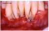
how does necrotizing periodontitis lesion look like under light microscopy?
identical to a necrotizing gingivitis lesion EXCEPT the destruction of the underlying periodontium.
under light microscopy what does the psuedomembrane look like
destroyed, replaced with fibrin, necrotic epithelium, PMNs and various types of microorganism
what does the linear erythema look like under light microscopy?
hyperemic with numerous engorged capillaries and dense infiltration of PMNs
body is trying to bring more WBC to fight it off, why it looks red and swollen.
primary S & S of NPD
and other S&S
- gingival nerosis
- gingival bleeding NOT active bleeding
- pain
common S&S
- pseudomembrane
- halitosis
- adenopathies
- fever
what are 4 dx of necrotizing periodontal ds
NG necrotizing gingivitis
NP necrotizing periodontitis
NS necrotizing stomatitis
Noma cancrum oris

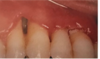
clinical S&S:
- necrosis and ulcer in the interdental papilla
- gingival bleeding
- pain
- pseudomembrane formation
- halitosis
- adenopathy
- fever
clinical characteristics of NG
clinical characterisics:
- not contagious
- age onset of 15-30 yo
- strong relationship between onset of ds and stress/anxiety
- responds to ab and non-sx periodontal therapy
- 75% pts exhibit a localized defect in neutrophil chemotaxis and/or phagocytosis

clinical S&S:
- necrosis and ulcer in the interdental papilla
- gingival bleeding
- pain
- pseudomembrane formation
- halitosis
- adenopathy
- fever (less than half % of the time)
microbio of NG
fusobacterium nucleatum
spirochetes

clinical S&S of NG:
- necrosis and ulcer in the interdental papilla
- gingival bleeding
- pain
- pseudomembrane formation
- halitosis
- adenopathy
- fever
NG differential dx (7)
- gingivitis
- herpetic gingivostomatitis
- linear gingival erythema
- mild or grade A/B periodontitis
- mucous membrane pemphigoid
- allergic rx (nickel)
- facticial injury
- gingivitis
- herpetic gingivostomatitis
- linear gingival erythema
- mild or grade A/B periodontitis
- mucous membrane pemphigoid
- allergic rx (nickel)
- facticial injury
- gingivitis has poor oral hygiene, gingiva red and inflammed at gingival margins
- herpetic gingivostomatitis, a viral infection with gingival erythema around the tooth
- linear gingival erythema has a really clear line around the teeth type of presentation that looks realted to HIV pt
- mild or grade A/B periodontitis, begins to have bone loss
- Mucous membrane pemphigoid more like erythematous blisters
- allergic reaction to nickle, just erythemous on the gingiva
- facticial injury can still see the interdental papilla
for NG you really see the necrotic interdental papilla as a clinical presentation.

how to differentiate NG and Herpetic gingivostomatitis?
age, body temp, lesion site, and clinical symptoms
age- NG: 15-30 yrs vs PHG: children
lesion site- NG: interdental papilla vs PHG: gingiva and entire oral mucosa
symptoms- NG: ulcerations, necrotic tissue and a yellowish-white plaque vs PHG: multiple vesicles, disrupt, leave small round fibrin-covered ulcerations.
how to differentiate between NG and HIV Association
HIV: has an intense linear gingival erythematous marginal gingvitis. May have profuse BOP.
NG tx
- improve OH an debridement (SRP)
- 0.12% chlorohexidine pre and post tx rinse
- Ab: metronidazole or amoxicillin