lecture 4: periodontal health gingival ds/conditions Flashcards
microscopic features of periodontal health
- gingival epithelium
- gingival CT
parts of gingival epithelium
- oral epithelium
- sulcular epithelium
- junctional epithelium
keratinized or non keratinized:
oral epithelium
sulcular epithelium
junctional epithelium
oral epithelium - keratinized
sulcular epithelium - non-keratinized, semi perm membrane, no rete pegs
junctional epithelium- non-keratinized attached to tooth with hemidesmosomes infiltrate by PMN.
supracrestal tissue attachment size
2.04
JE = 0.97
CTA = 1.07
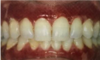
color of healthy
- coral pink on marginal/attached gingiva
- red smooth shiny on alveolar mucosa
- physiologic pigmentation
size of healthy
- size should correspond with the sum total of the bulk of cellular and intercellular elements and vascular supply
consistency of healthy
- firm and resilient (gingival fibers)
surface texture of healthy
- stippled on the attached gingiva BUT not always a sign of health, only 40% of pts will have it on attached gingiva.
contour of healthy
- scalloped and collar-like fashion
shape of healthy
- pyramidal towards the anterior, flattened towards the posterior
position of healthy
- the level at which the gingival margin is attached to the tooth
- continuous tooth eruption - active and passive eruption - altered passive eruption
definition of gingival health
absence of clinically detectable inflammation
3 determinants to initiate ds
- Microbiological determinants
- host determinants
- environmental determinants
microbiological determinants are
- supragingival plaque
- subgingival plaque
host determinants are
- local predisposing factors
(periodontal pockets, restorations, root anatomy, tooth position and crowding) - systemic modifying factors
(host immune function, systemic health, genetics)
environmental determinants are
- smoking
- medication
- stress
- nutrition and lack of vitamins
indicators for perio/gingival ds
- BOP
- Peridontal probing
- Radiographic features
- Tooth mobility
which is a reliable sign of ds for indicators
BOP
indicators - radiographic features
- if there is a well defined lamina dura = sign of healthy periodontium
- but if do not have, and this is the only sign it could be an xray prob
clinical features of intact periodontium
- no clinical attachment loss or bone loss
- inflammation is minimum <10%
- PD within 3mm
- no erythema and edema
- physiological bone levels range from 1-3mm (avg 2mm) apical to the CEJ
clinical feature of a reduced periodontium
- BOP more than 10%
- PD within 3mm
- no edema, erythema and pt symptoms in the presence of reduced clinical attachment and bone levels
two types of gingivitis
- dental plaque-induced gingival ds
- non-plaque-induced gingivial ds
how does smoking play a role in clinical findings of gingivitis
- smoking masks BOP by suppressing inflammatory response
clinical findings of gingival features pale color means what
reduced vascularization or increased keratinization