what is the vitreous made out of?
98% water with collagenous framework
how much of the total ocular volume is the vitreous?
about 75% - higher volume in myopic eyes
what are “floaters”?
any area of the vitreous that is dense enough to be detected by the patient or seen during DO
what is the most common entoptic phenomenon reported by patients?
floaters (most common reason for office visits)
where are the 4 vitreoretinal attachments?
vitreous base, around optic disc margins, around macula, and around peripheral retinal blood vessels
where is the strongest point of attachment for the vitreous?
vitreous base = 3-4mm wide zone straddling the ora errata
what is the vitreous attached to specifically in the retina in all areas?
the ILM (inner limiting membrane)
where are posterior vitreous detachments most likely to occur?
peripapillary = around the optic nerve (progressively weakens with age)
what are 3 ways you can view the vitreous?
DO (red reflex), fundus biomicroscopy before lens, and BIO (red reflex)
why is a dilated exam important?
to examine the peripheral retina in search for rhegmatogenous conditions (identifies risk for RD)
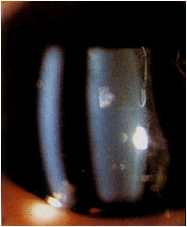
what is synchysis?
vitreous slowly undergoing liquefaction (slowly degrades) = seen as empty
what is syneresis?
shrinkage of the vitreous - secondary to separation of liquids and solids
which patients are more likely to get a PVD?
high myopia, post-surgical cataracts, trauma, and >40 y/o
what are some symptoms of a PVD?
floaters and may have flashes - flashes are due to traction stimulating photoreceptors
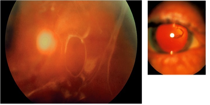
what are some signs of a PVD?
annular ring (Weiss’ Ring) in front of ONH, retinal hemorrhage and/or vitreous hemorrhage
what is Shaffer’s sign or tobacco dusting?
the presence of pigment granules in anterior vitreous and Berger’s space
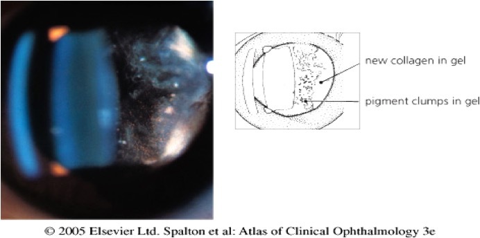
why is it important to look for Shaffer’s sign if the patient reported flashes and floaters?
98% will have a retinal detachment and 60% will have a retinal break (hole or tear)
when looking at Shaffer’s sign, you use a red-free filter, what should happen if the granules are RBCs?
they will disappear
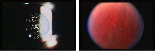
what is asteroid hyalosis?
common benign finding in >50 y/o (90% unilateral) = yellow-white spheres of calcium lipids adhering to vitreous framework
what does asteroid hyalosis look like?
seen as small yellow-white round opacities floating within vitreous - move freely in synchisis (snow globe)
what is asteroid hyalosis associated with?
aging collagen within vitreous and depolymerization of hyaluronic acid
what are 3 symptoms for a retinal detachment?
floaters, photopsia (flashes) and a visual field defect
what are some signs of a retinal detachment?
variable acuities, APD, lower IOP in affected eye, shaffer’s sign, retinal breaks, large holes or tears
