Lecture 4 Flashcards

1
Q

what is the magnification of the direct ophthalmoscope?

A

15x - magnification is dependent on patient and examiners refractive error

2
Q

when is the cobalt blue filter used?

A

with the 20D lens and fluorescein to evaluate the cornea

3
Q

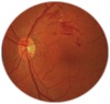
what does the red-free filter help to identify?

A

hemorrhages, choroidal nevus vs. retinal pigmentation, NFL loss and ON rim tissue

4
Q

where should your ending point be for direct ophthalmoscopy?

A

about 1.5 to 3mm from eye (middle finger touching cheek) and 15-20 degrees off visual axis

5
Q

why should you try and keep both eyes open during direct ophthalmoscopy?

A

to reduce the amount of accommodation

6
Q

what are some limitations of direct ophthalmoscopy?

A

lack of stereopsis, close working distance, refractive error dependent, and dependent on pupil size and lens

7
Q

what is the diagnostic code for an extended ophthalmoscopy?

A

CPT 92225 (first visit) and 92226 (subsequent visits)

8
Q

why is extended ophthalmoscopy one of the most audited codes by Medicare?

A

you cannot perform extended ophthalmoscopy and fundus photos in the same visit

9
Q

how many colors are preferred in extended ophthalmoscopy?

A

4-6 colors and all items must be identified

10
Q

what are the red colors used for in retinal mapping?

A

light red = attached retina

dark red = retinal arteries, pre-retinal or intraretinal hemorrhages

11
Q

what is light blue used for in retinal mapping?

A

retinal detachment

12
Q

what is dark blue used for in retinal mapping?

A

retinal veins, margins of retinal break and lattice is outlined in blue with inside crosslines

13
Q

what is black used for in retinal mapping?

A

chorioretinal pigmentation

14
Q

what is yellow used for in retinal mapping?

A

intraretinal or subretinal exudates, CWS

15
Q

what is brown used for in retinal mapping?

A

nevi, melanomas, choroidal detachments

16
Q

what is green used for in retinal mapping?

A

vitreous opacities

17
Q

what is the Gulstrand principle?

A

BIO narrows the users PD - need to place the illuminating/viewing beams within the patients pupillary aperture

18
Q

if your lens has a full view while looking at the patients superior temporal retina, what area are you actually viewing?

A

mid-periphery the periphery should be an elliptical/oval shape (not filling whole lens)

19
Q

how do you compensate for elliptical pupils in extreme gazes?

A

slight head tilt (superior/inferior views may require a chin tuck or lift)

20
Q

if you see something at the edge (not center) of your FOV in the lens, how do you bring it in the center?

A

scan towards it

21
Q

if the patient is looking up and nasal (OD) - you see the vortex veins in the center of your lens, how do you bring the ora into view?

A

scan/sweep the lens more to the far periphery = ora will be near your thumb

22
Q

what are “dark” retinal color changes?

A

Nevus, CHRPE, optic nerve choroidal ring, peripapillary atrophy, choroidal pigment changes near ampullae in tigroid and brunette fundi

23
Q

what are “white” retinal color changes?

A

CWS, infarction/retinal edema, myelination

24
Q

what are yellow-white retinal color changes?

A

drusen, exudates, emboli

25
what are "red" retinal color changes?
hemorrhages, holes and tears
26
what are 5 benign peripheral retinal conditions?
peripheral (equatorial retinal drusen), honeycomb (reticular degeneration), Pavingstone (degeneration) CHRPE, and post inflammatory scars
27
what does CHRPE stand for?
congenital hypertrophy of RPE
28
what does benign conditions of the retina mean?
they are non-rhegmatogenous conditions - not prone to tearing
29
what are 3 benign "dark stuff" seen on the retina?
pigment crescents, CHRPE/bear tracks, choroidal pigment changes near ampullae
30
what are 3 pathological "dark stuff" seen on the retina?
peripapillary atrophy when significant, nevus, and CR scars (chorioretinal)
31
what is an optic nerve choroidal ring?
benign condition - choroidal ring 360 or crescents that connect
32
what is peripapillary atrophy "PPA"?
potential pathological condition - appears as an irregular, hyper and hypopigmentation zone around the ONH
33
where is PPA most commonly seen and in which types of patients?
common on temporal side associated with myopia, glaucoma, and histoplasmosis
34
what is a choroidal nevus?
pathological - a flat or slightly elevated grayish-green lesion (disappears with red-free filter) may have overlying drusen and increase in size with age
35
what is a CHRPE (congenital hypertrophy of the RPE)?
benign - isolated, distinct edges, dark gray/black areas, anywhere and any size, flat, may be bear tracks
36
what happens to CHRPEs as they age?
they can become "halo nevus" - de-pigmented area surrounding the CHRPE (not a true nevus)
37
what causes CR scarring?
toxoplasmosis (protozoa) or histoplasmosis (fungi)
38
how do you document anything seen on the retina?
gauge size using disc diameters and location using DD from a certain structure - also clock location
39
what does hypoplasia of RPE on a fundus look like?
looks like ampulla on blond fundus - RPE drop out near periphery
40
what are cotton wool spots (CWS)?
white/fluffy appearance (soft exudates) = results from ischemia/hypoxia of arteriole in NFL bundles
41
where are CWS usually located?
within 3-5 DD of the disc (posterior pole) because this is where the NFL is the thickest
42
how long does it take CWS to resolve?
6-8 weeks (longer in diabetics)
43
who typically gets CWS?
HTN, DM, vein occlusions, vascular diseases (lupus, HIV/AIDS) and interferon therapy (hep C, melanoma/cervical cancers) and MS)
44
what are hard exudates made out of?
deposition of lipid and lipoproteins
45
what do hard exudates look like?
yellowish, well circumscribed, distinct margins and deep in the retina OPL (does not obscure blood vessels)
46
who typically gets hard exudates?
sign of abnormal vascular permeability = diabetic retinopathy, late stage HTN retinopathy and vein occlusions
47
where are drusen located?
in bruch's membrane (will not obscure blood vessels)
48
what may be present if there is "white stuff" on the ONH?
it is glial tissue (remnant of hyaloid artery) or myelination of the NFL
49
what does a myelinated nerve fiber layer look like?
white, feathery-edged that often obscures retinal vessels usually the optic nerve and peripapillary NFL (may develop in retina away from disc)
50
what is a hollenhorst plaque?
a branch retinal artery occlusion - causes an area hypoxia on retina
51
what is a cilioretinal artery?
some people have an extra vessel - origin lies in the choroid
52
what is a classic sign of a central retinal artery occlusion?
cherry red spot (macula is red because there are no vessels running across it)
53
what are the 4 basic types of hemorrhages?
pre-retinal (boat/D shaped), flame/feathered (NFL), dot and blot (round/intraretinal), and sub-retinal
54
what does a BRVO look like?
feathery or striated - leaks into the NFL
55
what does a CRVO look like?
mostly flame hemorrhages with some large blots
56
what is diabetic macular edema or CMSE?
clinically significant macular edema = hard yellow and thickening of the macula
57
what two things can be seen in proliferative (PDR)?
neovascularization of the disc (NVD) and neovascularization of the retina (NVE = neovascularization elsewhere)
58
What is seen in this photo?
choroidal crescent
59
what is seen in this photo? what cases it?
CR scarring - Toxoplasmosis (protozoa)
60
what is in this photo? what causes it?
CR scarring - Histoplasmosis (fungi)
61
what is seen in this photo?
macular drusen
62
what type of hemorrhage is seen here?
D shaped or Boat shaped (pre-retinal)
63
what type of hemorrhage is seen here?
Dot and Blot
64
what type of hemorrhage is seen here?
subretinal hemorrhage
65
what two landmarks are seen in this photo?
ampulla and short posterior ciliary nerve
66
what is seen in this photo?
choroidal nevus (will disappear with red-free filter)
67
what is seen in this photo?
remnant of hyaloid artery
68
what is seen in this photo?
macular edema
69
what is seen in this photo?
BRAO = branched retinal artery occlusion
70
what is seen in this photo?
hypoplasia of RPE
71
what is seen in this photo?
choroidal pigment near ampulla