Lecture 4: Spine & Trunk Flashcards
Name the parts of the sternum
How many cervical vertebrae are there, how many thoracic, how many lumbar, how many sacrum segments and how many coccyx segments?
7 cervical
12 thoracic
5 lumbar
5 sacral
4 coccyx
Name the bony prominences of the vertebrae
vertebral foramen- hole in the center
vertebral body - big piece
spinous process - big point posterior
transverse process - 2 lateral points
pedicle - from body to transverse process
facet - articulating surface
lamina - between spinous process and transverse process
What are the 4 functions of vertebrae and where are the located on the bone?
Support body weight
Muscle attachment and movement
Protection of spinal cord
Restriction of movement
What is the C1 and C2 vertebrae, how do you differentiate them?
C1 = atlas (holds the world, on top)
C2 = axis (pivot joint) has spine pointing ventrally (DENS)
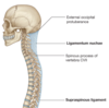
What are the 2 ligaments in the cervical and thoracic regions along the spine?
Ligamentum nuchae & supraspinous
How can you distinguish the 3 types of vertebrae?
Look at all from the sagittal view.
Cervical = mouse
Thoracic = giraffe
Lumbar = moose
What type of vertebrae has facets on the vertebral body and the vertebral foramen is large triangular shaped?
Cervical
What is the strongest cervical vertebra?
Axis
On what vertebra is the vertebral body heart-shaped?
Thoracic
Is the picture anterior or posterior? Name the bony processes of the sacrum.
S1-S5–> coccyx
Ala
1st sacral body
Sacral formina
Is the picture anterior or posterior? Name the bony processes of the sacrum.
Lateral, intermediate, median crests
Sacral canal, sacral hiatus
Sacral tuberosity
Facet and Articular surface
Sacral formina
Is this anterior or posterior this? Name the ligaments.
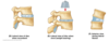
What are the two parts that make of the intervertebral disc?
Annulus fibrosus, nucleus pulposus
What are the 3 different views of the intervertebral disc under relaxed, erect and later flexion states?
What is a tear in the outer annulus fibrosus ring known as?
Slipped disc, intervertebral disc herniation
What is the superior and inferior attachments of the intercostal muscles (external, internal, innermost)
Superior = inferior border of ribs
Inferior = superior border of ribs below
What nerve innervates the intercostals?
Intercostal nerve
What is the main action of the intercostals (hint: 2 have the same action)
External intercostal: during forced inspiration: elevate ribs
Internal & innermost intercostal: During forced respiration, interosses part depresses rib, interchondral part elevates ribs
What is the superior and inferior attachment of the subcostal muscles and what is the action?
Superior = internal surface of lower ribs near angle
Inferior = superior border of 2nd or 3rd rib below
Action: act the same as internal intercostal muscles
What are these two muscles, what are there superior and inferior attachments?
Serratus posterior superior & serratus posterior inferior
Superior attachement: Superior = nuchal ligament, spinous process of c7-T3, Inferior = inferior border of ribs 8-12 near angle
Inferior attachment: Superior = superior border of 2nd and 4th ribs, Inferior = spinous process of T11-L2 vertebrae
What is the innervation and action of serratus posterior superior and serratus posterior inferior?
Innervation: Superior = 2-5th intercostal nerves, Inferior = 9-11 intercostal nerves, subcostal nerve
Action: Superior = elevate ribs, Inferior = depresses ribs
What is the origin, insertion, innervation, and action of rectus abdominis?
Proximal = pubic symphysis and pubic crest
Distal = Xiphoid process, 5-7th costal cartilages
Innervation = Thoracoabdominal and sub-costal nerves
Action = flex trunk, compress abdominal viscera, stabilizes and controls tilt of pelvis
What are the origin and insertions of the 3 oblique muscles?
External: Origin = external surface of 5-12th ribs, Insertion = linea alba, pubic tubercle, anterior half of iliac crest
Internal: Origin = Thoracolumbar fascia, anterior iliac crest, connective tissue deep to inguinal ligament, Insertion = Inferior border 10-12 ribs, linea alba, pubis via conjoint tendon
Transverse (abdominis): Origin = internal surface 7-12 costal cartilidge, thoracolumbar fascia, iliac crestand connective tissue deep to inguinal ligament, Insertion = lineal alba w/ aponeruosis of internal oblique, pubic crest, pubis via conjoint tendon
What is the innervation of the 3 oblique muscles?
External: Thoracoabdominal nerves and subcostal nerve
Internal and Transverse: Thoracoabdominal, subcostal and first lumbar nerves
What is the action of the 3 obliques?
External and Internal: Compress and support abdominal viscera; flex and rotate trunk
Transverse: compress and support abdominal viscera
Label the muscles, nerves, and blood flow of this cross section.
What are the three posterior wall muscles? What are there superior and inferior attachments?
Psoas major: Superior = transverse process of lumbar vertebrae; sides of bodies of T12-S1 vertebrae and intervening IV discs, Inferior = Strong tendon to lesser trochanter of femur
Illiacus: Superior = superior two-thirds of iliac fossa, ala of sacrum, anterior sacroiliac ligaments, Inferior = lesser trochanter of femur and shaft inferior to it, psoas major tendon
Quadratus lumborum: Superior = medial of inferior border of 12th rib and tips of lumbar transverse process, Inferior = iliolumbar ligament, internal lip of iliac crest
What is the innervation of Psoas major, iliacus and quadratus lumborum?
Psoas major: lumbar plexus via anterior branches of L2-L4
Iliacus: femoral nerve
Quadratus lumborum: anterior branches of T12 and L1-L4 nerves
What is the action of psoas major, iliacus, and quadratus lumborum?
Psoas major: flexes thigh, flexes vertebral column, laterally balance trunk, while sitting; w/ iliacus to flex trunk
Iliacus: flex thigh, stablize hip joint, acts w/ psoas major
Quadratus lumborum: extends and laterally flex vertebral column; fixes 12th rib during inspiration
Label the structures in this image.
Where does the diaphragm attach? What are the sensory and motor nerves it is innervated by?
Inferior margin of thoracic cage superior to lumbar vertebrae
Phrenic nerve (motor), subcostal (sensory)
What is the superficial muscle of the spinal column, where is it located, what is its action?
Splenius: Acting alone = laterally flex neck and rotate head to the side of active muscles, Acting together = extend head and neck
What are the 3 muscles comprised of the intermediate layer of the spinal column and what is there action?
Iliocostalis, Longissimus, Spinalis
Action: Bilateral = extend vertebral column, and head as back is flexed, control movement by gradually lengthening fibers Unilateral = laterally flex column
What are the deep muscle layers of the intrinsic back?
Semispinalis (capitis, cervicis, and thoracis)
Multifundis
Rotares (brevis and longus)
What are the actions of the deep muscle of the intrinsic back?
Semispinalis: extends head and thoracic and cervical regions of vertebral column and rotates them contralaterally
Multifundus: Unilateral contraction = rotates contralateral side; stabilizes vertebrae during the local movements of vertebral column
Rotatores: stabilize vertebrae and assist w/ local extension and rotary movements of vertebral column
What are these 3 muscles of the deep layer of the intrinsic back and their actions?
Interspinalis: Aid in extension and rotation of vertebral column
Intertransverssi: aid in lateral flexion of vertebral column, acting bilaterally, stabilize vertebral column
Levatores costarum: elevate ribs, assisting respiration; assist w/ lateral flexion of vertebral column
Name the vasculature of this thoracic vertebra.
What is the vasculature of the thoracic cavity?
What are the nerves that are located at the lower end of the spinal cord in the lumbosacral spine.
Cauda equina
What are the 3 meninges layers of the spinal cord?
Outer = Dura
Middle = Arachnoid
Inner = Pia