Lecture 2 - Respiratory tests Flashcards
What is low PaO2 called, and what is high PaCO2?
Hypoxia and hypercapnia, respectively
What are the ways we can assess ventilation?
- Blood gases
- PaO2
- PaCO2
- Lung volumes/flows
- Spirometry
- PEFR
- Exhaled Nitric Oxide (eNO)
What does spirometry measure?
How much and how fast
In spirometry, how is the air in the lungs divided into volumes?
- Tidal volume (~500ml) (VT) - Volume of air that moves in and out during normal quiet ventilation.
- Inspiratory reserve volume ~3L. An extra 3L can be inspired if the external intercostal muscles are contracted too.
- Expiratory reserve volume ~1.5L. An extra 1.5L can be expelled if internal intercostal muscles and abdominal muscles are contracted for maximal active expiration.
- Residual volume ~1L. Even after maximal expiration, our lungs are still partially inflated.

The volumes of the air in the lungs can be grouped in capacities.
List them.
- Vital capacity ~5L - Maximal breath in to maximal breath out (ERV + VT + IRV)
- Total lung capacity ~6L. If you breath all the way in, you hold about 6L in your lungs (VC + RV).
- Inspiratory capacity - Tidal volume + IRV
-
Functonal residual capacity ~2.5L. Volume in lungs at end of tidal expiration (RV + ERV)
- Represents “equillibrium point” for respiratory to change volume from FRC need to do work
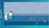
Label


How is residual volume measured?
Can’t be measured directly by spirometry, need to use helium dilution
Process: Breathe on spirometer, then add known amount of He.
Measure conc. at TLC (=VC + RV)
- Calculate volume He has distributed into = spirometer + VC + RV
- Measure spirometer & VC, RV = difference
- RV can also be measured by body plethysmography
In spirometry, how do we measure “how fast”?
- Forced measurements give us information about flow
- We use a forced vital capacity (FVC)
- Or a forced expiratory volume in one second (FEV1)
When does the FEV1 become affected?
It gets reduced in diseases which cause resistance to airflow (airways obstruction) OR Small lungs (scarred or fibrotic lungs)
What FEV1/FVC ration defines obstruction?
Value <0.70 defines obstruction
What are the three variables affected the “Normal”/Predicted values
- Height
- Age
- Gender
NOT WEIGHT - however will be effected if morbidly obese
The FVC, FEV1 will be ≥80% predicted

What determines the quality of a spirometry measurement
- Technician dependant/Subject dependent
- Need an acceptabe effort
- Sharp peak
- Gradual return to 0 flow
- At least 4 seconds
- 3 acceptable attempts within 5% of each other
- Often more easily seen on Flow-volume tracings
What value to volume loops have?

May show the presence of specific patterns of upper airway obstruction. Specific patterns in other disease processes confirm but add little to spirometry numbers.
What is the units for PEFR
L/min (NOT VOLUME) it’s a rate
It gives the peak flow rate for expiration
Is an individual absolute value for someones PEFR useful?
Not really useful if its just one value, since there’s a wide range of normal. But it’s the changes in PEFT from a persons normal value which are useful for assessing disease progresson. e.g. worsening asthma, there’s a decrease in PEFR.
Can use changing PEFR to change/assess therapy.
What are the indications for pulmonary function tesing
- Objective assessment of pulmonary symptoms
- Categorization of the type and severity of physiologic abnormalities
- Documentation of progression of disease
- Documentation of the patient’s response to therapy.
- Preoperative assessment
- Screening for sub clinical disease
How are lung conditions categorised
As either obstructve or restrictive
- In restrictive conditions their is an inability to stRetch the lungs due to increased lung stiffness, this decreases lung volume
- e.g. Lung compliance-related disease (pulmonary fibrosis/edema; chest wall compliace (kyphoscoliosis); pleural and respiratory muscle disease.
- In obstructive conditions there is a resistance to airflOw
- e.g. Chronic obstructive lung disease (chronic broncitis and emphysema), asthma
What happens to FVC, and FEV1, and the FEV1/FVC in restrictive lung disease?
- FVC Decreased
- FEV1 often decreased proportionate to FVC
- FEV1/FVC Normal or Increased
- May need lung volume measurements (RV, FRC, TLC) to confirm.

Describe the changes seen in FEV1/FVC, FEV1 and FVC in obstructive lung disease
- FEV1/FVC <0.7 defines obstructive disease
- FEV1 usually decreased
- FVC may be decreased
- e.g. if expiration incomplete due to air trapping
- Can have simulataneous obstruction and restriction
Pic of common patterns

