Kodas Flashcards
1
Q

A
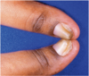
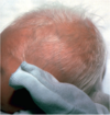
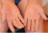
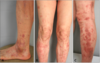
elastosis perforans serpiginosa
2
Q

A
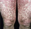
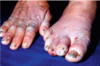
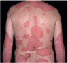
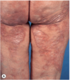
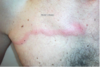
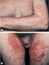
PRP
3
Q

A
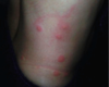
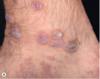
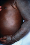
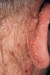
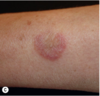
CARP
4
Q

A
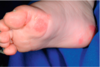
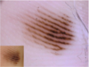
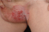
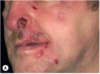
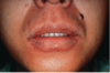
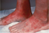
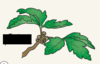
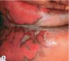
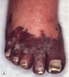
phytophotoderm
5
Q

A
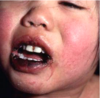
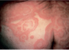
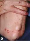
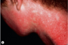
EM
6
Q

A
TEN
7
Q

A
pityriasis lichenoides
8
Q

A
pityriasis lichenoides, PIH
9
Q

A
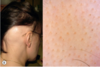
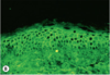
chronic GVHD
epidermal lichen planus-like changes on the posterior neck and upper aspect of the back
10
Q

A
lichen nitidus, isomorphic phenomenon
11
Q

A
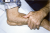
Flaccid blisters and erosion as a result of a ruptured bulla
12
Q

A
pemphigus foliaceus
13
Q

A
synblepharon in cicatricial pemphigoid
14
Q

A
Chronic bullous disease of childhood
15
Q

A
Epidermolysis bullosa acquisita
IgG against NC1 domain of Type 7 collagen (anchoring fibrils)
16
Q

A
Darier’s
- ATPA2 –> SERCA2 in endo reticulum*
- typical seborrheic distribution of brown, keratotic papules*
- also see: palmar pits; longitudinal red/white nail streaks w/”V “ notching; hard palate cobblestoning*
17
Q

A
Hailey Hailey
ATP2C1 –> hSPCA1 in Golgi
18
Q

A
DLE
19
Q

A
tumid lupus
20
Q

A
acute cutaneous lupus
presence of small erosions can aid in clinical ddx
21
Q

A
papulosquamous SCLE
- Anti-Ro/SSA*
- Complement deficiencies (C1, C2, C4)*
- HLA-B8 and HLA-DR3 assoc*
22
Q

A
Bullous SLE
- Ab to NC1 & NC2 domains of Type 7 collagen*
- dramatic response to dapsone in a few days*
23
Q

A
Lymphocytic infiltrate of Jessner
CD8 predominance
24
Q

A
dermatomyositis
25

mechanic's hands - dermatomyositis
26

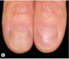
Cuticular hypertrophy, splinter hemorrhages, periungual telangiectasias - dermatomyositis
27

relapsing polychondritis
## Footnote
* autoab to Type 2 collagen*
* HLA-DR4*
28
rheumatoid nodules
29

Still's disease
## Footnote
* increased IL-1 production*
* episodic high fevers + rash corresponding w/fever spikes*
30

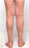
morphea
31

linear morphea
32

linear morphea
33

Marked atrophy of subcutaneous structures, including bone - Parry Romberg Syndrome
34

Hemiatrophy of the tongue - Parry Romberg Syndrome
35

Early edematous phase of systemic sclerosis
36

The “salt and pepper” sign. Leukoderma with retention of perifollicular pigmentation in systemic sclerosis.
37

Hyperpigmented sclerotic plaques of nephrogenic fibrosing dermopathy
## Footnote
*2-4 weeks post-gadolinium contrast exposure in pt w/AKI or CKD*
38

Scleral plaques in a patient less than 45 years of age - minor criterion for nephrogenic systemic fibrosis
39
disseminated GA
40

Necrobiosis lipoidica
41

Necrobiotic xanthogranuloma
## Footnote
*in a patient with a paraproteinemia*
42

Cutaneous Crohn's disease
43

Sarcoidosis
44

* Sarcoidosis – lupus pernio*
* note the notching of the nasal rim*
45

Langerhans cell histiocytosis
## Footnote
*seborrheic dermatitis-like eruption with hemorrhage*
46

Juvenile xanthogranuloma
47

Benign cephalic histiocytosis
48

Multicentric reticulohistiocytosis
49

Xanthoma disseminatum, sclerotic form
50

Eruptive xanthomas due to hypertriglyceridemia
51

Xanthomas of palmar striae
52

plane xanthoma
53

Neutrophilic dermatosis
54

Sweet's syndrome
55

Behcet's
56

Erythema gyratum repens
57

Lupus miliaris disseminatus faciei
58

Fox-Fordyce disease
59

PCT
60

Hepatoerythropoietic porphyria
## Footnote
*Hypertrichosis and severe scarring, resulting in clinical appearance similar to congenital erythropoietic porphyria*
61

Congenital erythropoietic porphyria
## Footnote
*Vesicles, bullae, and crusts on sun-exposed areas*
62

Lichen amyloidosis
## Footnote
*Keratotic, hyperpigmented plaques on the legs*
63

Acute hemorrhagic edema
64

strawberry gingiva of Wegener's
65

cutaneous PAN
## Footnote
*Livedo reticularis of the abdomen and lower extremities with multiple small “punched out” ulcers*
66
Kawasaki disease
## Footnote
*Lip erythema, fissuring, and bleeding*
67

Atrophie blanche from livedoid vasculopathy
68

Dissecting cellulitis of the scalp
69

Lichen planus pigmentosus of the face
70

Linear and whorled nevoid hypermelanosis
71

Subcutaneous fat necrosis
72

Transient neonatal pustular melanosis
73

Dermal melanosis
74

Membranous aplasia cutis with a subtle hair-collar sign
75

Neonatal herpes simplex virus
76

Congenital candidiasis
77

Lacy eruption on extensor arm of child with parvovirus B19 infection
78

Hand-foot-and-mouth disease
79

Gianotti-Crosti syndrome
## Footnote
*Grouped red papules on leg of child with EBV infection*
80

McCune-Albright syndrome
## Footnote
*Large, segmental café-au-lait macules with “coast of Maine” border*
81

Peutz-Jeghers
82
EB simplex, localized type
## Footnote
*Note superficial blistering w/both intact bullae and denuded skin. Tends to be limited to palms/soles*
83

Recessive dystrophic epidermolysis bullosa in a neonate
84

LUMBAR (SACRAL) syndrome
85

Sturge-Weber syndrome
## Footnote
*diffuse capillary hemangioma in the distribution of the ophthalmic, nasociliary, and maxillary branches of the trigeminal nerve*
86

Microcystic lymphatic malformation
## Footnote
*irregularly-grouped, translucent, and red papules*
87

Cutis marmorata telangiectatica congenita
88
Pachyonychia congenita
89

hypohidrotic ectodermal dysplasia
## Footnote
*Note the flat nasal bridge, depressed nasal tip, sparse hair (scalp, eyebrows, and eyelashes), peg-shaped teeth, full lips, and sebaceous hyperplasia.*
90

Trichorrhexis nodosa
91

Trichorrhexis invaginata in Netherton syndrome
92

acrodermatitis enteropathica
93

Cutis laxa / generalized elastolysis
94

Pseudoxanthoma elasticum
95

Goltz syndrome / focal dermal hypoplasia
## Footnote
*(usually lethal in males - this one probably mosaic)*
96

von Recklinghausen's neurofibromatosis
97

axillary freckling / Crowe's sign in von Recklinghausen's neurofibromatosis
98

Facial angiofibromas in tuberous sclerosis
99

Periungual fibrous nodules (Koenen's tumors) in tuberous sclerosis
100

Incontinentia pigmenti
101

Progeria syndrome
102

Xeroderma pigmentosum
103

regularly-spaced giant melanosomes in the hair shaft of Chediak-Higashi syndrome
104

irregularly-spaced giant melanosomes in the hair shaft of Griscelli syndrome
105

Collodion baby
106

Lamellar ichthyosis phenotype of ARCI
107

Netherton Syndrome
## Footnote
*Erythroderma, hypotrichosis, and areas with ichthyosis linearis circumflexa*
108

Conradi-Hünermann syndrome
## Footnote
*pattern of scale along Blaschko's lines*
109

CHILD Syndrome
## Footnote
*Unilateral erythema and scale with ipsilateral limb defects*
110

Conradi-Hünermann syndrome
## Footnote
*Thick, psoriasiform scaling overlying erythema; also w/chondrodysplasia punctata*
111

Unna–Thost syndrome
112

Vohwinkel's
## Footnote
*Mutilating keratoderma*
113

urticaria pigmentosa
114

Eczema herpeticum
115

Staphylococcal scalded skin syndrome
116

Erysipelas
117

Superficial infection of the skin with Pseudomonas
118

meningococcemia
## Footnote
*stellate purpura with a central gunmetal-gray hue*
119

Secondary syphilis
120

Mycetoma (N. brasiliensis)
121

Mycetoma (N. brasiliensis)
122

Bartonella henselae
Warthin-Starry stain
123

Chromoblastomycosis
124

Histoplasmosis
## Footnote
*PAS-positive intracellular yeast*
125

Blastomycosis
## Footnote
*Direct microscopy: budding yeast w/wide fusion base*
126

Coccidioidomycosis
127

Coccidioidomycosis
## Footnote
*giant cell containing small spherules*
128

Paracoccidioidomycosis
129

aspergillus
130

Zygomycosis (mucor)
131
Tungiasis
132

Smooth nodular cutaneous leishmaniasis
133

Leishmaniasis
134

Romaña sign in acute Chagas disease
135

seabather's eruption
## Footnote
*larvae of Linuche unguiculata*
136
Cimex spp. (bedbugs)
137

Loxosceles reclusa (brown recluse spider)
138

Linear plaques, erythema, and edema following contact w/Portuguese man-of-war tentacles
139

porokeratosis
140

Porokeratotic eccrine ostial and dermal duct nevus
## Footnote
*Markedly hyperkeratotic spines arise from dilated eccrine ostia*
141

Verrucous carcinoma
142

MAC
143

Leiomyoma
144

MF, tumor stage
145

Alopecia mucinosa
146

Lymphomatoid papulosis
147

Congenital dermatofibrosarcoma protuberans
148

Collagenomas
149

angiosarcoma
150

Seborrheic keratosis
## Footnote
*milia-like cysts (white dots), comedo-like openings (black targetoid circles)*
151

Dermatofibroma
## Footnote
*central white patch (asterisk) and subtle pigment network (arrows)*
152

Basal cell carcinoma
## Footnote
*arborizing blood vessels, blue-gray blotches (asterisks), and ulceration (circle)*
153

Basal cell carcinoma
## Footnote
*Leaf like areas (3 o'clock to 6 o'clock at periphery)*
154

Pyogenic granuloma
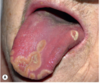
## Footnote
*homogenous red color with red lacunae*
155

Keratoacanthoma
## Footnote
*classic hairpin-shaped vessels (arrows), white background (hyperkeratosis of keratinizing tumor), central crust*
156

Pigmented Bowen's disease
## Footnote
*well-circumscribed glomeruloid vessels (circle) with closely-packed tiny brown dots (arrows)*
157

Clear cell acanthoma
## Footnote
*this pattern of dotted vessels is rather typical*
158

Typical acquired nevus
159

Reed nevus
## Footnote
*classic starburst pattern (regular streaks at periphery of heavily pigmented and symmetric small macule)*
160
Acral nevus
## Footnote
*parallel furrow pattern*
161

Acral melanoma
## Footnote
*parallel ridge pattern*
162

Melanoma
## Footnote
*annular-granular structures make up rhomboidal structures (arrow); confluent rhomboidal structures make up the gray pseudonetwork (circle)*
163

Melanoma
## Footnote
*atypical pigment network (circle), irregular dots and globules (asterisks), irregular streaks (black arrows), irregular blotches (white arrows), blue-white structures centrally*
164

Trichodiscomas and fibrofolliculomas and acrochordons in Birt-Hogg-Dubé
165

HHT
## Footnote
*multiple telangiectasias on tongue and lip*
166

Bullous diabeticorum
167

NLD
168

Pretibial myxedema
169

PXE
## Footnote
*Lax skin and redundant folds on the anterior neck*
170

Pyoderma gangrenosum
171

pyostomatitis vegetans
## Footnote
*erythematous oral mucosa with overlying yellow pustules*
172

scurvy
## Footnote
*perifollicular purpura and follicular hyperkeratosis*
173

Bazex syndrome
## Footnote
*nail dystrophy and erythematous to violaceous plaques*
174

erythema gyratum repens
175
Extramammary Paget disease
176

Increased lanugo hair on the nose and face of a man with underlying metastatic prostate cancer
177

necrolytic migratory erythema
178

NXG
179

paraneoplastic pemphigus
180

Large, thin, yellow-orange plaque in a patient with monoclonal gammopathy
181

Tripe palms
## Footnote
* alone --\> lung CA*
* + acanthosis nigricans --\> gastric CA*
182

epidermolytic ichthyosis
183

lymphocytoma
## Footnote
*borrellia*
184

alternaria
"beaver tail"
185

erythrasma
186

nevus sebaceous w/trichoblastomas
187

DH
188

post-efudex reaction
189

pretibial myxedema
190

SCLE
191

SCLE w/secondary hemorrhage
192

SCLE
193

sarcoid
194

dissecting cellulitis
195

metastatic melanoma w/possible primary on back
196

BCC
197

MF
198

zoster
199

sarcoid (lupus pernio)
200

BP
201

SK
202

psoriasis
203

sarcoidosis
204

antiphospholipidd syndrome
205

lichen planus
206

lichen planus
207

necrosis 2/2 vasculopathy from levamisole-contaminated cocaine
208

melasma
209

argyria
210

zoster
211

calciphylaxis
212

secondary excoriation
213

acute ACD
214

atypical mycobacteria w/sporotrichoid spread
215

koebnerized lichen planus
216

papular mucinosis
217

epidermal nevus
218

ACD to PPD
219

erythema migrans
220

EM
221
lichen planus
222

acute ACD
223

psoriasis
224

pemphigus vulgaris
225

annular pustular psoriasis
226

bullous pemphigoid
227

cutaneous small vessel vasculitis
228

polyarteritis nodosa
229

cutaneous lymphoid hyperplasia
230

eruptive xanthomas
231

lichen planopilaris
232

linear morphea
233

erythema nodosum
234

pancreatic panniculitis
235

Small superficial melanoma
asymmetry of color and structure, atypical network, and blue-white structures
236

Larger, thick melanoma
prominant blue-white veil and few irregular black globules/areas
237

small facial melanoma in situ (lentigo maligna)
typified by gray granules around hair follicles
238

Acral melanoma in situ
characteristic parallel-ridge pattern
239

Melanoma in situ
asymmetry of color/structure, atypical network, blue-white structures intermingled with dotted vessels
240

Melanoma 0.5 mm in depth
atypical pigment network, blue-white structures
241

Melanoma 0.8 mm in depth
asymmetry of color/structure, irregular dots and globules, blue-white structures, and peripheral irregular streaks
242

Melanoma 1.3 mm in depth
blue-white veil, atypical vascular pattern (signs of increased tumor thickness); remnants of atypical pigment network and irregular brown globules
243

acquired nevus
244

congenital nevus
245

reed nevus
246

blue nevus
247

Small superficial melanoma
asymmetry of color/structure, atypical network, blue-white structures, and irregular streaks at periphery
248

Large thick melanoma
prominent blue–white veil
*Combination of blue color + irregular black-brown dots, globules, and blotches is highly specific for thick melanoma*
249

Small facial melanoma in situ (lentigo maligna)
gray color and rhomboidal structures
250

Melanoma in situ
asymmetry of color/structure, atypical network, blue-white structures, irregular black dots and globules (at the upper side of the lesion)
251

Melanoma 0.5 mm thick
atypical pigment network and regression structures (areas of pigment loss and bluish pepper-like granules corresponding to melanophages)
252

Melanoma 0.75 mm thick
asymmetry of color and structure, atypical network, irregular streaks at the periphery, irregular dots and globules (upper side), and blue-white structures especially in center
253

Melanoma 0.9 mm thick.
palpable area / blue-white veil (increased tumor thickness), irregular dots and globules (upper side), irregular streaks at periphery, and uneven brown to black pigmented areas (blotches)
254

acquired nevus
255

congenital nevus
256

reed nevus
257

pigmented BCC
258

Angiokeratoma
red–black lacunae, well-demarcated roundish structures
259

dermatofibroma
central white patch, peripheral delicate pseudo-network
260

amelanotic melanoma
central ulceration, polymorphic vascular structures (combination of dotted + linear-irregular vessels), and milky-red color in background
261

Nodular basal cell carcinoma
striking arborizing vessels
262

Spitz nevus
dotted vessels and typical negative pigment network (reticular depigmentation) at periphery
263

Bowen disease
clusters of glomerular vessels; in combination with superficial scales, are highly specific for the dx
264

Pigmented basal cell carcinoma
typical leaf-like areas (islands of brown-gray) and blue-gray globules
265

Seborrheic keratosis
typical milia-like cysts (white shining dots/globules) and comedo-like openings (black targetoid globules)
266

Angiokeratoma
red-blue lacunae, well-demarcated roundish structures
267

dermatofibroma
characteristic central white patch and peripheral delicate network
268

amelanotic melanoma
polymorphic vascular structures, milky-red color in background
269

Nodular basal cell carcinoma
arborizing vessels and blue-gray globules
270

Spitz nevus
dotted vessels and typical reticular depigmentation
271

Bowen disease
clusters of dotted/glomerular vessels + superficial scale
272

intradermal melanocytic nevus
typical comma-shaped vessels
273

Clear cell acanthoma
dotted vessels, linearly arranged as strings of pearls
274

Well-differentiated squamous cell carcinoma
white keratin masses, white circles and clods, and polymorphic vessels
275

Molluscum contagiosum
peripheral arborizing vessels superimposed on white background
Amorphous yellow clods are typically seen in the center
276

Scabies
characteristic “jet with contrail” structure, corresponding to anterior part of mite (arrow) and burrow behind it
277

psoriasis
regular dotted vessels
278

lichen planus
dotted vessels at the border of typical whitish lines and clods, which closely resemble Wickham striae found in oral mucosa
279
alopecia areata
typical follicular yellow dots
280

infantile pustular psoriasis
281

periungual wart
282

zoster in V1 and V2 distribution
283

pemphigus vulgaris
284

bullous pemphigoid
285

epidermolytic ichthyosis
286

TEN
287
oculocutaneous albinism
288

cutaneous small vessel vasculitis
289

vitiligo
290

alopecia areata
291

TEN
292

albinism
293

insect bites
294

dermographism
295

atopic derm
296

prurigo simplex, prurigo nodularis, and angulated ulcerations
297

prurigo nodularis
298

prurigo nodularis
299

prurigo nodularis
300

pemphigoid nodularis
301

lichen simplex chronicus
302

lichen simplex chronicus
303

Xerosis and scratching in a patient with ESRD undergoing hemodialysis
304

brachioradial pruritus

305
trigeminal trophic syndrome
306

trigeminal trophic syndrome
307

Complex regional pain syndrome
with scaling and erosions of the fingertips
308

prurigo nodularis superimposed on atopic derm
309

linear excoriations and xerosis
310

prurigo nodularis
311

Prurigo nodularis
312

Prurigo nodularis
313

Lichen simplex chronicus of scrotum
314

Xerosis and scratching in a patient with renal insufficiency undergoing hemodialysis
315

Extreme xerosis a/w pruritus in HIV-infected patient
316

Notalgia paresthetica
317

brachioradial pruritus
318

Trigeminal trophic syndrome
319

Trigeminal trophic syndrome
320

excoriations and scars in delusions of parasitosis
321

nail-biting and cuticle-picking
322

Trichotillomania
323

acne excoriee
324

Dermatitis artefacta
325

Dermatitis artefacta
326

Dermatitis artefacta
327

Dermatitis artefacta
328

Dermatitis artefacta
329

Non-suicidal self-injury
330

Bite fibroma from years of cheek chewing
331
Habit-tic deformity of the thumbnail
332

Dermatitis artefacta
333

Excoriation (skin-picking) disorder
334
scars from excoriation (skin-picking) disorder
335

annular psoriasis plaques
336

psoriasis
337

psoriasis
338
psoriasis
339

Palmoplantar psoriasis
340

Palmoplantar psoriasis
341

psoriasis
342

psoriasis
343

Guttate psoriasis
344

Guttate psoriasis w/koebnerization
345

Guttate psoriasis w/sunburn-related koebnerization
346

Generalized pustular psoriasis
347

Annular pustular psoriasis
348

Annular pustular psoriasis
349

Pustulosis of the palms
350

Acrodermatitis continua of Hallopeau
351

Scalp psoriasis
352

Inverse psoriasis
353

Nail psoriasis
354

Psoriatic arthritis
355

keratoderma blennorrhagicum in reactive arthritis
356

keratoderma blennorrhagicum in reactive arthritis
357

Sneddon-Wilkinson disease (subcorneal pustular dermatosis)
358

Sneddon-Wilkinson disease (subcorneal pustular dermatosis)
## Footnote
*significant overlap w/pustular psoriasis*
359

papulosquamous penile lesions in reactive arthritis
360

psoriasis
361

psoriasis
362

psoriasis
363

psoriasis
364
generalized pustular psoriasis
365

Annular pustular psoriasis
366

Sneddon-Wilkinson disease
367

Small plaque parapsoriasis
368

Small plaque parapsoriasis
369

Large plaque parapsoriasis
370

Large plaque parapsoriasis
371

PLEVA
372

PLEVA
373

PLEVA
374

PLC
375

PLC (can appear to overlap w/PLEVA)
376

PLC
377

Pityriasis rubra pilaris
378

Pityriasis rubra pilaris
379

Pityriasis rubra pilaris
380

Pityriasis rubra pilaris
381

Pityriasis rubra pilaris
382
Pityriasis rubra pilaris
383

Circumscribed juvenile PRP (type IV)
384

Pityriasis rosea
385

Pityriasis rosea
386

Follicular accentuation of pityriasis rosea in darkly pigmented skin
387

Pityriasis rosea
388

Pityriasis rosea
389

Pityriasis rosea
390

Granular parakeratosis involving the axilla
## Footnote
*Coalescing brown papules w/hyperkeratosis, slight maceration*
391

Digitate small plaque parapsoriasis
392

PLEVA / PLC
393

PRP
394

PRP
395

PRP
396

Pityriasis rubra pilaris-like presentation in Wong variant of juvenile dermatomyositis
397

Pityriasis rosea
398

Pityriasis rosea
399

Inverse pityriasis rosea
400

Cowden's
401

steroid atrophy
402

poikilodermatous MF
403

chemo nails
404

erythroderma with desquamation
405

Ectropion in the setting of erythroderma
406

Psoriatic erythroderma
407

Erythroderma due to atopic dermatitis
408

Idiopathic erythroderma
409

Erythroderma due to pityriasis rubra pilaris
410

Erythroderma secondary to pityriasis rubra pilaris
411

erythroderma due to Sezary syndrome
412

Erythroderma due to pemphigus foliaceus
413

Erythroderma with desquamation
414

psoriasis
## Footnote
*pitting and onycholysis w/proximal rim of inflammation*
415

Psoriatic erythroderma
416

Lichen planus
417

Lichen planus
418

Koebnerization of lichen planus into the site of excision of saphenous vein
419

Lichen planus with postinflammatory hyperpigmentation
420

Lichen planus with postinflammatory hyperpigmentation
421

Annular lichen planus
422

Annular lichen planus
423

Exanthematous lichen planus
424
Atrophic lichen planus of the lower extremities
425

Bullous lichen planus on the shin
426

Lichen planus pemphigoides
427

Hypertrophic lichen planus
428

Hypertrophic lichen planus
429

Inverse lichen planus
430

Lichen planus pigmentosus – intertriginous variant
431

Lichen planopilaris
432

Lichen planopilaris
433

Lichen planopilaris
434

Linear lichen planus
435

Nail lichen planus
436

Nail lichen planus
437

Nail lichen planus
438

Oral lichen planus
439

Oral lichen planus
440

Lichenoid drug eruption
441

Lichenoid drug eruption
442

lichen striatus
443

lichen striatus
444

Lichen nitidus
445

Lichen nitidus
446

Erythema dyschromicum perstans
447

Keratosis lichenoides chronica
448

Keratosis lichenoides chronica
449

Keratosis lichenoides chronica
450

Lichen striatus
451

Postinflammatory hypopigmentation in atopic dermatitis
452
Infantile atopic dermatitis on the extensor arms
453

Infantile atopic dermatitis
454

Infantile atopic dermatitis
455

Flexural atopic dermatitis
456

atopic dermatitis
457

Chronic atopic dermatitis
458

Chronic atopic dermatitis
459

Chronic atopic dermatitis
460

Chronic atopic dermatitis
461

severe atopic dermatitis
462

atopic dermatitis
## Footnote
*Chronic papular lesions resulting from habitual rubbing and scratching in the setting of longstanding disease*
463

atopic dermatitis
## Footnote
*Prurigo lesions presenting as firm, dome-shaped papules and nodules with central hemorrhagic crust*
464

nummular atopic dermatitis
465

Atopic cheilitis
466

Nipple eczema
467

Numerous punctate and a few linear excoriations in an area of papular eczema
468

Keratosis pilaris
469

Keratosis pilaris
470

infected hand dermatitis in a patient with atopic dermatitis
471

Pityriasis alba
472

Eczema herpeticum
473

Eczema herpeticum
474

Postinflammatory pigmentary alteration in atopic dermatitis
475

Infantile atopic dermatitis
476

Infantile atopic dermatitis
477

Infantile atopic dermatitis
478

Childhood atopic dermatitis
479

Childhood atopic dermatitis
480

Childhood atopic dermatitis
481

Childhood atopic dermatitis
482

Atopic dermatitis in childhood exacerbated by Malassezia
483

Keratosis pilaris rubra
484

Keratosis pilaris rubra
485

Severe chronic hand dermatitis in atopic dermatitis
486

Infantile seborrheic dermatitis
487

Infantile seborrheic dermatitis
488

seborrheic dermatitis
489

seborrheic dermatitis
490

seborrheic dermatitis
491

inverse psoriasis
492

tinea cruris
493

candidiasis
494

erythrasma
495

granular parakeratosis
496

Darier's
497

cutaneous crohns
498

langerhans cell histiocytosis
499

extramammary paget
500

Asteatotic eczema
501

Asteatotic eczema
502

Id reaction due to ACD to nickel
503

Id reaction due to ACD to nickel
504

Nummular dermatitis
505

Autosensitization dermatitis in venous ulceration
506

stasis dermatitis
## Footnote
*microangiopathy and chronic inflammation*
507

stasis dermatitis
508

Dyshidrotic eczema
509

Juvenile plantar dermatosis
510

seborrheic dermatitis
511

Seborrheic dermatitis-like eruption due to dermatomyositis
512

Asteatotic eczema
513

Asteatotic eczema
514

Nummular dermatitis
515

Nummular dermatitis
516

dyshidrotic eczema
517

Infectious eczematous dermatitis
518

irritant dermatitis
519

irritant dermatitis
520

candidiasis
521

psoriasis
522

Streptococcal perianal dermatitis
523

Bullous impetigo
524

Acrodermatitis enteropathica
525

langerhans cell histiocytosis
526

Allergic contact dermatitis
527

Allergic contact dermatitis
528

Chronic allergic contact dermatitis
529

chronic allergic contact dermatitis
530

Acute allergic contact dermatitis to shoes
531

Chronic allergic contact dermatitis to shoe
532

Acute vesiculobullous allergic contact dermatitis
533

Acute vesiculobullous allergic contact dermatitis
534

Acute allergic contact dermatitis w/prominent edema
535

Acute allergic contact dermatitis w/prominent edema
536

chronic allergic contact dermatitis
537

chronic allergic contact dermatitis
538
Allergic contact dermatitis due to aloe-containing cream
539

Allergic contact dermatitis due to cashew nut shell oil
540

eyelid dermatitis (ACD)
541

Allergic contact dermatitis to fragrance found in cologne
542

Allergic contact dermatitis to nickel
543

Allergic contact dermatitis to p-phenylenediamine in a temporary tattoo
544

Systemic contact dermatitis
545

Airborne contact dermatitis
546

Airborne contact dermatitis
547

Chronic allergic contact dermatitis leading to hand dermatitis
## Footnote
*This golfer wore one leather glove and had + patch tests to potassium dichromate and a piece of his glove*
548

chronic allergic contact dermatitis
## Footnote
*In this bowler, allergen was colophony contained within a tackifier*
549

Acute allergic contact dermatitis
## Footnote
*Obvious edema of shaft plus subtle crusts of glans due to tiger balm*
550

Acute vesiculobullous allergic contact dermatitis
## Footnote
*caused by the plant Grevillea which grows in Australia*
551

Acute vesiculobullous allergic contact dermatitis
## Footnote
*due to compound benzoin tincture; note the geometric shape*
552

Chronic allergic contact dermatitis due to glutaraldehyde
553

Facial dermatitis due to nickel in cell phone
## Footnote
*demonstrated by + dimethylglyoxime test*
554

Allergic contact dermatitis to neomycin
555

Allergic contact dermatitis to p-phenylenediamine in a temporary tattoo
556

Airborne contact dermatitis
557
Bilateral irritant contact dermatitis of the feet and ankles due to chronic occlusive footwear
558

Bilateral irritant contact dermatitis of the palms secondary to repeated contact with paint solvents
559

Irritant contact dermatitis of the hands due to chronic exposure to disinfectants
560

Irritant contact dermatitis
lip-licking habit; note involvement of vermilion and cutaneous lips as well as perioral region
561

Allergic contact dermatitis to oxybenzone with involvement of the upper and lower lips
562

Moderately severe irritant contact dermatitis of the hands due to chronic exposure to disinfecting solutions and antiseptics
563

dyshidrotic (pompholyx) pattern of eczema
## Footnote
*--\> allergic contact dermatitis to chromate found in cement and contact urticaria to latex gloves*
564

Cement burns
## Footnote
*Ulcerations on the fingertips of a construction worker exposed to wet cement*
565

Fiberglass dermatitis
566

Positive prick test with commercial latex extract (L)
## Footnote
*Histamine (H) and saline (C) controls are appropriately positive and negative*
567
Chloracne
## Footnote
*primary ddx: folliculotropic MF*
568

Vibration White Finger
* vasoconstriction & endothelial damage 2/2 to exposure to vibration @ 30 - 300 Hz (chainsaws, pneumatic tools)*
* Associated transient loss of sensation; possible permanent finger neuropathy & pain in affected limb*
569

Stinging nettle (Urtica dioica)
## Footnote
*Note trichomes on both stem and leaf surfaces*
570

Broken trichome from stinging nettle (Urtica dioica)
## Footnote
*A bubble is visible where urticating chemicals are found (histamine, serotonin, acetylcholine)*
571

Microscopic appearance of glochids from Opuntia microdasys (prickly pear)
## Footnote
*mechanical irritant*
572

Prickly pear (Opuntia ficus-indica)
573

Prickly pear (Opuntia ficus-indica) fruit
## Footnote
*Fine spines and glochids are visible*
574

Bullous phase of phytophotodermatitis
575

Giant hogweed (Heracleum mantegazzianum) -- apiaceae
## Footnote
*phytophotodermatitis*
576

Compound umbel of giant hogweed -- apiaciae
## Footnote
*phytophotodermatitis*
577

Phytophotodermatitis
578

poison ivy
579

western poison oak
580
eastern poison oak
581

poison sumac
582

Shrub-type poison ivy
583

Climbing vine of poison ivy
584

Shrub-type poison ivy
## Footnote
*Leaf trauma allows urushiol to reach surface, oxidize, and turn black*
585

Leaves and seeds of Ginkgo biloba
## Footnote
*only the fresh seed covering contains the allergen, ginkgolic acid, which cross-reacts with urushiol*
586

Typical composite flower heads of members of the family Asteraceae (Compositae)
## Footnote
*Allergens: sesquiterpene lactones*
587

Poison ivy (Toxicodendron radicans) dermatitis
588

Poison ivy dermatitis
589

Widespread erythema and edema a/w intense pruritus after carrying logs of poisonwood tree (Metopium toxiferum) of family Anacardiaceae
590

“Black-spot” poison ivy dermatitis
## Footnote
*polymerized plant oleoresin*
591

Weed-whacker dermatitis - widespread spotted pattern
592

Airborne contact dermatitis
## Footnote
*farmer from Gujarat, India allergic to sesquiterpene lactones in Parthenium hysterophorus*
593

chronic, lichenified, hyperkeratotic dermatitis caused by allergy to Peruvian lily and tulipalin
594

Wheals (urticaria)
595

Wheals (urticaria)
596

Wheals (urticaria)
597

Angioedema
598

Symptomatic immediate dermographism
599

Delayed pressure urticaria
600

Cold urticaria
## Footnote
*Wheals developed after placement of an ice cube for 10 minutes, followed by rewarming*
601

Cholinergic urticaria after hot bath provocation
* small, monomorphic wheals that are symmetrically distributed*
* vs adrenergic: blanched vasoconstricted skin surrounding small pink* *wheals induced by sudden stress*
602

Urticarial vasculitis
## Footnote
*Lesions look like spontaneous urticaria but last longer and may bruise*
603

Urticaria in a young child
## Footnote
*This is sometimes misdiagnosed as EM d/t dusky centers - some clinicians call it urticaria multiforme. Unlike EM, central clearing can occur as lesions expand*
604

Urticaria
605

Erythema annulare centrifugum
606

Erythema annulare centrifugum
607

Erythema annulare centrifugum, deep form
608

Erythema annulare centrifugum
609

urticaria
610

erythema marginatum (rheumatic fever)
611

Annular erythema of infancy
612

erythema migrans
613

erythema migrans
614

erythema migrans
615

Disseminated erythema migrans
616
erythema gyratum repens
617

Resolving pityriasis rubra pilaris
## Footnote
*can resemble erythema gyratum repens*
618

EM - edematous/urticarial
619

EM - urticarial with central crusting
620

EM - erythematous plaques with dusky centers
621

EM - classic targetoid
622

EM - classic targetoid
623

EM - isomorphic response with crust formation
624

EM
## Footnote
*the dusky or crusted centers help to differentiate from morbilliform drug eruption*
625

Mucosal involvement in EM major
626

Mucosal involvement in *mycoplasma-*associated EM major
627

Acute annular urticaria / urticaria multiforme in an infant (misdiagnosed as EM)
628

Mucosal involvement in SJS
629

Mucosal involvement in SJS
630

Characteristic dusky red color of the early macular eruption in TEN
631

TEN
632

TEN
633

epidermal detachment of palmar skin in TEN
634

TEN
635

SJS/TEN
636

SJS/TEN
## Footnote
*close-up of epidermal detachment, likened to wet cigarette paper*
637

TEN
638

sequelae of TEN
## Footnote
*Symblepharon, erosion of lower lateral eyelid margin and sparse eyelashes; the patient also had entropion with an ingrowth of eyelashes and pebble-like scarring of facial skin*
639

sequelae of TEN
## Footnote
*Larger irregular areas of hypopigmented scarring*
640

sequelae of TEN
## Footnote
*Nail dystrophy: longitudinal ridging and fissuring, fragility, and distal notching*
641

early SJS
642

SJS/TEN
643

Childhood SJS secondary to TMP-SMX
644

TEN
645

TEN
646

resolving TEN
647

Methotrexate toxicity
## Footnote
*Increased serum MTX levels 2/2 decreased renal excretion can cause epidermal necrosis*
648
Methotrexate toxicity
## Footnote
*Increased serum MTX levels 2/2 decreased renal excretion can cause epidermal necrosis*
649

Urticaria secondary to penicillin
650

Morbilliform (exanthematous) drug eruption induced by amoxillin
651

Morbilliform (exanthematous) drug eruption
## Footnote
*lesions on lower extremities can become petechial or purpuric due to dependency*
652

Morbilliform (exanthematous) drug eruption induced by phenobarbital
653

DRESS due to carbamazepine
654

DRESS due to carbamazepine
655

Serum sickness due to antithymocyte globulin
656

AGEP - secondary to cephalosporin
657

AGEP - secondary to amoxicillin
658

Iododerma
659

Neutrophilic eccrine hidradenitis
660

Fixed drug eruption
661

Fixed drug eruption
662

Fixed drug eruption
663

bullous fixed drug eruption
664

generalized bullous fixed drug eruption
665

fixed drug eruption
666

fixed drug eruption
667

PIH after fixed drug eruption
668

Phototoxic reaction 2/2 methotrexate
669

Photolichenoid drug eruption due to HCTZ
670

Horizontal melanonychia due to 5-fluorouracil
671

Toxic erythema of chemotherapy due to cytarabine, with acral erythema involving plantar surface
672
Inflammation surrounding SK in pt receiving paclitaxel
673

Raynaud phenomenon and digital necrosis due to systemic bleomycin
674

flagellate erythematous urticarial plaques due to systemic bleomycin
675

Toxic erythema of chemotherapy (fludarabine + busulfan)
676

Papulopustular (acneiform) reactions to EGFR inhibitors
677

Papulopustular (acneiform) reaction to EGFR inhibitors
678

Exanthematous (morbilliform) reaction to lenalidomide with accentuation at sites of bortezomib injections
679

SJS/TEN overlap due to ipilimumab
680

Inflammation of AKs due to IV interleukin-2
681

Heparin-induced thrombocytopenia with thrombosis
682

Heparin-induced thrombocytopenia with thrombosis
683

Cutaneous discoloration due to amiodarone
684

Psoriasiform eruption due to TNF-α inhibitor
685

Local reaction to vitamin K injections
686

Local reaction to vitamin K injections
687

Exanthemaotus drug eruption due to a cephalosporin
688

Exanthemaotus drug eruption due to phenobarbital
689

AGEP 2/2 metronidazole
690

SJS
691

Bullous fixed drug eruption
692

Ulceration due to extravasation of doxorubicin
693

Papulopustular (acneiform) reaction to an EGFR inhibitor
694

Round to oval petechiae, \<3 mm in diameter
695

Solar (actinic) purpura in sites of actinic damage + trauma
696

Palpable purpura due to cutaneous small vessel vasculitis
697

Non-inflammatory (bland) retiform purpura and hemorrhagic bullae in DIC
698

Schamberg disease
## Footnote
*oval yellow-brown patches containing pinpoint "cayenne pepper" petechiae*
699

Purpura annularis telangiectodes of Majocchi
## Footnote
*1-3 cm annular plaques containing punctate petechiae/telangiectasias; may slowly expand*
700

Pigmented purpuric lichenoid dermatitis of Gougerot and Blum
## Footnote
*Schamberg-like lesions + purpuric lichenoid papules*
701

lichen aureus
702
Pigmented purpuric lichenoid dermatitis of Gougerot and Blum
703

Granulomatous pigmented purpura
704

Schamberg disease
## Footnote
*Discrete yellow-brown and -pink patches with superimposed petechiae*
705

stasis purpura
## Footnote
*Petechiae superimposed on more diffuse hemosiderin deposits in venous HTN*
706

Purpura annularis telangiectodes of Majocchi
## Footnote
*annular plaques w/cayenne pepper petechiae in border*
707

Eczematid-like purpura
## Footnote
*often presents with pruritus*
708

Linear pigmented purpuric dermatosis
## Footnote
*linear array of yellow-brown macules and patches w/superimposed petechiae and small, red-brown purpuric papules*
709

Hypergammaglobulinemic purpura of Waldenström
710
Mondor Syndrome of Superficial Thrombophlebitis
## Footnote
* classically: anterolateral thoracoabdominal wall*
* up to 10% have underlying breast carcinoma*
711

Linear pigmented purpura
712

Heparin necrosis at site of SQ heparin injection
713

retiform purpura of Type I cryoglobulinemia in multiple myeloma (IgG type)
714

Areas of necrosis within the areas of retiform purpura of Type I cryoglobulinemia in multiple myeloma (IgG type)
715

classic acral purpura of Type I cryoglobulinemia in multiple myeloma (IgG type)
716

Ecthyma gangrenosum
## Footnote
*syndrome of occlusion by organisms (usually bacterial, classically pseudomonas) proliferating in adventitia of SQ blood vessels*
717

Livedo reticularis in cholesterol embolus
718

Livedo reticularis and retiform purpura in cholesterol embolus
719

Purpura of the digits in warfarin blue toe syndrome
720

Several irregularly shaped ulcers with eschars surrounded by retiform purpura, caused by cholesterol embolus
721

Protein C deficiency in an infant
## Footnote
*cutaneous microvascular occlusion; presents hours to 5 days after birth and fatal if untreated*
722

warfarin necrosis
723

warfarin necrosis
724
Antiphospholipid antibody syndrome
725

Antiphospholipid antibody syndrome
726

Antiphospholipid antibody syndrome with atrophie blanche-like scarring
727

livedo racemosa in Sneddon Syndrome
## Footnote
*neurocutaneous syndrome w/persistent and usually widespread livedo racemosa, labile hypertension, and CNS disease*
728

Livedo racemosa
## Footnote
*Sneddon syndrome; also AR adenosine deaminase 2 deficiency*
729

Livedoid vasculopathy
## Footnote
*primary idiopathic OR secondary to vascular / thrombotic disease*
730

Malignant atrophic papulosis / Degos disease
## Footnote
*rare, chronic, thrombo-obliterative vasculopathy --\> erythematous papules with central porcelain-white atrophy and surrounding teleangiectatic rim; + GI and CNS involvement*
731

retiform purpura and necrosis with livedo reticularis in the setting of intravascular B-cell lymphoma
732

Cutaneous small vessel vasculitis
733

Cutaneous small vessel vasculitis
734

Cutaneous small vessel vasculitis
735

Cutaneous small vessel vasculitis
736

Cutaneous small vessel vasculitis, targetoid form
737

Cutaneous small vessel vasculitis with hemorrhagic vesicles
738

Cutaneous small vessel vasculitis with annular morphology
739
740

Cutaneous small vessel vasculitis
741

Cutaneous small vessel vasculitis with morbilliform appearance
742

Cutaneous small vessel vasculitis a/w Sjogren's
743
Cutaneous small vessel vasculitis a/w RA
744

Cutaneous small vessel vasculitis a/w relapsing polychondritis with myelodysplasia
745

Cutaneous small vessel vasculitis a/w *strep mitis* endocarditis
746

Henoch-Schonlein purpura / IgA vasculitis
747

Henoch-Schonlein purpura / IgA vasculitis
748

Henoch-Schonlein purpura / IgA vasculitis
749

vesiculobullous variant of Henoch-Schonlein purpura / IgA vasculitis
750

Henoch-Schonlein purpura / IgA vasculitis
751

Acute hemorrhagic edema of infancy (CSVV)
752

Acute hemorrhagic edema of infancy (CSVV)
753

Urticarial vasculitis
754

Urticarial vasculitis
755

Erythema elevatum diutinum
## Footnote
*immune complex deposition*
756

Erythema elevatum diutinum
757

Erythema elevatum diutinum, late-stage
758

Cryoglobulinemic vasculitis
759

Cryoglobulinemic vasculitis
760

Microscopic polyangiitis
## Footnote
*no granulomas*
761

Microscopic polyangiitis
## Footnote
*no granulomas*
762

PG-like ulcer in granulomatosis with polyangiitis (Wegener's)
763

tongue ulceration in granulomatosis with polyangiitis (Wegener's)
764

subungual infarcts in granulomatosis with polyangiitis (Wegener's)
765

palpable purpura in granulomatosis with polyangiitis (Wegener's)
766

palpable purpura in eosinophilic granulomatosis with polyangiitis (Churg–Strauss)
767

purpuric (vasculitic) palmar plaques in eosinophilic granulomatosis with polyangiitis (Churg–Strauss)
768

crusted papules in eosinophilic granulomatosis with polyangiitis (Churg–Strauss)
769

retiform purpura in systemic polyarteritis nodosa
770
nodules and livedo racemosa in cutaneous polyarteritis nodosa
771

livedo reticularis in cutaneous polyarteritis nodosa
772

Temporal arteritis
773

digital cutaneous small vessel vasculitis
774

Granuloma faciale (w/eosinophilia)
775

papuloerythroderma of Ofuji
## Footnote
*a/w malignancy (especially T-cell lymphoma and gastric carcinoma)*
776

Wells syndrome / eosinophilic fasciitis
777

Sweet syndrome
778

Sweet syndrome
779

Sweet syndrome, pseudocellulitic
780

Sweet syndrome with pseudomammillated plaques
781

Sweet syndrome – ocular involvement
782

Neutrophilic dermatosis of the dorsal hands
## Footnote
*Clinically and histologically, there is overlap with Sweet syndrome and bullous PG*
783

Neutrophilic dermatosis of the dorsal hands
## Footnote
*Clinically and histologically, there is overlap with Sweet syndrome and bullous PG*
784

Bullous pyoderma gangrenosum
785

Pyostomatitis vegetans in ulcerative colitis
786

vegetative PG following skin trauma
787

Pyoderma gangrenosum
788

Pyoderma gangrenosum
789

pustular/nodular pyoderma gangrenosum
790

inflammatory pyoderma gangrenosum
791

pyoderma gangrenosum
792

healing pyoderma gangrenosum with cribriform scarring
793

Peristomal pyoderma gangrenosum
794

postsurgical pyoderma gangrenosum
795

Pyoderma gangrenosum in a patient with PAPA syndrome
796
Behcet disease
797

Behcet disease
798

Behcet disease
799

pathergy in Behcet disease (IV site)
800

Iritis and cutaneous pustular vasculitis in Behcet disease
801

Bowel-associated dermatosis-arthritis syndrome
802

Bowel-associated dermatosis-arthritis syndrome
803

Sweet syndrome
804

Pathergy in a patient with pyoderma gangrenosum (IV site)
805

Sweet syndrome
806

Neutrophilic dermatosis of the dorsal hands
807

pyoderma gangrenosum
808

Peristomal pyoderma gangrenosum
809

Behcet disease
810

Bowel-associated dermatosis-arthritis syndrome
811

Onychotillomania w/physiologic melanonychia
812

DSAP
813

pesudocyst of auricle
814

periungual fibromas (TS)
815

trigeminal trophic syndrome
816

acquired acrodermatitis enteropathica
817

keloid
818

ochronosis
819

fungal melanonychia
820

Darier's
821

multicentric reticulohistiocytosis
822

Pemphigoid gestationis
823

Pemphigoid gestationis
824

Polymorphic eruption of pregnancy
825

Polymorphic eruption of pregnancy
826

Polymorphic eruption of pregnancy
827

Polymorphic eruption of pregnancy
828

Polymorphic eruption of pregnancy
829

Polymorphic eruption of pregnancy
830

Polymorphic eruption of pregnancy
831

Pemphigoid gestationis – histologic features of early lesion
Focal subepidermal vesicle, superficial and mid-dermal perivascular and interstitial mixed inflamm infiltrate. Eos seen w/i blister cavity and epidermis.
832

Pemphigoid gestationis – DIF
Linear deposits of C3 along BMZ
833

Polymorphic eruption of pregnancy – histologic features.
In this early lesion: superficial and deep perivascular and interstitial lymphohistiocytic infiltrate w/i dermis. Also w/numerous eos.
834

Intrahepatic cholestasis of pregnancy
Marked pruritus leads to secondary skin lesions that vary based on disease duration--subtle linear excoriations and prurigo simplex early on
835

Intrahepatic cholestasis of pregnancy
Marked pruritus leads to secondary skin lesions that vary based on disease duration--pronounced prurigo nodularis
836

Atopic eruption of pregnancy
837

Atopic eruption of pregnancy
838

Atopic eruption of pregnancy
839

Atopic eruption of pregnancy
840

Well-demarcated linea nigra in a patient with polymorphic eruption of pregnancy
841

Palmar erythema (physiologic in pregnancy)
842

Gingival pyogenic granuloma (physiologic in pregnancy)
843

Pemphigoid gestationis
844

Pemphigus vulgaris sera containing anti-desmoglein 3 (anti-Dsg3) IgG alone stain the cell surfaces in the lower epidermis.
845
Pemphigus vulgaris sera containing both anti-Dsg3 IgG and anti-Dsg1 IgG stain the cell surfaces throughout the epidermis.
846

Pemphigus foliaceus sera, which contain only anti-Dsg1 IgG, stain cell surfaces throughout the epidermis, but more intensely in the superficial layers
847

Pemphigus vulgaris – oral involvement
848

Pemphigus vulgaris – oral involvement
849

Pemphigus vulgaris – oral involvement
850

Pemphigus vulgaris
851

Pemphigus vulgaris
852

Pemphigus vulgaris
853

Pemphigus vulgaris
854

Pemphigus vulgaris -- rare dyshidrosiform variant
855

Pemphigus vegetans
856

Pemphigus foliaceus
857

Pemphigus foliaceus
858

Pemphigus foliaceus
859

Pemphigus foliaceus -- cornflake scale
860

Pemphigus erythematosus.
Erythematous plaques with scale-crust and erosions on the nose and malar area of the face
861

Paraneoplastic pemphigus
## Footnote
*characteristic clinical feature: severe intractable stomatitis w/multiple erosions; may resemble erosive oral lichen planus*
862

paraneoplastic pemphigus
863

IgA pemphigus – subcorneal pustular dermatosis type
864

IgA pemphigus – subcorneal pustular dermatosis type
865

Pemphigus vulgaris
Blisters show suprabasilar acantholysis with a few acantholytic cells in the blister cavity
866

Pemphigus vulgaris
## Footnote
*Blisters in the mouth rarely remain intact, so blister roof is often not seen. Diagnosis can be made by the location of the split w/i epithelium, presence of acantholytic cells, and “tombstone” basal cells*
867

Pemphigus foliaceus
## Footnote
*B/c of blister fragility, roof may become detached. The split is in the upper spinous layer of epidermis and acantholytic cells are readily apparent.*
868

Paraneoplastic pemphigus
## Footnote
*Areas of suprabasilar acantholysis as well as interface dermatitis w/basal cell vacuolar change, necrotic keratinocytes, and lymphocytes w/i epidermis*
869

IgA pemphigus
## Footnote
*Intraepidermal pustule w/neutrophils subcorneally in the subcorneal pustular dermatosis type (A), and w/i entire epidermis in intraepidermal neutrophilic type (B)*
870

Bullous pemphigoid
871

Bullous pemphigoid
872

Bullous pemphigoid
873

Bullous pemphigoid – urticarial presentation
874

Bullous pemphigoid – eczematous presentation
875

Bullous pemphigoid – nonspecific lesions d/t pruritus
876

dyshidrosiform BP
877

dyshidrosiform BP
878

pemphigoid vegetans
879

TEN-like BP
880

Childhood bullous pemphigoid
881

Childhood bullous pemphigoid
882

Childhood bullous pemphigoid
883

Bullous pemphigoid localized to a psoriatic plaque
884

Urticarial phase of bullous pemphigoid
Eos in dermis + epidermis (eosinophilic spongiosis) Some eos lined up @ DEJ, which is a typical finding in urticarial stage of BP
885

Bullous pemphigoid
## Footnote
*Subepidermal blister containing fibrin, eos, and mononuclear cells*
886

IIF of BP
## Footnote
*Circulating IgG autoantibodies bind to epidermal side (roof ) of salt-induced split (arrows); artificial separation indicated by asterisk*
887

IgG autoantibodies in EBA, anti-laminin γ1 pemphigoid (AKA anti-p200 pemphigoid), and certain forms of MMP (Ab to laminin 332) react w/dermal side (floor) of blister (arrows)
888

Mucous membrane (cicatricial) pemphigoid
## Footnote
*Desquamative gingivitis w/erythema and erosions of gingival margins. Note sloughing of mucosa w/shaggy margins*
889

Mucous membrane (cicatricial) pemphigoid
## Footnote
*Chronic erosions on hard palate w/irregular borders; sloughing of mucosa superiorly*
890

MMP progression
## Footnote
* A. Erosion/erythema of lower medial eyelid margin + scale-crust of inner canthus & lower eyelid*
* B. 3mo later: ectropion & thickening of the lower eyelid + erosions*
* C. 6mo later: smaller erosions but + scarring and milia*
* D. 7yr later: more scarring w/significant shortening of inferior fornix d/t symblepharon*
891

MMP
## Footnote
*Typical ocular involvement: fibrous tracts, rep. partial or incomplete symblepharon*
892

MMP, Brunsting–Perry variant
## Footnote
*Crusted erosion on the lower cheek within an oval area of inflammation and scar*
893

MMP, Brunsting–Perry variant
## Footnote
*Involvement of scalp w/scarring and atrophy resulting in alopecia*
894

Epidermolysis bullosa acquisita – mechanobullous presentation
## Footnote
*Milia and scarring that favor sites of trauma overlying joints, a/w skin fragility*
895

Epidermolysis bullosa acquisita – inflammatory bullous pemphigoid-like presentation
896

Epidermolysis bullosa acquisita – oral involvement
897

Bullous pemphigoid – progression of disease
898

Bullous pemphigoid
899
Bullous pemphigoid
900

urticarial BP
901

eczematous BP
902

MMP
903

MMP w/scalp involvement
904

Dermatitis herpetiformis
905

Dermatitis herpetiformis
906

Dermatitis herpetiformis
907

Dermatitis herpetiformis
908

Dermatitis herpetiformis
909

Dermatitis herpetiformis
## Footnote
*Subepidermal clefts; collections of neuts w/i dermal papillae. Scattered eos.*
910

Dermatitis herpetiformis, DIF
## Footnote
A. Granular IgA deposition along DEJ of normal-appearing skin adjacent to lesion
B. Granular deposition of epidermal transglutaminase (TG3) w/i dermal papillae, which co-localizes w/IgA
911

Linear IgA bullous dermatosis
912

Linear IgA bullous dermatosis, DIF
## Footnote
*linear pattern of IgA present perilesionally*
913

Drug-induced linear IgA bullous dermatosis
914

Linear IgA bullous dermatosis
915

Linear IgA bullous dermatosis
916

Linear IgA bullous dermatosis
917

Linear IgA bullous dermatosis
*Subepidermal blister filled w/neuts**. Neuts & few eos also present w/i underlying dermis*
918

Linear IgA bullous dermatosis
919

Linear IgA bullous dermatosis
920

Linear IgA bullous dermatosis
921

Linear IgA bullous dermatosis
922

Linear IgA bullous dermatosis
923

localized EB simplex
924

localized EB simplex
925

Generalized intermediate epidermolysis bullosa simplex
926

Generalized intermediate epidermolysis bullosa simplex
927

Dominant dystrophic epidermolysis bullosa
928

Dominant dystrophic epidermolysis bullosa
929

Dominant dystrophic epidermolysis bullosa
930

Acral peeling skin syndrome
931

Generalized severe epidermolysis bullosa simplex
932

Generalized severe epidermolysis bullosa simplex
933

Generalized severe junctional epidermolysis bullosa
934

Generalized severe junctional epidermolysis bullosa
935

Dystrophic epidermolysis bullosa, pruriginosa
936

Generalized severe recessive dystrophic epidermolysis bullosa
937

Generalized severe recessive dystrophic epidermolysis bullosa
938

Large squamous cell carcinoma on the ankle of a yougman with generalized severe recessive dystrophic epidermolysis bullosa
939

Large acquired melanocytic nevus at a site of blistering in a teenager with junctional epidermolysis bullosa
940

Skin fragility-ectodermal dysplasia syndrome due to plakophilin 1 deficiency
941

SAVI (STING-associated vasculopathy w/onset in infancy).
## Footnote
*This vasculopathic genetic disorder can result in digital resorption resembling that of recessive dystrophic EB*
942
Kindler syndrome
## Footnote
*Poikiloderma of the face and neck with “skip” areas*
943

Kindler syndrome
## Footnote
*Erythema and atrophy on the dorsal hand*
944

Kindler syndrome
## Footnote
*Wrinkling d/t atrophy and fusion btwn 4th & 5th toes. This pt also had ectropion and urethral strictures.*
945

Generalized severe epidermolysis bullosa simplex
946

Skin fragility-ectodermal dysplasia syndrome due to plakophilin 1 deficiency
947

Palmar keratoderma in skin fragility-ectodermal dysplasia syndrome due to plakophilin 1 deficiency
948

Bullosis diabeticorum
## Footnote
*Note the absence of erythema or edema in the surrounding tissue*
949

Coma bullae
## Footnote
*Blisters developed in an area of pressure in a previously comatose patient*
950

Neurologic blisters
Tense blisters on dorsal fingers on the hemiplegic side in a patient with hx CVA
951

Bulla due to prolonged immobilization
## Footnote
*Following a hip fracture, pt was immobilized on the floor for 2 days. Detection of characteristic sweat gland necrosis aided in dx*
952

Bullous variant of cutaneous small vessel vasculitis
953

Ofloxacin-induced linear IgA bullous dermatosis
954

Bullous insect bite reaction
955

Exaggerated insect bite-like reactions in a patient with chronic lymphocytic leukemia
956

Edema bullae on the thigh of an infant
957

Bullous bug bite reaction
## Footnote
*In addition to intense papillary dermal edema, there is a prominent perivascular mononuclear cell infiltrate in the upper and mid dermis. Note the basket-weave pattern of the stratum corneum, suggesting an acute process*
958

Fracture blisters
959

Friction blisters
## Footnote
*Tense blisters that developed on the shin of the author after continuous rubbing against a ski boot*
960

Bullous variant of cutaneous small vessel vasculitis
961
