Knee Joint Flashcards

1
Q

The knee joint is the most important joint in the lower limb. It is composed of two joints that share one single capsule.

What are the two joints?

A
  1. Tibiofemoral joint (femur to the tibia)
  2. Patellofemoral joint (femur with patella)
2
Q

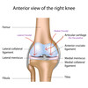
The knee joint capsule is composed of 3 articular surfaces.

What are they?

A
  1. Medial condyle of the femur with the tibia plateau
  2. Lateral condyle of the femur with the tibia plateau
  3. Articulation with the patella (a sesamoid bone)
3
Q

The knee joint is a modified hinge joint.

What are the implications of this on its range of movement?

A
  • It still has the flexor and extensor movements
  • But it also has rotation movements that can occur during flexion.
4
Q

The knee joint has a relatively large range of movement in the knee joint.

What is the most stable position of the knee? Why?

A

The most stable position of the knee joint is during extension (where it is in a close packed position) and has the support from ligaments.

5
Q

There is an incongruency in the knee joint.

Describe This.

A

There is a lack of fit of the articular surfaces. The femoral chondyles are round while the tibial articular facet is flat (plateau).

6
Q

Compare the length medial condyl to the lateral condyl in the anterior-posterior (AP) plane for both the tibia and the femur

A

The tibia and femur are longer in the medial condyles compared to lateral

The medial condyle is longer by about a centimetre

(medial condyle is also longer in the vertical plane)

7
Q

The medial femoral condyle projects further distally than the lateral. What is the significance of this?

A

The medial condyl bears most of the weight (75%) – concentrating it on the medial aspect of the knee.

This causes the femur to have an normal inward angle

8
Q

There are 2 major pathologies of bone that occur as a result of an abnormal femorotibial articulation causing the centre of gravity to be shifted across the knee.

What are the two types? Describe them

A

Genu valgum

The weight of the limb is more lateral than the knee joint causing the knee to take a lateral displacement of the bone (compared to the long axis of the femur) – excess inward angle

Genu varum – “Bow leg”

Where the line of gravity passes medial to the knee joint resulting in knees that angle outwards.

9
Q

Describe the location and shape of the tibial plateau

A

The tibial plateau is the proximal most part of the tibial bone. It is where the femur ends and articulates with it.

The plateau is made up of two flat (with only a slight concavity) articular surface – the medial is larger than the lateral in the AP direction

10
Q

A very important structure lies between the two major parts of the tibial plateau.

What is it and why is it so important?

A

Between the two tibial condyles is an intercondylar eminence or notch which contains attachments for very key structures:

  • The anterior cruciate ligament (ACL) and posterior cruciate ligament (PCL) attachments
  • Attachments for the horns of the cartilaginous menisci
11
Q

Describe the attachment of the medial horns (of the medial menisci) vs. the lateral horns (of the lateral menisci)

A

The medial horns are quite far apart from one another while the lateral are close together (concentrated in the centre of the plateau)

12
Q

The articular surfaces provide little support to the knee joint. It thus requires reinforcement from other structures.

What structures provide support to the knee joint? [4]

A
  1. Powerful muscles (and their tendons)
  2. Pair of cruiciate ligaments
  3. Pair of collateral ligaments
  4. Two Cartilaginous menisci
13
Q

What are the main positions in which the knee is injured?

A

Flexion and rotation

14
Q

Describe the knee joint capsule

A

The capsule is attached around the articular margins of the knee joint encircling the tibiofemoral articulations and incorporates the patellofemoral joint.

It is lined by synovial membrane and contains synovial fluid.

15
Q

Describe the synovial membrane of the knee joint

A

The synovial membrane lines the bones in the knee joint but it doesn’t cover any articulating surface.

Other extrasynovial structures inclue the menisci.

16
Q

The menisci are intracapsulr but extrasynovial. Why aren’t they lined by synovium? - the synovium is located at the margins only.

A

The menisci are articulating directly with the femur condyles. Thus they recieve lots of sheering stress (protect the bones)

It they were lined by synovial membrane, it risks tearing, damage and bleeding into the joint (haemarthritis). Thus is because synovium has a dense net of small blood vessels that provide nutrients.

17
Q

Describe how the cruicate ligaments (ACL and PCL) are lined by synovial membrane

A

The ACL and PCL are lined on their anterior and lateral aspects by synovial membrane (more of ACL is lined than is PCL)

During development, the cruciate ligaments start posteriorly and migrate forwards into the joint . As they migrate forwards they push the synovial membrane in front of them.

18
Q

The joint capsule is reinforced around all sides of the joint by muscles and/or the tendinous insertions of the muscles.

Describe the anterior support of muscles

A

The quadricepts muscle terminates in the quadricepts tone that lines the top of the patella ending in the patellar tendon with retinacular fibres running along side it (anteriolateral aspects)

19
Q

Describe the lateral support of the knee joint by muscles and muscular structures

A
  • Popliteus muscle (that originates on the lateral side of the knee wrapping around the posterior part)
  • Biceps femoris attachment
  • Iliotibial tract
  • Also the lateral retinaculum of the quadricepts tendon
20
Q

What is the pes anserinus?

A

The medial muscle support of the knee

It refers to the conjoined tendons of 3 muscles that insert onto the anteromedial surface of the proximal extremity of the tibia “goose foot”

  1. Sartorius muscle
  2. Gracilis muscle
  3. Semitendinosus muscle
21
Q

Describe the posterior reinforcement support of the knee joint

A
  • Oblique popliteal ligament
  • Ligament from the smimembranosus muscle
22
Q

Describe the Pes Anserinus (Goose’s Foot) structure of the knee

A

Describes muscle insertions into the medial part of the knee.

In order from top to bottom (from front to back): “Say Grace Before Tea”

Sartorius

Gracilis

Bursa

SemiTendinousus

23
Q

Describe the attachments of the two cruciate ligaments

A

These are intracapsular, extrasynovial structures that attach from the tibia to the femur.

They cross within the joint

  • Anterior: starts from the anterior part of the tibia and pass laterally to attach to the interior part of the lateral condyle of the femur
  • Posterior: comes from the posterior aspect of the tibia and passes medially to the medial condyl of the femur
24
Q

Describe the function of the anterior cruciate ligament

A

It prevents forward displacement of the tibia on the femur.

It is a primary stabiliser in the anterior-posterior direction
25
Q

Describe the structure of the ACL

A

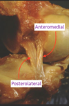
It is made up of 2 bands of fibres

  • Anteriomedial
  • Posteriolateral
26
Q

Describe the ACL position in ectension compared to flexion

A

In extension and flexion the ACL is completely taut

It is loosed packed in semi flexion (midway between flexion and extension). This position is the likely position where injury occurs

27
Q

What is meant by the “scewing home” mechanism of the knee? (locking”

A

In the last 15-20 degrees of extension, the femur rotates medially relative to the tibia as the knee locks.

This is permitted by tightening of the ACL causing weight bearing on the tibia and limits backwatd movement of the lateral condyl.

(during flexion there iis lateral rotation back of the femur)

28
Q

Describe an ACL injury

ACL injuries often lead to haemarthiosis

A

Occurs when rotation of the two bones (femur and tibia) occurs in opposite directions.

ie. instead of the screw home mechanism of medial rotation in extension, there is lateral extension instead.

When an external force causes a lateral rotation of the femur causing the ACL to tear

Most often occurs when there is a planted foot and a deceleration of movement

29
Q

Describe the Posterior Lateral Ligament in terms of structure and function

A
  • It is larger (2x the thickness) and stronger then the ACL.
  • It is reinforced by meniscofemoral ligaments (commonly 2 of them)

Function:

To limit posterior displacement of the tibia in relation to the femur (especially when there is a lot of weight on the knee preventing the tendancy for the femur to fall off the front of the tibia)

30
Q

In what position is the PCL likely to be injured?

A

It is prone to injury in the flexed position

  • Falling onto the flexed knee (causing anterior blow forcing the tibia to be pushed too far backwards)
  • Bumper bar impact
  • Hyperextension of the knee
31
Q

What are the anterior drawer test and the posterior draw test?

A

With the knee in flexion at 90 degrees

Displacing the tibia anteriorly to test the ACL and posteriorly to test the PCL.

32
Q

Unlocking of the joint from extension requires an active component.

Describe what is meant by this

A

Unlocking the joint from extension into flexion requires an active contraction of the popliteus muscle

The popliteus attaches from the lateral part of the femur to the medial part of the tibia.

Contraction of the muscle rotates the femur laterally on a fixed tibia

This is why further rotation is only enabled by the knee joint in the flexed position

33
Q

Describe the medial collateral ligament in terms of structure and function

A

Structure

  • It is a long, flat superficial muscle.
  • It has a depp part the blends with the capsule attaching to the medial meniscus.

Function

  • Resists valgus forces (abduction or lateral) and resists lateral rotation of the tibia
34
Q

Describe injury to the medial meniscus

A

Occurs when there is a large valgus force (from the lateral side) on the knee.

it is often injured together witht the ACL

35
Q

Describe the lateral collateral ligament in terms of structure and function

A

Structure

  • It has a round cord shape
  • It is saparated from the lateral meniscus by the popliteus tendon

Function

  • Resists varus forces (adduction or forces coming from medially)
  • It is not injured as commonly as the MCL
36
Q

What are the functions of the fibro-cartilaginous menisci in the knee? [4]

A

The are structures in the synovial cavity sitting on top of the tibia that move with the femur in rotation.

  • They shock absorb forces
  • They are important to weight distribution around the knee
  • They separate the joint into 2 cavities (one above the menisci and one below it)
  • The are gap fillers: They are wedged shape and thus increase the area of contact (by about a third)
37
Q

The menisci have considerable range of movement, particularly the lateral meniscus.

In flexion, the lateral meniscus slides to the back of the tibia. What is the implication of this in terms of injury?

A

In the flexed position when there is lots of weight on the joint, the lateral meniscus slides back to overlap the tibial plataeu posteriorly.

Here it is suscptible to entrapment injury

38
Q

The lateral meniscus cover more of the articular surface and has an attachment to the PCL (meniscofemoral ligaments) and not to the LCL. What does this mean?

A

It means, in comparison to the medial meniscus, the lateral meniscus bears more force

39
Q

The medial meniscus is more commonly injured than the lateral is.

Why?

A
  • The medial meniscus is longer
  • The horns are further apart
  • It is less mobile than the lateral (it is attached to the MCL)
40
Q

What are the types of meniscus injury?

A

It may be nipped or torn or crushed.

  • Longitudinal tears
  • Radial tears
  • Horizontal tears

(The fragements glip up into the joint space leading to a locking of the knee in flexion)

41
Q

What part of the menisci are most capable of surviving/repair after injury?

A

The outer third of the menisci because it is closer to the capsule of the joint.

This capsule delivers the blod supply that penetrates into this outer third part of the meniscus

42
Q

Describe the bursae of the knee joint

What are they?

A

There are several bursae in the knee: communicating and non-communicating

  1. Suprapatellar bursa
  2. Semimembranous bursa
  3. Prepatellar bursa
  4. Superficial infrapatellar bursa
  5. Deep infrapatellar bursa

They protect the ligaments and the tendons against damage from bone

43
Q

Which of the two bursae of the knee communicate with the joint? What is the significance of this communication?

A
  1. Suprapaterallar communicating anteriorly
  2. Semimembranosus/popliteus bursa communicating posteriorly

They are important as fluid, blood and pus can project from thecaity into the bursa (communicating) leading to infection, effusion and haemarthosis.

44
Q

What is the purpose of the infrapatellar fat pad and plicae?

A
  • They fill out irregularities of the joint by filling out spaces
  • It lies extrasynovially with its margins pushing out synovial fluid.

The plicae are folds of synovial membrane at the corners

45
Q

Describe the patello-femoral joint

A

It has articular surfaces between the patella sesamoid bone and the patellar surface of the femur.

  • The patella is a triangular sesamoid bone that is slightly irregular in shape. The lateral facet of the patella is larger than the medial
  • The articular surfaces on the femur: The lateral articular surface is raised while the medial is slightly lower.
46
Q

What is a barpartite patella?

A

It is a common anomaly seen in individuals where it looks like a fracture in scans.

It is actually a separate ossification of the patella bone separated by a growth plate. It is not a fracture.

47
Q

Describe the movement of the patella during flexion and extension of the knee

A

The patella slides up and down the femoral groove

48
Q

The normal alignment of the patella in the femoral groove is maintained by the major structures. What are these?

A
  1. Strong Vastus medialis muscle (active mechanism of stabilisation) - a component of the quadricepts
  2. Medial patellar retinaculum (semi-passive) - the retinacular fibres from the quadricepts.
  3. Raised lip on the lateral femoral condyl (passive) this depth in the groove stops the patella from displacing in the lateral direction.
49
Q

What is the significance of the Q angle (quadricepts angle)?

A

The Q angle is the angle between the line of force of the quadriceps to the mid-point of the Patella.

There is a natural tendance for the patella to be dragged in the lateral direction by the quadricepts due to the width of the pelvis as the femur is pulled laterally to the knee. This predisposes the patella to subluxation/dislocation