Introduction to opthalmology Flashcards
- the globe. eye has three coats/layers, three compartments and contains three fluids
- what are the three coats(3)
-three coats
>outter fibrous layer (cornea, scelera, lamina cribrosa)
>middle vascular layer aka uvea (iris, ciliary body, choroids)
>inner nervous layer (epithelium of retina, retinal photreceptors, retinal neurons)

- what are the three compartments(3)
- clinically how can the eye be devided (2)
-three compartments
>ant chamber = space between cornea and iris diaphragm
>post chamber = triangular space between iris anteriorly, lens posteriorly and ciliary body
>vitreous chamber = space behind lens
-clinically -
>ant segment = all structures ant to and inc lens
>post segement = all structures post to lens
-what are the three fluids of the eye
- aqueous humour = a watery clear solution of water and electrolytes. normally low protein
- vitreous humour = transparent gel consisdting of network of collagen fibres with interspaces filled with water and molcs. fills space between post lens, ciliary body and retina
- blood - most located within choroid. supplies eye and also conbtributes to pressure.
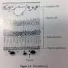
- outer layer. the anterior 1/6th of the outer layer is the cornea, it is transparent. the posterior 5/6th of outer layer is formed by scelera and lamina cribosa, which are white. what and where is conjuctivia and episclera.
- -*middle layer. pigmented and vascular. is called the uvea. ant part of uvea forms bulk of iris body hence inflammation of ant uvea called iritis or ant uveitis. post part is called the choroid. the iris is a thin circular disc perforated centrally by pupil. contration of iris sphincter mm constricts pupil, contraction of dilator pupillae mm dilates pupil. ciliary body aka intermediate uvea. continuous ant with iris and post with choroid
- inner layer. forms retina posteriorly. contains rods + cones, bipolar cells, ganglion cells. the rods and cones sense light. nerves of retina “invaginate” to form the optic cup. the retinal vessels are not show in the image. they sit on the very inside against the vitreous humour nb different to choroid vessels.
the conjuntivia is a mucous membrane on top of outer layer. it starts at the edge of the cornea and covers the anterior portion of the scelera nb not over cornea. it also covers post portion of eyelid. there is a mucocutaneous junction on the lid margin where it becomes skin. there is also a layer of cose connective tissue deep to conjuctivia on top of sclera called episclera
-blood supply of globe derived from 3 sources (3) all derived from (1) which is a brance of (1)
-central retinal aa, ant cilliary arteries, post cilliary arteries. all derived from opthalmic aa which is a brance of int carotid

central retinal aa runs with optic nn
-optic nn / cranial nn 2
-meets post globe slightly nasal to post pole and slightly sup to horizontal meridean. -when seen it is referred to as optic disk. -nerve fibres sweep across whole retina to reach optic disc where they join and “invaginate” to form optic cup. - there are no light sensitive cells within hence the blind spot. - retinal vessels also converge at location of disc
image showing visual pathway of optic nn

- extraocular mms
- there are 6 extraocular mms (6) and their functions and nns
- sup rectus - elevation - cn3
- inf rectus - depression - cn 3
- medial rectus - adduction - cn3
-lat rectus - abduction - cn6
-sup oblique -depression and adduction - cn4
-inf oblique -elevation and adduction - cn3

what to ask in ocular hx
- background (1)
- HPC (5)
- Assx to ask about other that HPC in ocular Hx (15)
- PMH (5)
- DH
- FH
- SH
- background - age is important
- macro and micro timing, nature, exac, reliev
- Assx
>subnormal vision i assume this refers mainly to VA - duration, degree, differences between eyes.
>disturbances in vision - distortion, haloes, floaters, flashing lights, momentrary losses of vision, field defects, diplopia
>pain and discomfort
>discharge - appearance
>change in lacrimation, any swellings
-PMH - diabetes, HTN, COPD, thyroid, autoimmune diseases
-pt may chose to be registered visually impaired. what are two options (2)
- sight impaired (previously “partially sighted”)
- severely sight impaired (previously “blind”).
