Interactive cases: Prof Bain and Prof McDonald Flashcards
How to distinguish between thymoma and ALL?
In thymoma you wouldn’t get high WCC unlike in ALL
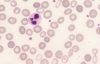
ALL:
- high WCC count
- Blast cells
- possible anaemia/thrombocytopaenia due to bone marrow infiltration
- possible enlagment of organs i.e. Testes, thymus
**NB: T-cell ALL can cause a thymoma**
Immunophenotyping vs cytochemistry
Immunophenotyping: used to distinguish between AML and ALL. uses flow cytometry.
Cytochemistry: looks for specific things in cells using stains. not used much anymore.
What does high WCC in acute leukamiea signify in terms of prognosis?
Bad prognosis that reduces the chances of cure
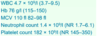
What does a coagulation screen include?
APTT
PT
Bleeding time
*does nOT include platelet count - this is done in FBC*
Coagulation screen in DIC
- APTT:
- initially SHORT - because there’s so much activation of the clotting factors
- then later prolonged as clotting factors get used up
- PT
- prolonged
- Bleeding time
- prolonged
- Fibrinogen
- low
- FDP (D-dimer)
- elevated
What test confirms DIC?
D-dimer
Which leukamiea is associated with DIC?
APML
Which ethnicities tend to have lower neutrophil counts?
Afrocarribbeans and Africans
ethnic neutropenia whereby Africans and Afro-Caribbeans could have lower neutrophil count and this might be NORMAL.
Name a key haemoatological cause of macrocytic non-megaloblastic anaemia?
Myelodysplastic syndrome
Also multiple myeloma and myeloproliferative disorders
What causes megaloblastic anaemia?
Vitamin B12 or folate deficiency or cytotoxic drugs
there are no haematological malignancies that cause megaloblastic anaemia
Anti IF vs anti parietal cell antibodies
anti-IF: more specific for pernicious anaemia
anti-parietal cell: more sensitive
Platelet count and RBC in essential thrombocythaemia vs PCV
PCV:
- erythrocytosis
- thrombocytosis
ET:
- isolated thrombocytosis
PCV treatment
- venesection: lower the Hb
- hydroxycarbamide: lowers platelet count
- aspirin to reduce thrombosis risk
Testicular swelling, 3-5 year old
ALL
Auer rods
AML
Philadelphia chromosome
t (9:22)
BCR-ABL-1
Left shift
CML
*left shift means that you seei mmature myeloid cells in the bloodstream*
Smear cells/smudge cells
CLL
or small cell lymphoma
JAK2+
High haematocrit
flushed appearance
strokes/budd chiari
PCV
Budd chiari: Hepatic veins (veins that drain the liver) are blocked or narrowed by a clot (mass of blood cells).
high platelets
strokes
may have jak2 mutation
ET
Dry tap, teardorp cells (dacrocytes), massive splenomegaly
Myelofibrosis
Painful LNs with alcohol
Reed0sternberg cells
EBV+
Hodgkin’s
t(14,18); centroblasts
Follicular lymphoma
*low grade B cell NHL
t(11,14)
mantle cells
mantle cell lymphoma
*aggressive B cell NHL
t (8,14)
starry sky appearance
EBV+
HIV+
Burkitt’s
CRAB+
Bence jones protein\IgG/A > 30
Multiple myeloma
**rmb IgM - waldernstrom’s
No CRAB
Paraprotein < 30 (but elevated)
MGUS
WBC count normal range
Females: 4.5 to 11.0 × 10⁹/L
Males: 4.5 to 11.0 × 10⁹/L
Hb normal range: males and females
Females: 12.0 to 15.5 grams per deciliter (120-155 grams/litre)
Males: 13.5 to 17.5 grams per deciliter (135-175 grams/ litre)
MCV normal range
Females: 80-96 femtolitres per cell
Males: 80-96 femtolitres per cell
**basc above 100 is marocytosis
Platelet count normal range
Males + females: 150-400 x 109/L
How to distingiush between PCV and chronic hypoxia induced polycythaemia (secondary polycthaemia)?
PCV:
- thrombocytosis + erythrocytosis
- low erythropoietin
- Jak 2 mutation
In secondary polycythaemia you would get:
- isolated erythrocytosis
- high erythropoietin
- no Jak 2 mutation
Test to confirm PCV
FBC:
- High HB + Hct
- high platelets
- high MCV
- WBC normal (or moderately raised)
JAK2 mutation - DIAGNOSTIC- this is the first one to do; usually this is enough
Bone marrow aspirate + trephine biopsy
- Increased cellularity- i..e no fat spaces, all being occupied by mature cells
- moderate reticulin fibrosis
serum EPO - low
Reactive neutrophilia vs CML vs AML
Reactive
- neutrophillia i.e. 15-20)
- band cells (neutrophils without segmented nucleus)
- toxic granuloation
- vacuolation
- no immature cells
CML
- neutrophilia i.e. (50-500 x 109/L)
- basiphilia
- immature myelocytes BUT NO excess myeloblasts (< 5%) in the bone marrow
- eosinophilia
AML
- neutrophilia i.e. > 300)
- myeloblasts (even more immature than myelocytes)

high WCC, RBC count, Hb is upper limit of normal
neutrophilia
basophilia
eosinophilia
CML
**you can have high RBC*
What causes gangrenes?
Abnormlaities of the blood vessels - atheroscleorsis
Abnormalities of the circulating blood-
- polycythaemia:
- cold agglutinin: which causes the RBCs to stick together in the cold
- cryoglobulinaemia:protein that precipitates in the cold which blocks blood vessels
Isolated thrombocytopaniea in an elderly man
Autoimmune thrombocytopaniea purpura
Differentials for bruising in a child
- NAI
- thrombocytopaenia
- autoimmune thrombocytopaenia
- alloimmune thrombocytopaenia
- ALL - coagulation disorders
- haemophilias- most likely
- VWD
What does high RCDW mean?
RDW= red cell distribution width
RDW= red cell distribution width (which is high) – this tells you there is anisocytosis: diff sized cells present
What haematological abnormalities can be seen in patients with rheumatoid arthritis?
- anaemia of chronic disease
- iron deficiency anaemia
- haemorrhage due to aspirin and NSAIDS
- neutropaenia or thrombocytopaniea
- due to drugs
- Felty syndrome
- due to the inflammation
What is felty syndrome?
Triad of
- neutropaenia
- splenomegaly
- raised ESR
Osteomyelitis x-ray changes
- patchy changes in bone
*distinguishes it from septic arthritis
Red herrings in osteomyelitis
- fever may not be present
- WCC may not be raised
CRP is usually always raised
—> can still be osteomyelitis
DDx of MAHA
- HUS
- TTP
- DIC
TTP pentad
+ defect
+ treatment
Pentad:
- MAHA
- thrombocytopaenia
- renal failure
- fever
- neurological symptoms eg altered consciousness
Defect:
deficiency of ADAMSTS13 enzyme: von Willebrand factor cleaving protease
Causes multimers of VWF to form and cause platelet clots
How to treat TTP?
DO NOT GIVE PLATELET TRANSFUSION BECAUSE THIS MAKES THE CONDITION WORSE!!
tx: plasma exchange
Blood film features of hyposplenism
Howell–Jolly bodies - nuclear remnants
Target cells
Occasional nucleated red blood cell
Lymphocytosis
Macrocytosis
Acanthocytes
Normal myeloblast and lymphoblast counts in bone marrow vs myelodysplasia + AML and ALL
myeloid cells
<5%: normal
5-20%: myelodysplasia
>20%: AML
lymphoid cells
<5%: normal
>20%: ALL
Key marker of lymphoblasts
TDT positive
What may myeloblasts have in their cytoplasm?
Aeur rods
Which cell markers are expressed on mature B cells?
CD19: receptor for CAR T cells
CD20: receptor for rituximab
Cell surface markers of peripheral blood lymphoid T cells
CD3
CD4- helper T cells
CD8- cytotoxic T cells
CD5
How to distinguish between immature and mature lymphoid cells?
Immature: TDT positive
Mature: TDT negative
**true for T and B lymphocytes
How to distinguish between mature and immature B cells?
Mature B cells and plasma cells express surface Ig
Which receptor on B cells to CART cells bind to?
CD19
If you see myeloblasts in the blood film what does that suggest?
1) AML
2) leukoerythroblastic anaemia
What single test distinguishes autoimmune from inherited haemolytic anaemia?
DAT test
Positive: autoimmune haemolytic anaemia
If you see spherocytes what two diagnoses are suggested?
1) autoimmune haemolytic anaemia
2) hereditary spherocytosis
What type of anaemia is haemolytic anaemia?
Can be macrocytic- because of reticulocytes are larger than normal rbc
Give causes of non-immune acquired haemolytic anaemia
- malaria!!!
- MAHA- HUS, TTP, DIC
- paroxysmal nocturnal haemoglobinuria
How can SLE affect the platelets?
Can cause idiopathic thrombocytopaenic purpura
Causes of autoimmune haemolytic anaemia: key
Warm: SLE, drugs, CLL (evan’s syndrome)
Cold: mycoplasma, other infections
ACD vs iron deficiency anaemia
The key is transferrin!!!
Iron deficiency anaemia: high transferrin
ACD: low/normal transferrin

What protein mediates anaemia of chronic disease
hepcidin
*this is raised in ACD as it’s an inflammatory protein
it sequesters iron in macrophages so it’s low in the blood
Can you get IDA with normal ferritin?
Yes!! If you have co-existent ACD
Isolated single lineage cytopaenia: causes

If you have very high MCV (higher than 120)- what are the top differentials?
B12 or folate deficiency
What does pancytopaenia with tear drop cells, nucleated RBC and immature granulocytes in blood film suggest?
Leukoerythroblastic anaemia- i.e. bone marrow infiltration
- haematological cancer: leukaemia, lymphoma, myeloma
- metastatic: prostate, breast, lung
Chronic vs acute leukemia: white cell count
Chronic always has high white cell count (Acc to Prof McDonald)
Classification of pancytopaenia

- normal Hb
- VERY HIGH WCC
- normal lymphocytes
- raised neutrophils
Confirmatory test?
CML
BCR-ABL1 gene fusion i.e philadelphia chromosome
What type of protein is ABL?
Tyrosine kinase
Treatment for CML
Tyrosine kinase inhibitor- imatinib
Do you see myeloblasts or myelocytes in CML?
Myelocytes - in chornic phase
if you see >20% blasts –> blast crisis
Which cell surface marker is aberrently expressed in CLL?
CD5 and CD23 POSITIVE markers
- NOTE: CD5 and CD23 should never be seen in a normal B cell
Main prognostic factor for CLL
TP53 mutation status
Link between CLL and anaemia
CLL can cause haemolytic anaemia
as it is a disease of the immune system
Can you exclude myeloma if you have normal plasma imuunoglobulin?
No- can have light chain only myeloma
Need to check serum free light chains or urine bence jones protein
What is the cause of renal failure in myeloma?
Cast nephropathy
- deposition of light chains in the basement membrane precipitating renal failure
Name a JAK1 inhibitor
Ruxolitinib
What causes raised ESR?
raised acute phase proteins such as fibrinogen
**promotes rouleaux formation
Causes of isolated raised ESR and normal CRP
SLE
Multiple myeloma
lymphoma
pregnancy
anaemia
translocation : t(11,14) associated to which malignancny
mantle cell lymphoma- agressive high grade NHL
- A 5y/o boy of Indian ethnic origin presented with lymphadenopathy and a mediastinal mass on chest radiology
- WBC: 180 x 109/L
- Hb: 93g/L
- Platelet count: 43 x 109/L
what is the diagnosis

ALL
- there are blast cells present on blood film
- there was a VERY HIGH WCC
- The low Hb and platelet count are the result of bone marrow infiltration
- mediastinal mass is the thymus, which is infiltrated by T lymphoblasts
The VERY HIGH WCC in a child means a diagnosis of leukaemia is almost CERTAIN
48y/o male- railway engineer
- 2 week history of bleeding gums
- Attended dentist- severe bleeding
- 1 episode of haematuria
- Minor bruising
- Attended A&E
O/E
- Left subconjunctival haemorrhage
- Small bruises over the abdomen
- No enlarged lymph nodes
- No hepatosplenomegaly
QUESTION 3: What test is most likely to reveal the cause of the problem?
- Blood count, film and coagulation screen
would also do LFTs and creatinine as this sounds like a very ill patient
How could you explain a SHORT APTT and a low fibrinogen?
- DIC
- The APTT is short is because there are activated coagulation factors in the circulation which is making it short
- will also have severe thrombocytopaenia and low fibrinogen
what does this blood film show

granules AND Auer rods
would suspect these cells are MYELOID
- Granules strongly suggest which lineage
myeloid
What diagnosis do you suspect on a background of abnormal clotting

hyper-granular (very granular) promyelocytes (blast looking)
Acute promyelocytic leukaemia
Which tests would be the most useful to confirm the APML

- Cytogenetic analysis/ FISH/ molecular genetic analysis
- Do NOT need cytochemistry as you can see the brightly stained granules.
- Immunophenotyping can help but it is less specific than cytogenetic analysis to show the translocation or FISH to show the 15;17 fusion gene
Fish: FISH showed PML-RARA fusion
what is treatment for APML + prognosis
- Platelets
- Chemotherapy
- All-trans-retinoic acid (ATRA)
Complete remission can be achieved
- 68 y/o woman, retired secretary
- Gradual onset of fatigue, lethargy and exertional dyspnoea
- Non-smoker, not much alcohol, good diet
O/E:
- Pallor (conjunctival and nail bed)
- Mild ankle oedema
which one test should you do next :
- Blood film
- Bone marrow aspirate
- Liver function tests
- Thyroid function tests
- Serum Vitamin B12 and red cell folate

Blood film
In reality, with a macrocytosis you would also do Vitamin B12 and Cell folate and LFTs
what does this show

- There are macrocytes but NO oval macrocytes nor hypersegmentation of neutrophils
- There are hypochromic microcytes
- There is also an elliptocyte
- This is a dimorphic film
- This is NOT associated with B12/ folate deficiency, hypothyroidism or liver disease
what does this show of bone marrow aspirate

- shows increased blast cells (12%)- the normal is < 5%
- ring sideroblasts (can see the ring of iron-containing granules around the nucleus)
DIAGNOSIS: myelodysplastic syndrome with excess blasts
- 72y/o Indian woman
- Vegetarian, teetotal, non-smoker
- SOBOE
- Fatigue
- Painful gums and tongue
- Unable to eat spicy food
O/E: pallor only
FBC:
- low Hb
- normal platelets
- high MCV
- normal WCC
what does this show + What is the most important test

hyper-segmented neutrophils
macrocytes and oval macrocytes too
Vitamin B12 and folate assays
what is the next investigation to do if there is unexplained macrocytes (normal B12 + folate) and elevated LDH
Bone marrow aspirate
if anaemia was caused by vitamin B12 and folate what would you see in the blood film
oval macrocytes
macrocytosis
hypersegmented nuclei

what is the diagnosis of this blood film

- Giant metamyelocytes are present
- Megaloblasts are also present
- Hyper-segmented neutrophils can also be seen
This is CLASSICAL for a vitamin B12 and folate deficiency
+ What test would you do next to try and confirm the diagnosis?

- Polycythaemia vera
- With polycythaemia vera, you also often have a:
- High platelet count
- High WCC
Note: Essential Thrombocythemia, there is an isolated high platelet count
- Molecular analysis for JAK2 mutation
what does this show

- The RBC, Hb and Hct are HIGH
- The patient appears to be polycythaemic
This can be true or pseudopolycythaemia- canno tell from these results
identify which is normal/abnormal + most likely diagnosis given this is seen on a background of low Hb and MCV

- The left film= normal, right film= abnormal
hypochromia (increased pallor in the centre of the RBCs)- confirms anaemia
some poikilocytes and anisocytosis
- Iron deficiency anaemia
what is the likley diagnosis

- Osteomyelitis
- The abnormality in the bone (patchy changes) points you to osteomyelitis more so than septic arthritis
Note can get osteomyleitis in the absecne of fever, raised WCC. but CRP would almost always be high
- 21-year-old woman presented with abdominal pain, bruising and altered level of consciousness
- She had a low-grade fever
- Her platelet count was 15 x 109/L
- Her bilirubin was increased and LDH was gently increased
Her creatinine was marginally increased
QUESTION 8: What is the most likely diagnosis?

- There are RBC fragments - indicative of MAHA
- Microspherocytes are present
- Thrombotic thrombocytopaenic purpura
most common complication of infectious mononucleiosis
- splenomegaly occurs in 50% of patients with infectious mononucleosis
- Splenic rupture occurs in 0.1-0.5% of patients
risks of hyposplenism
- Overwhelming bacterial sepsis (particularly pneumococcal or H. influenzae
- Fatal malaria
- Fatal Capnocytophaga canimorsus infection (typically occurs from dog bites)
- Babesiosis (parasitic infection
follow up for hyposplenism
- Vaccinate for:
- Pneumococcus
- Meningococcus
- Haemophilus influenzae
- Vaccinate against influenza
- Prescribe life-long penicillin
- Advise the patient on:
- Dog bites
- Travel to malaria zones
- Prompt treatment of infection
- Issue a splenectomy card and information sheet